
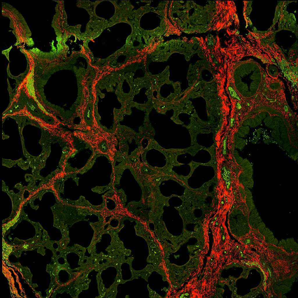
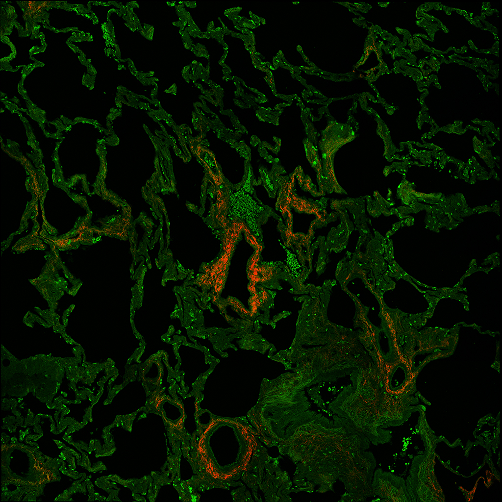
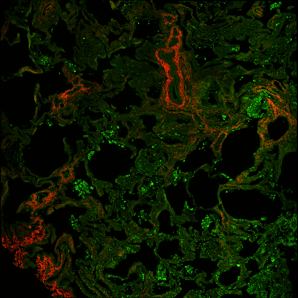
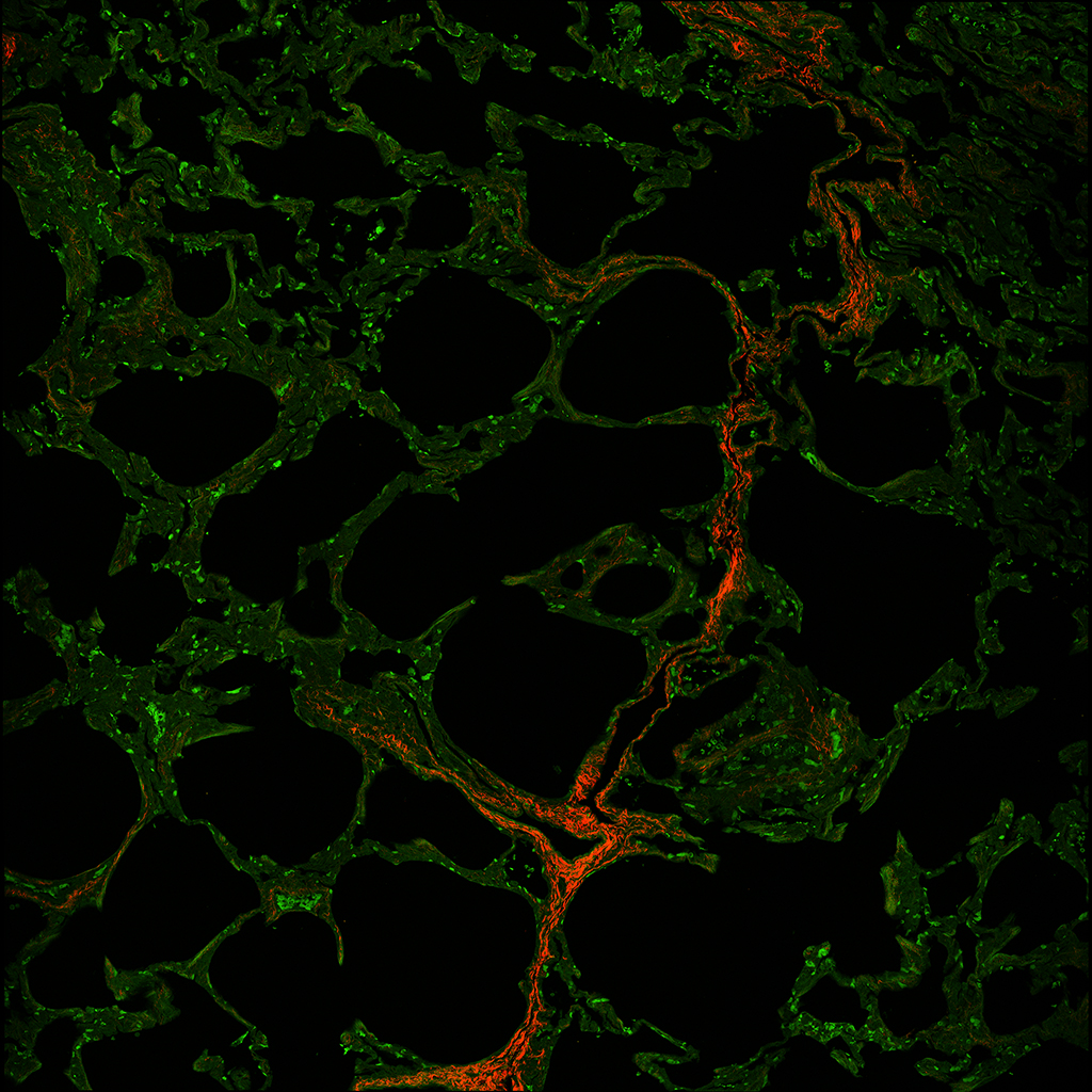
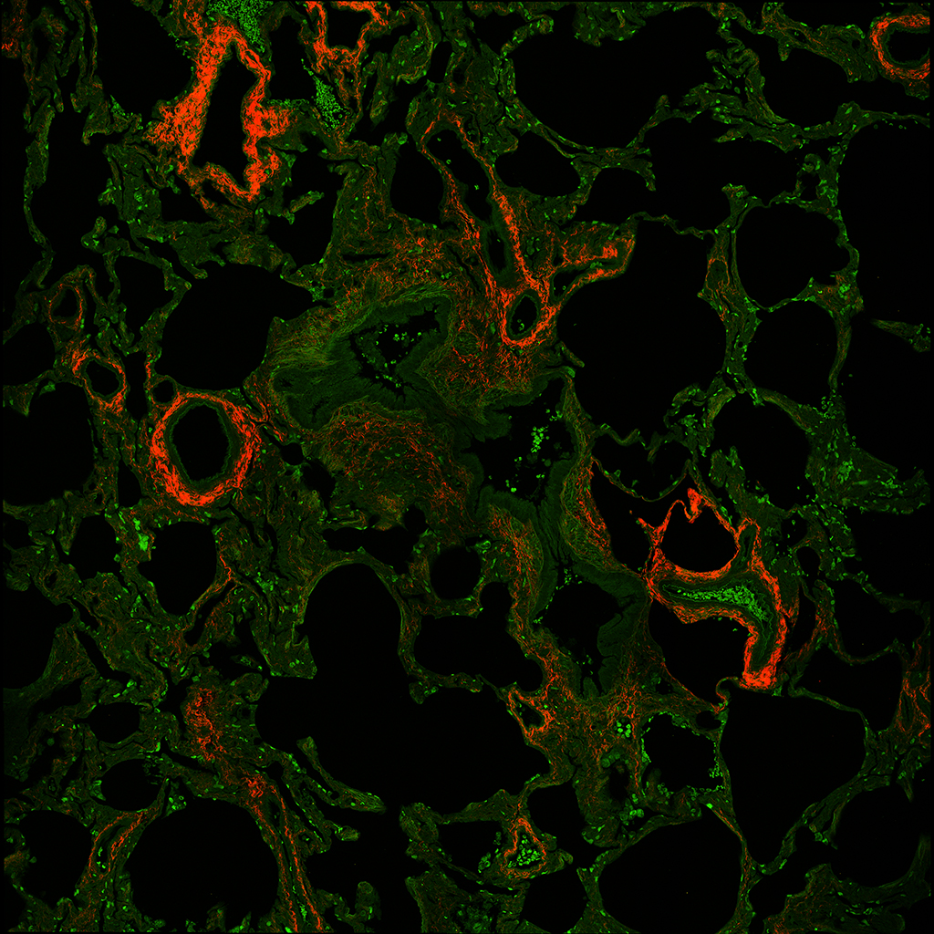
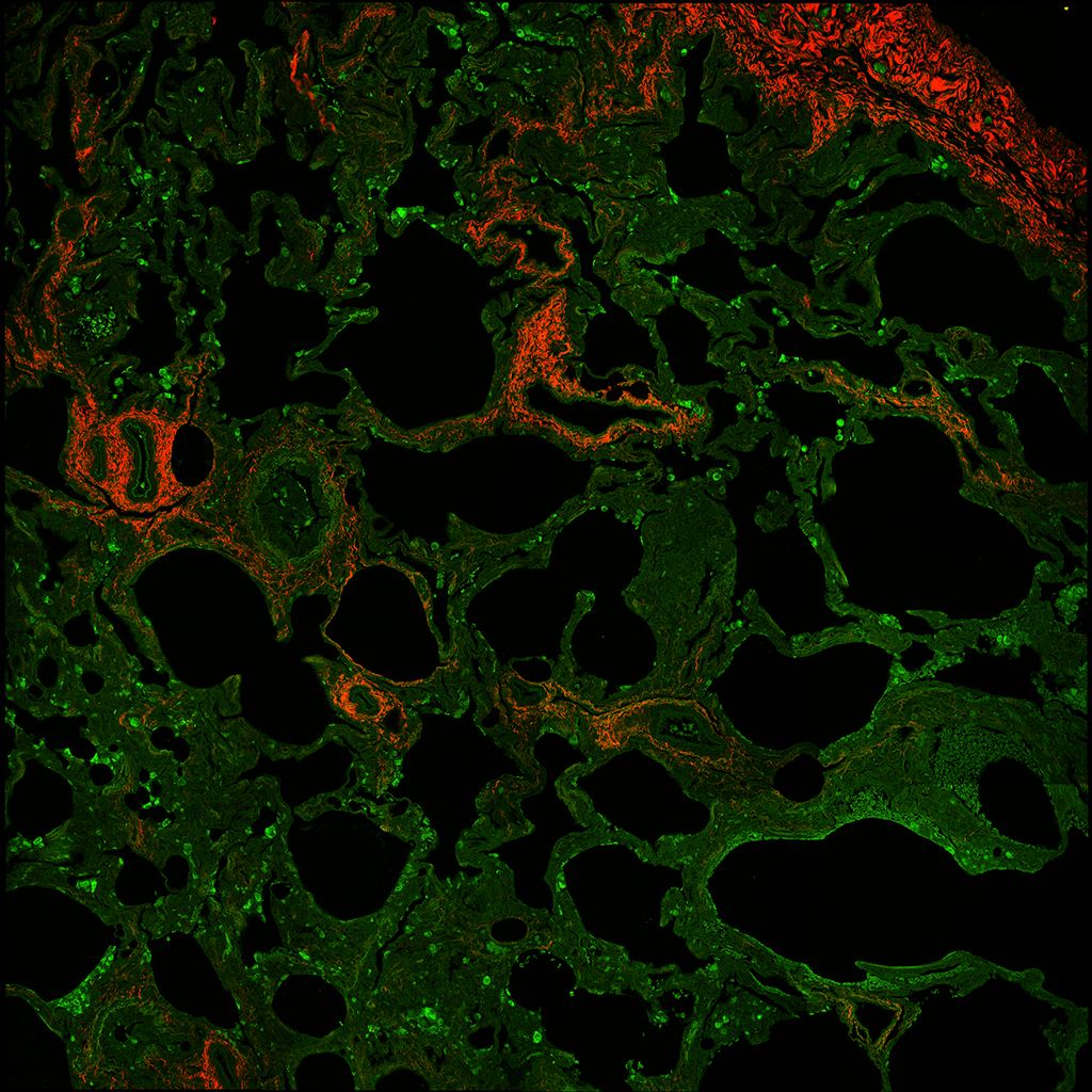
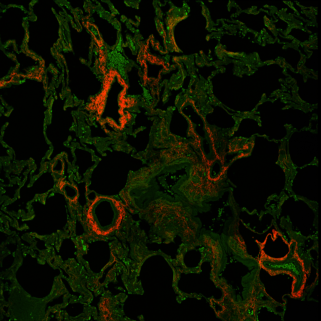
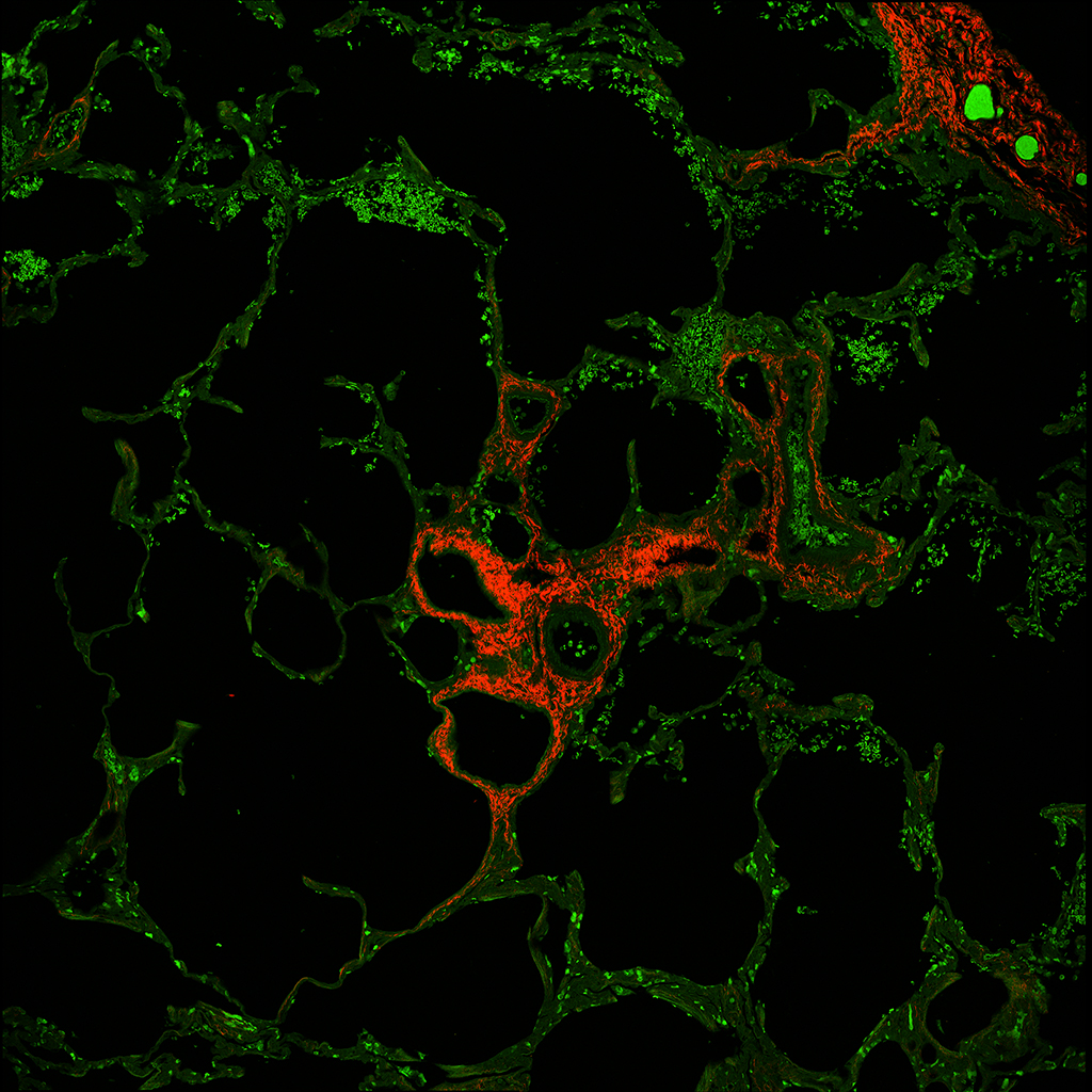
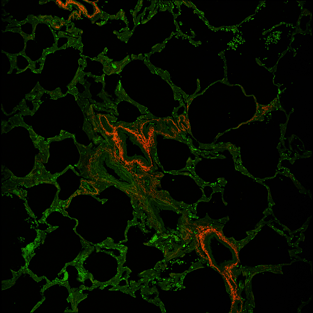
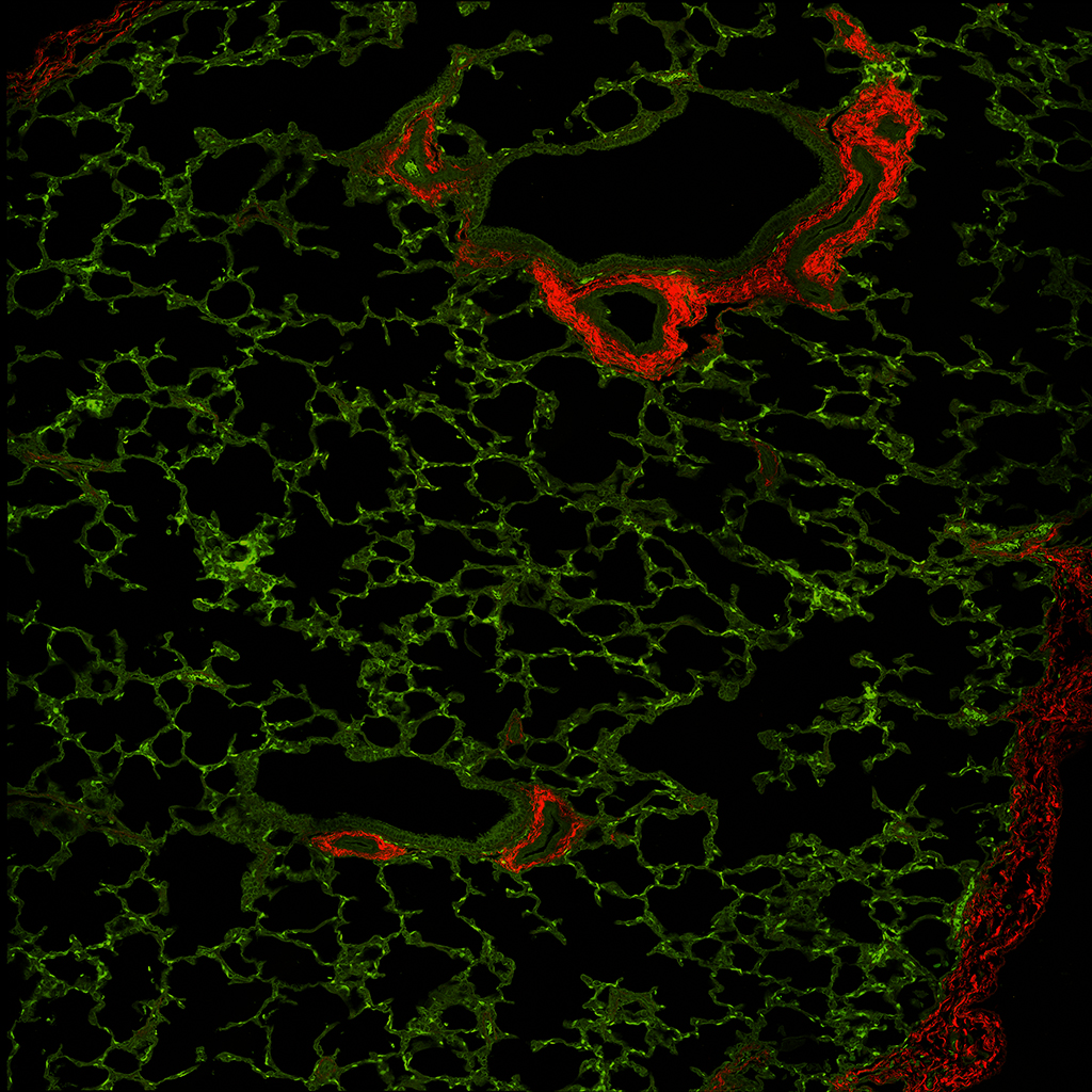
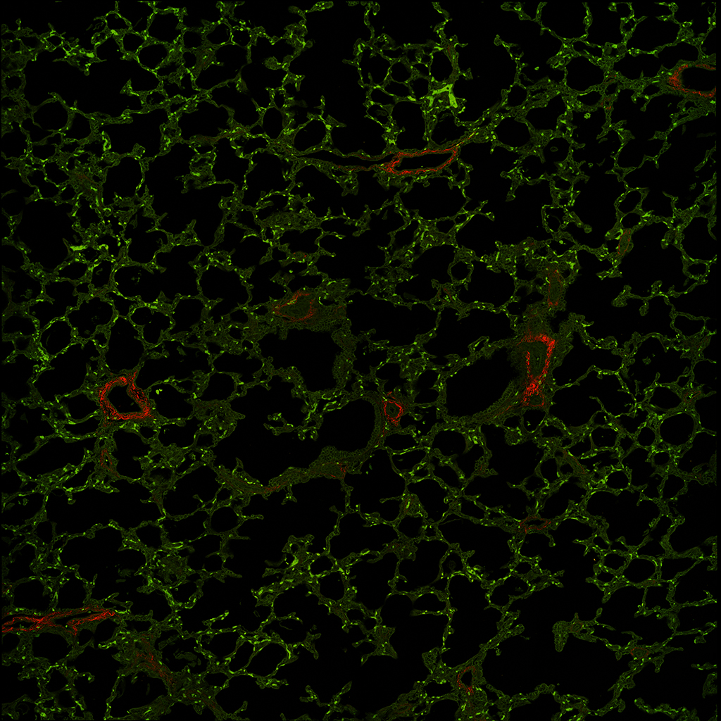
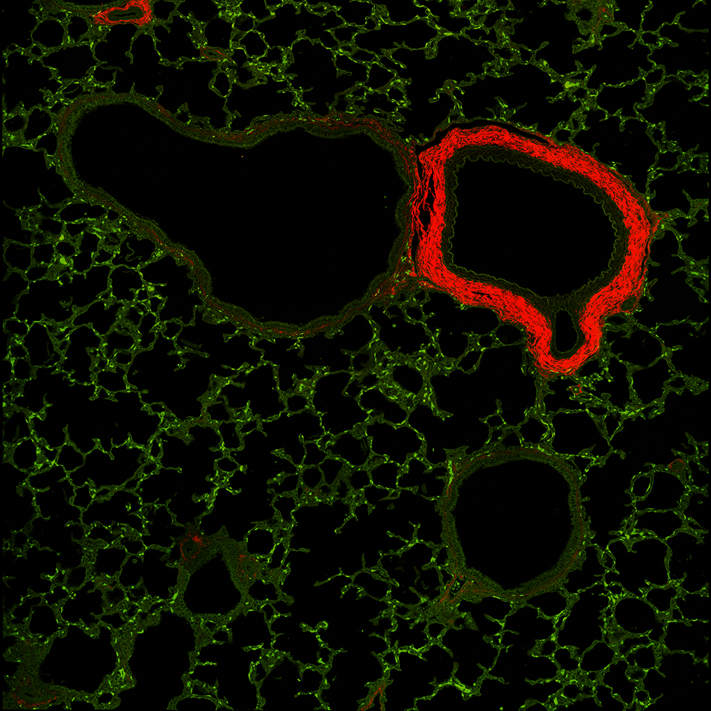
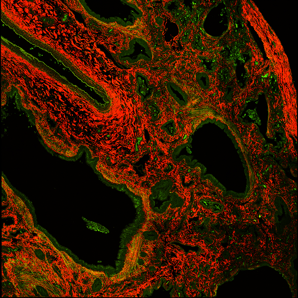
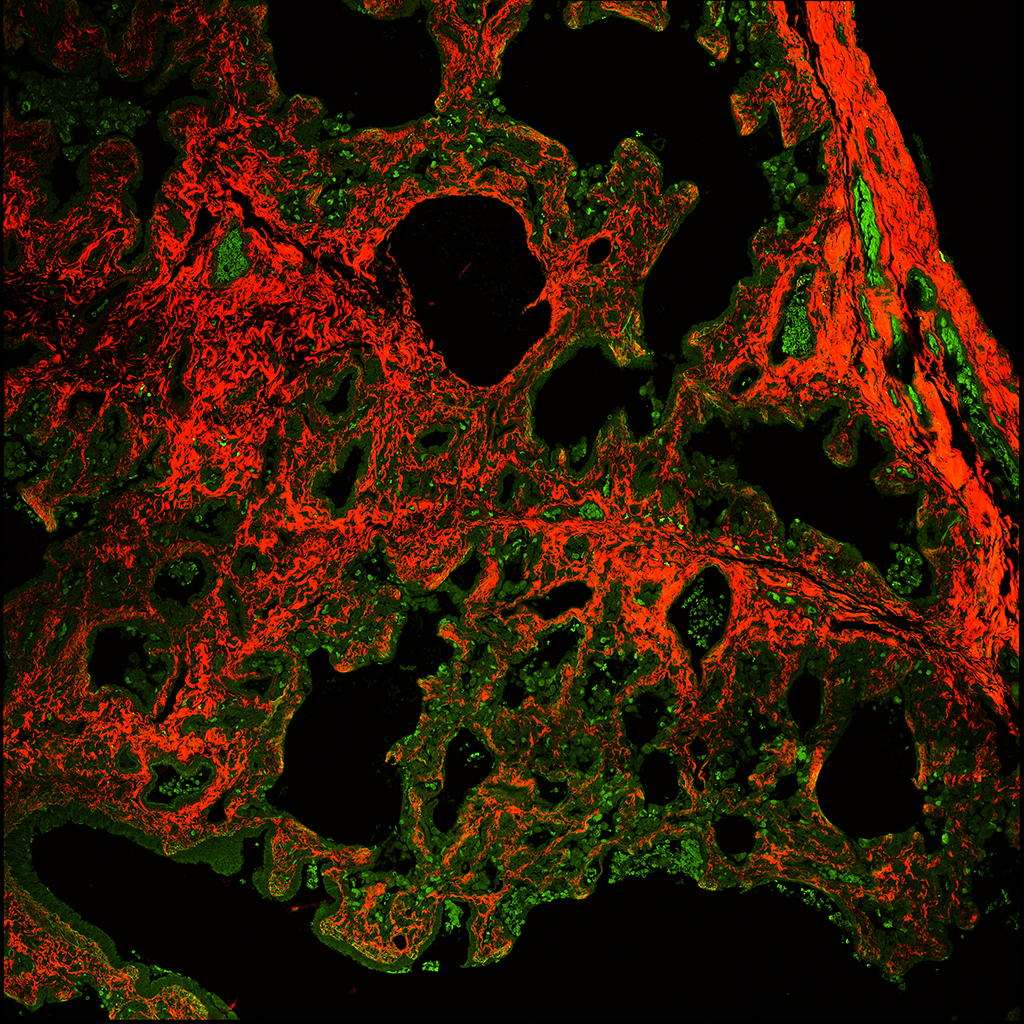
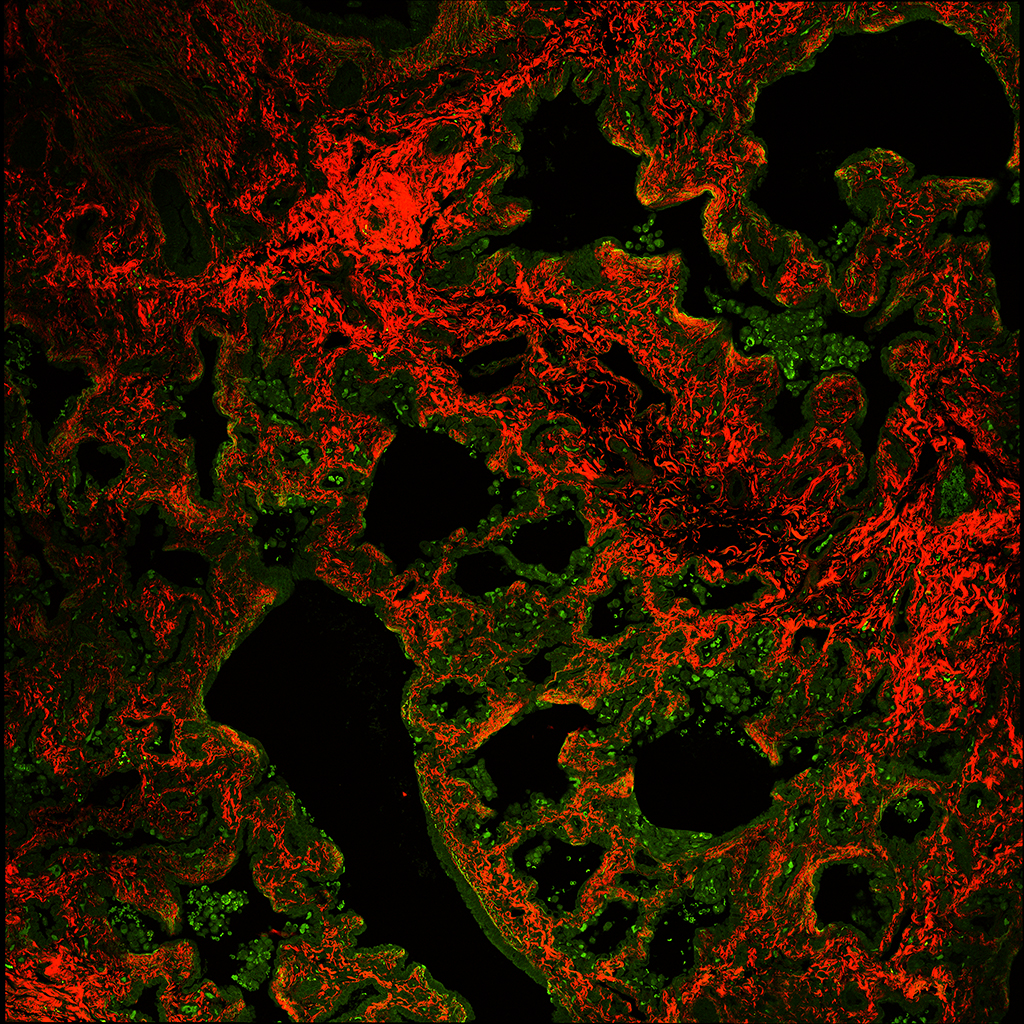
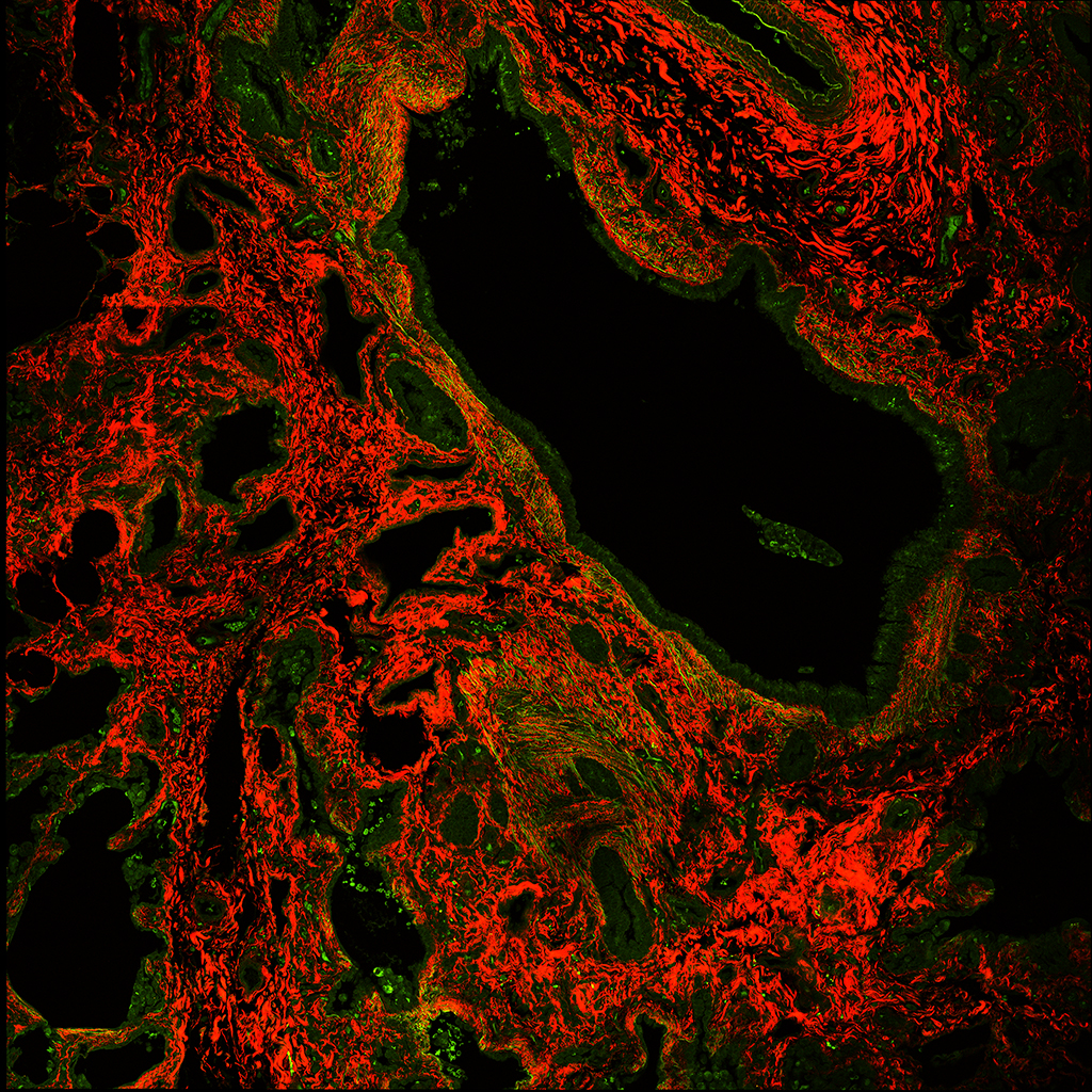
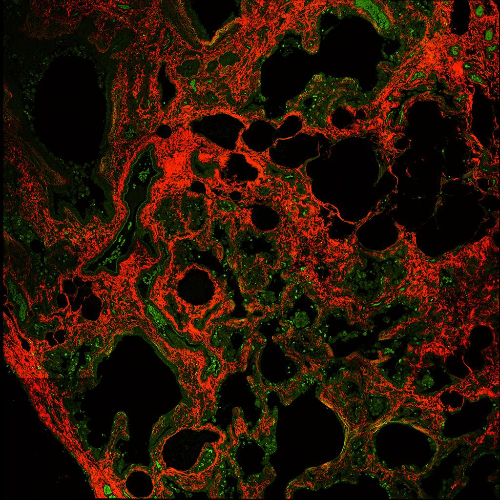
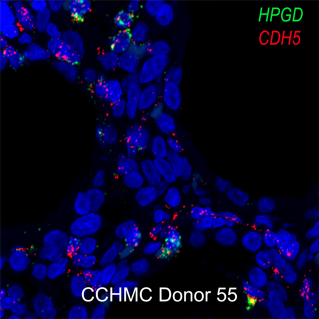
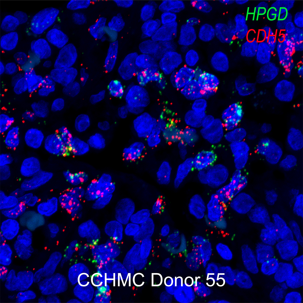
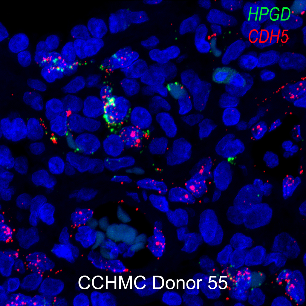
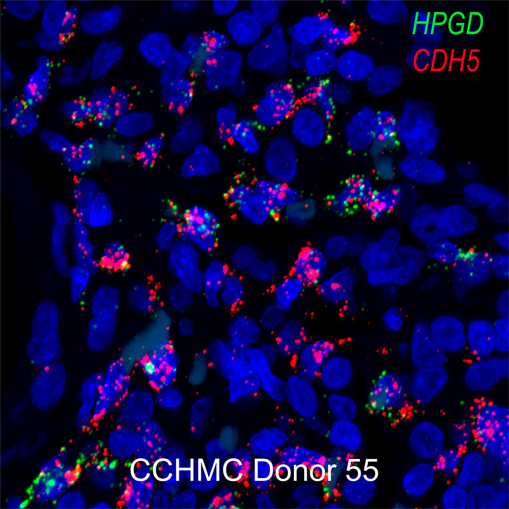
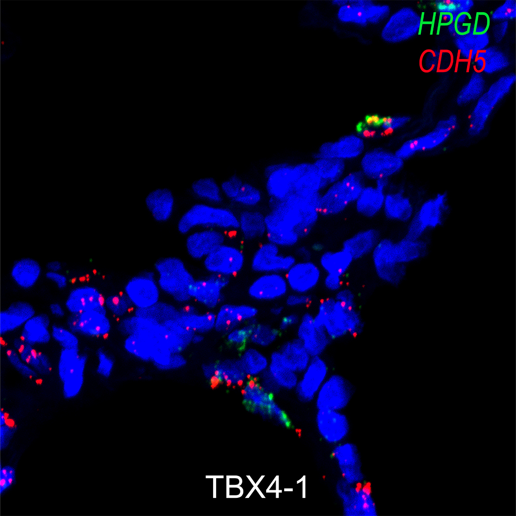
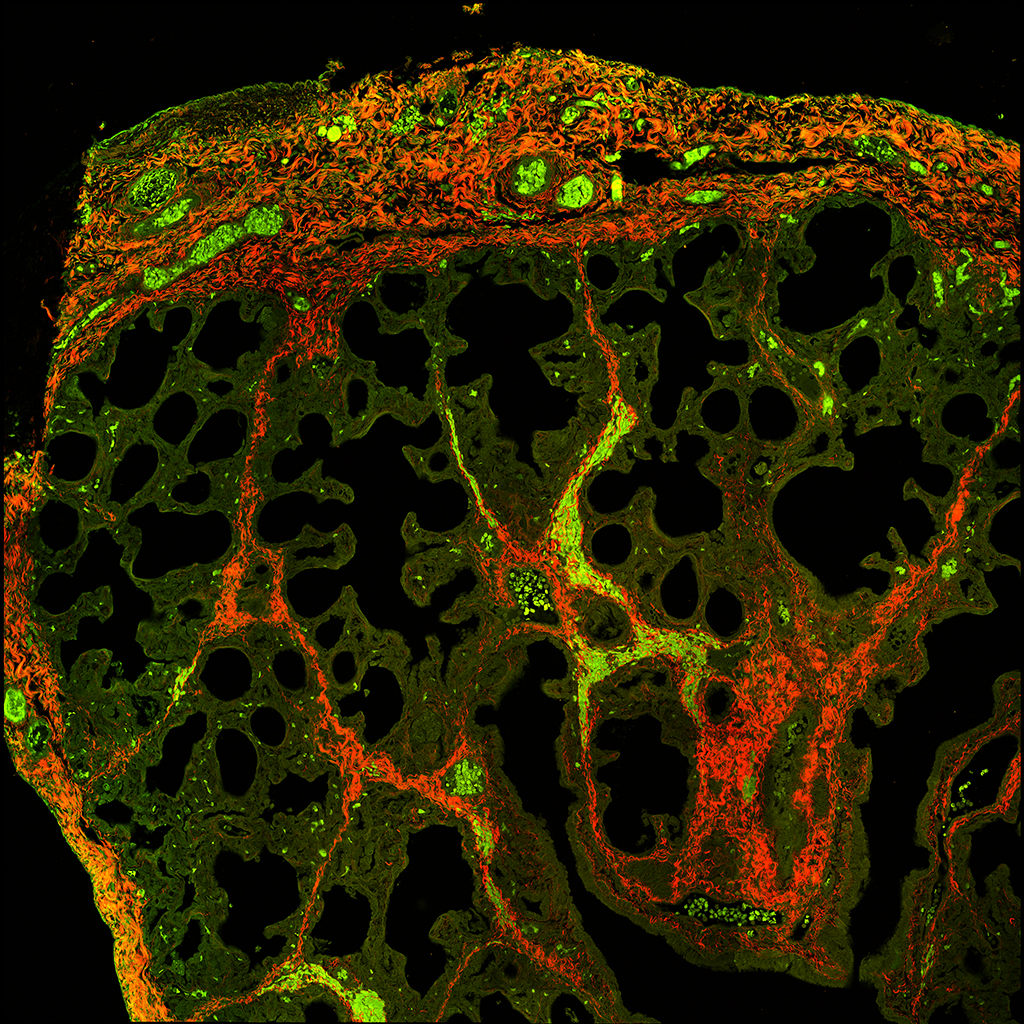
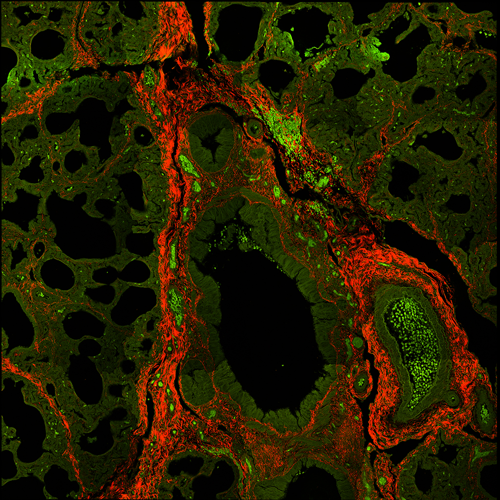
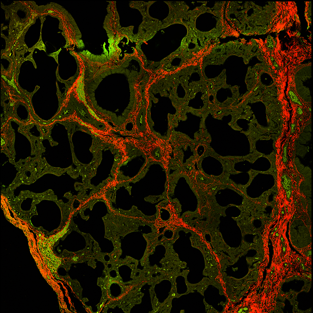
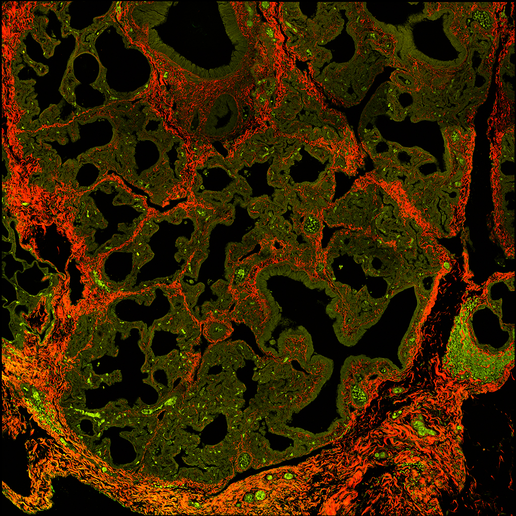
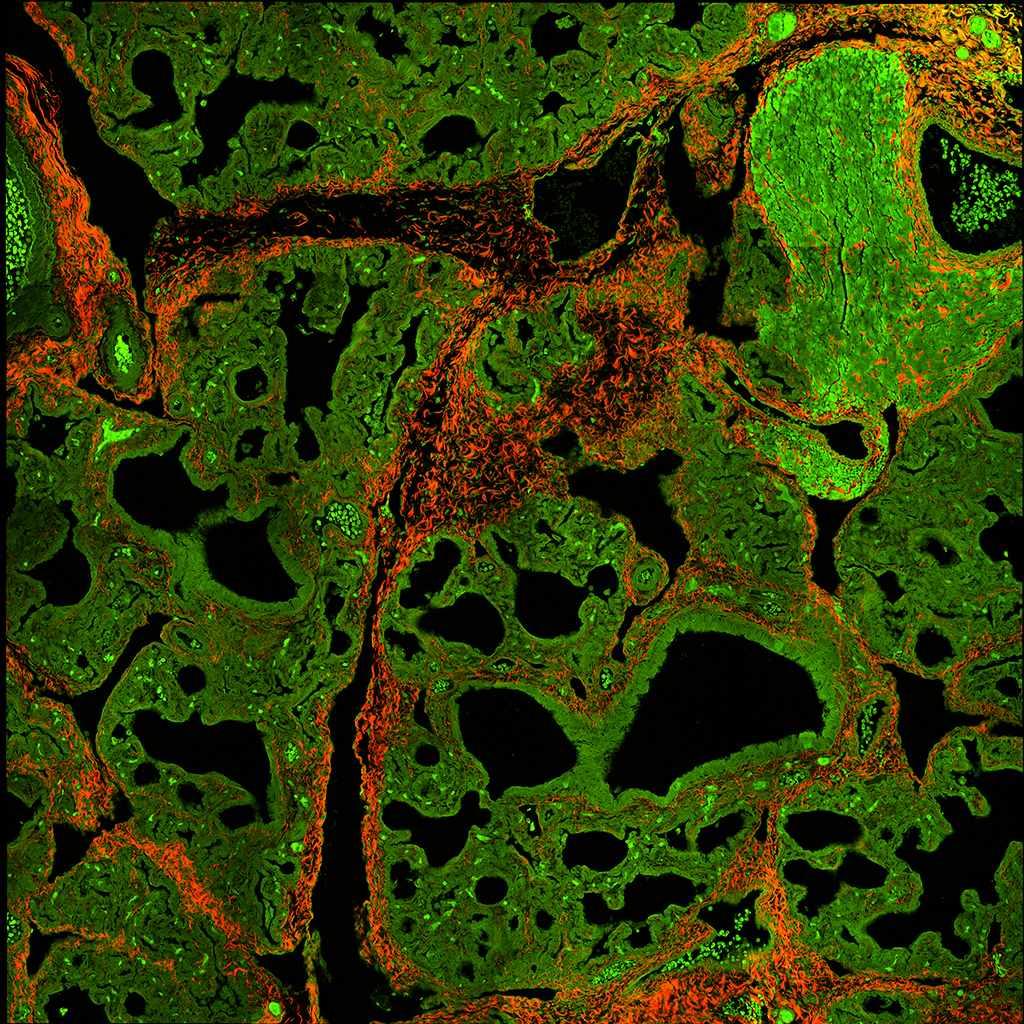
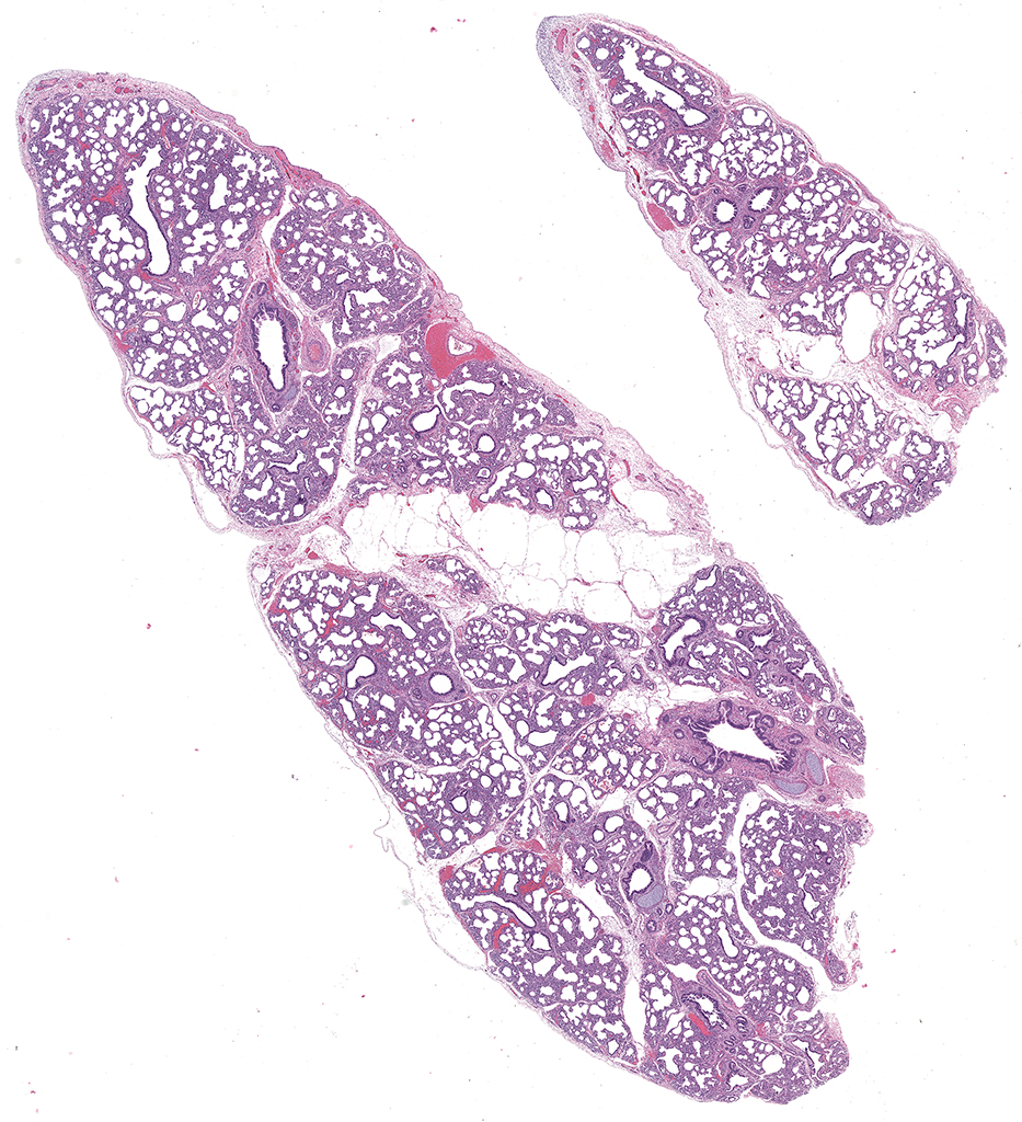
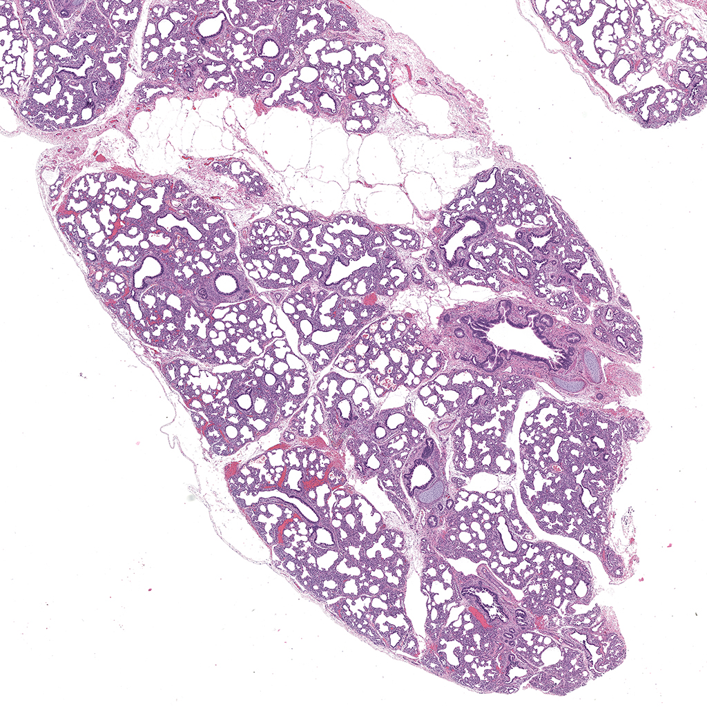
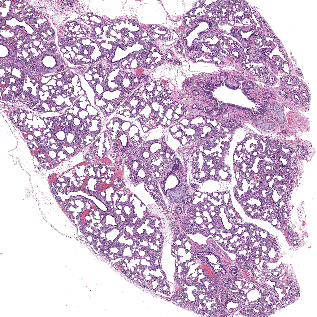
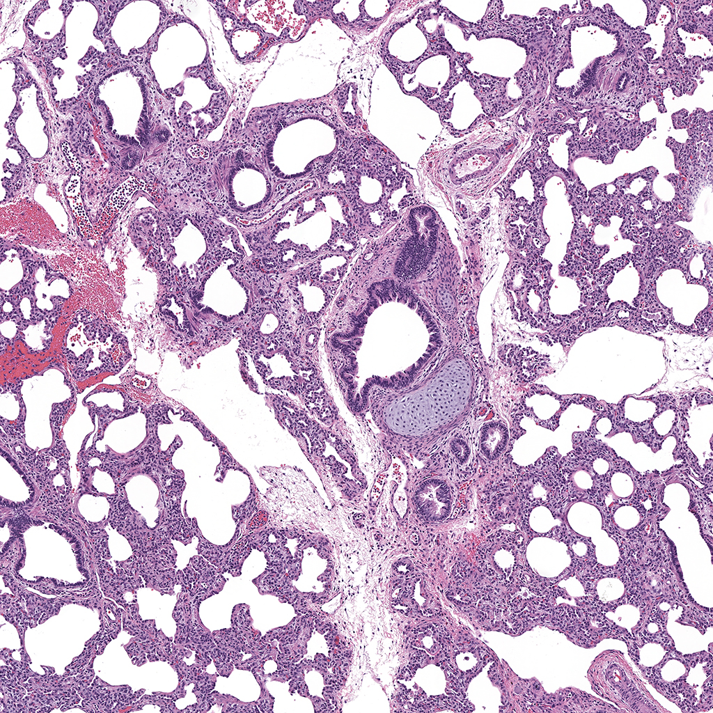
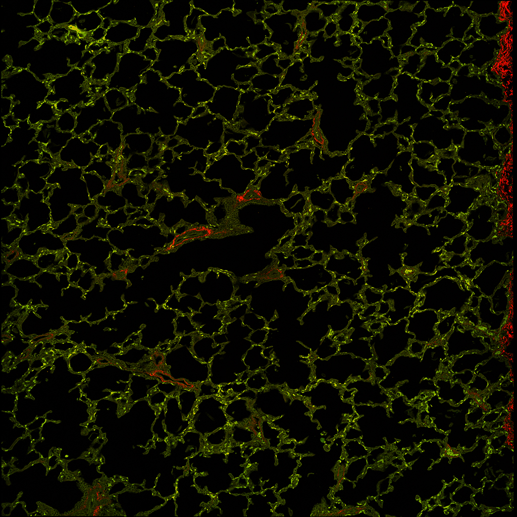
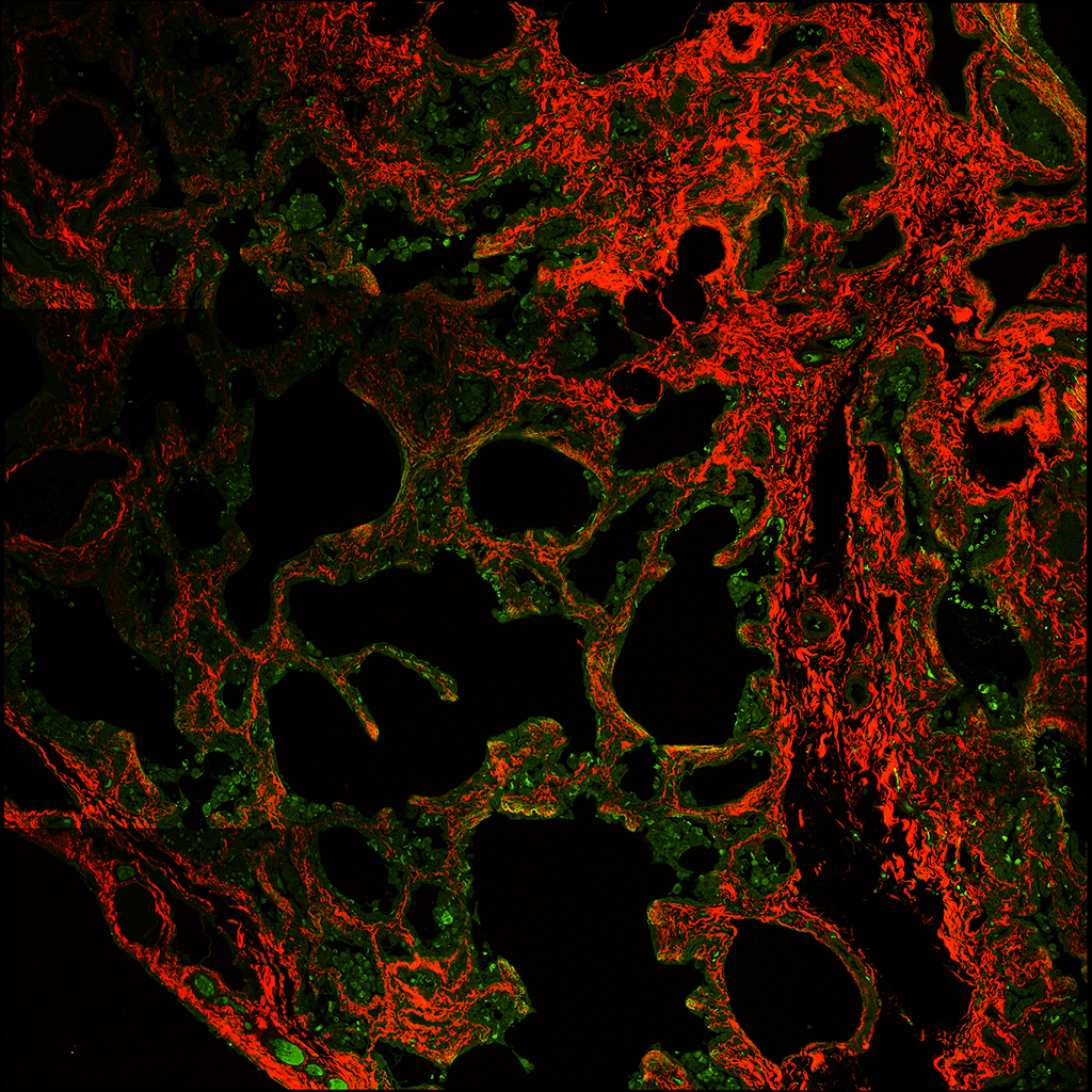
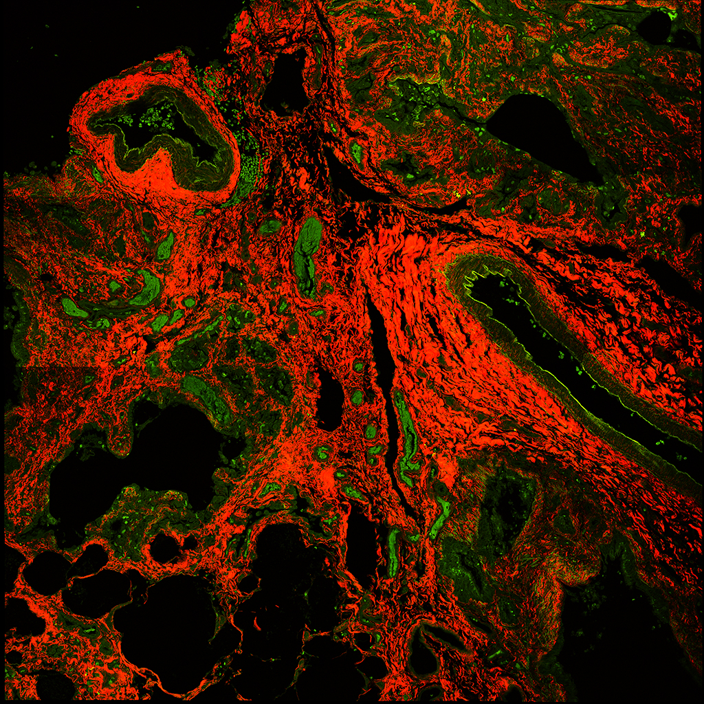
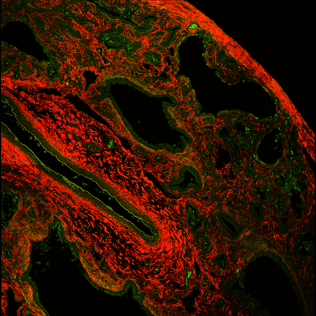
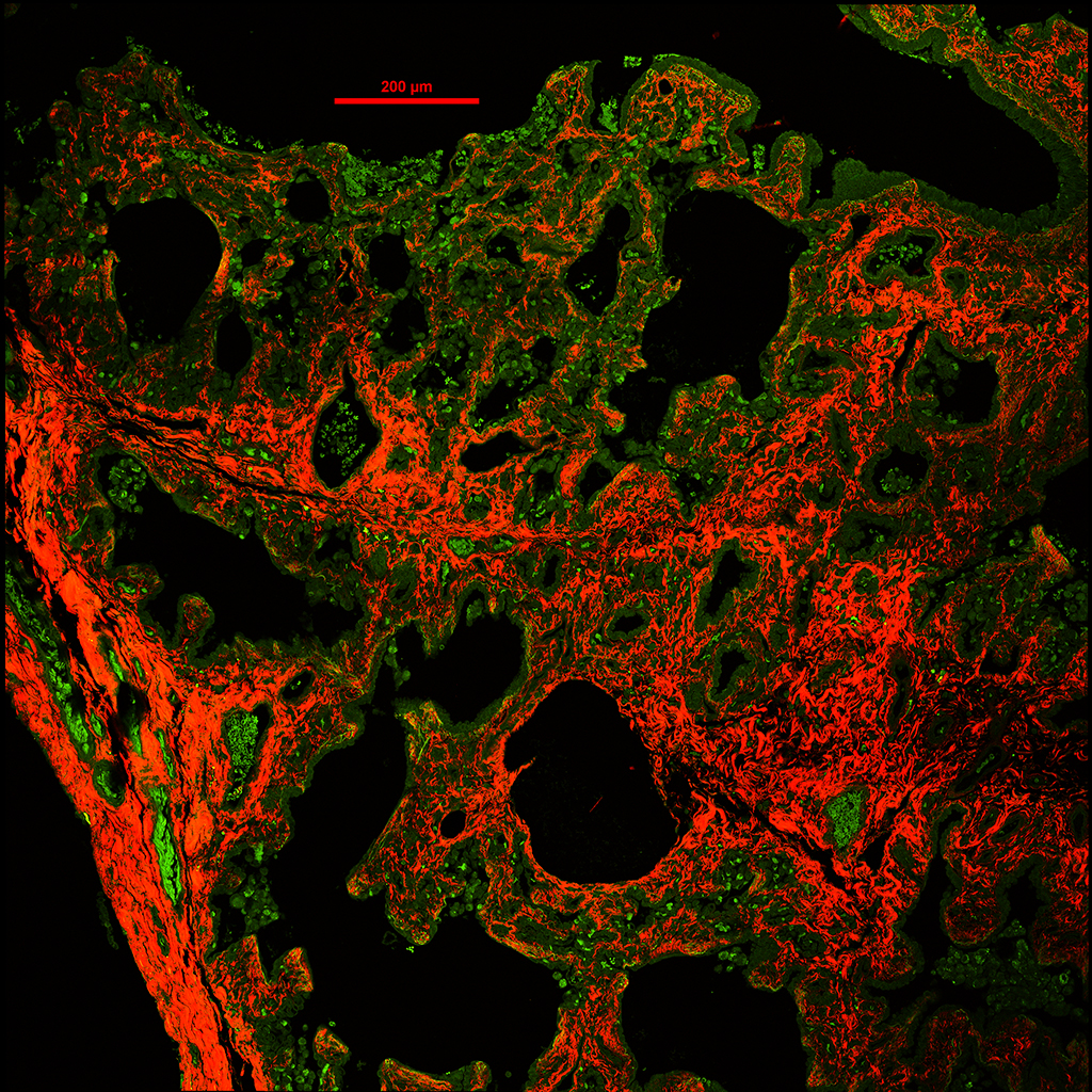
2D rendering of the Z-stacks showing the MUC5B expressing goblet cells (red) , ACTA2 expressing myoepithelial cells (green), and collagen from the second harmonics (white) confocal imaging.

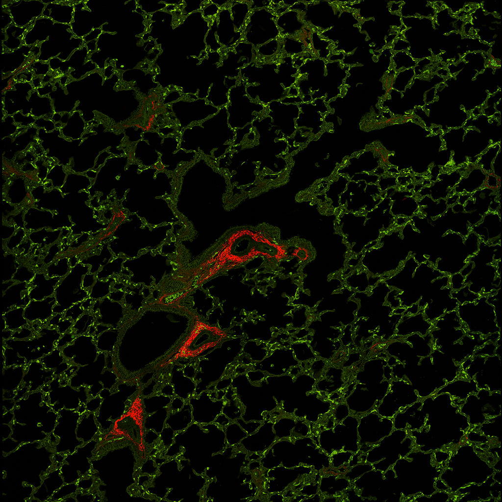
3D surface rendering of the MUC5B expressing goblet cells (red), the ACTA expressing myoepithelial cells (green), and collagen from the second harmonics (white) confocal imaging.


