


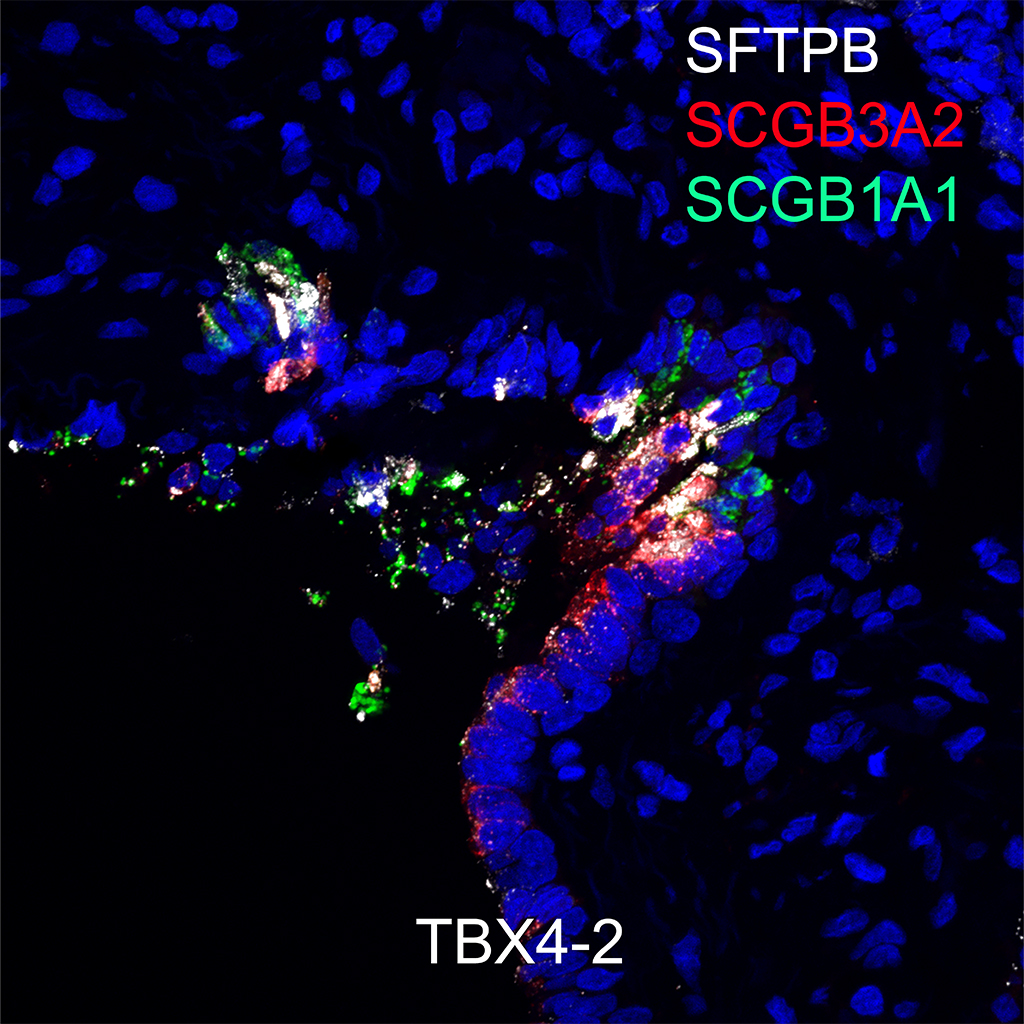
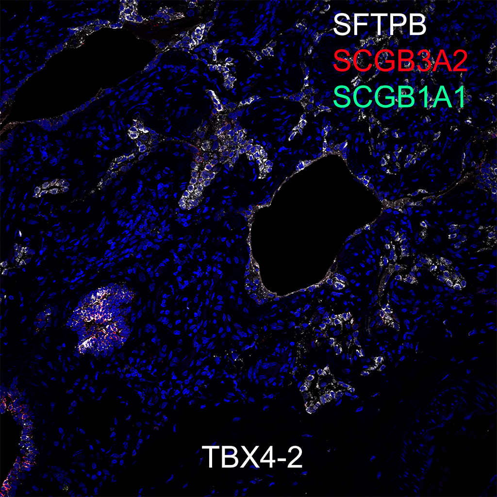
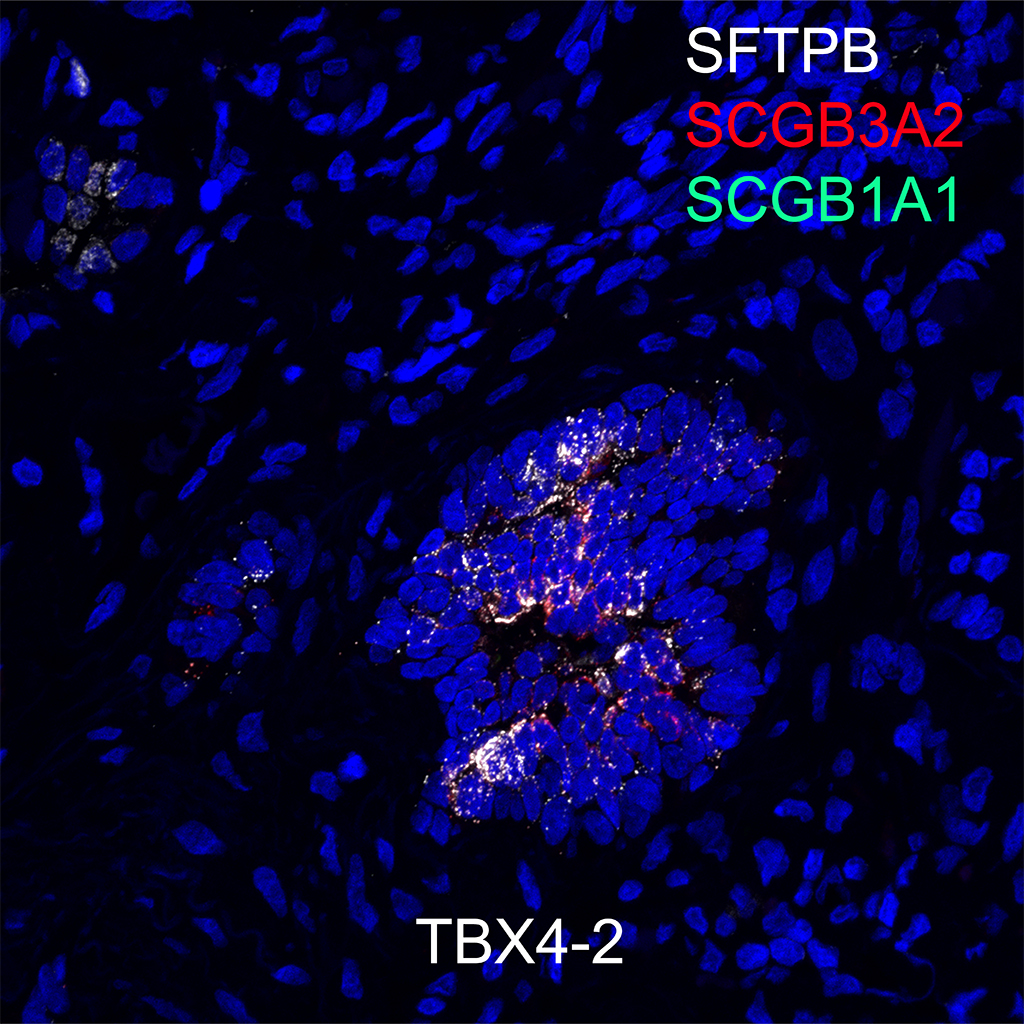
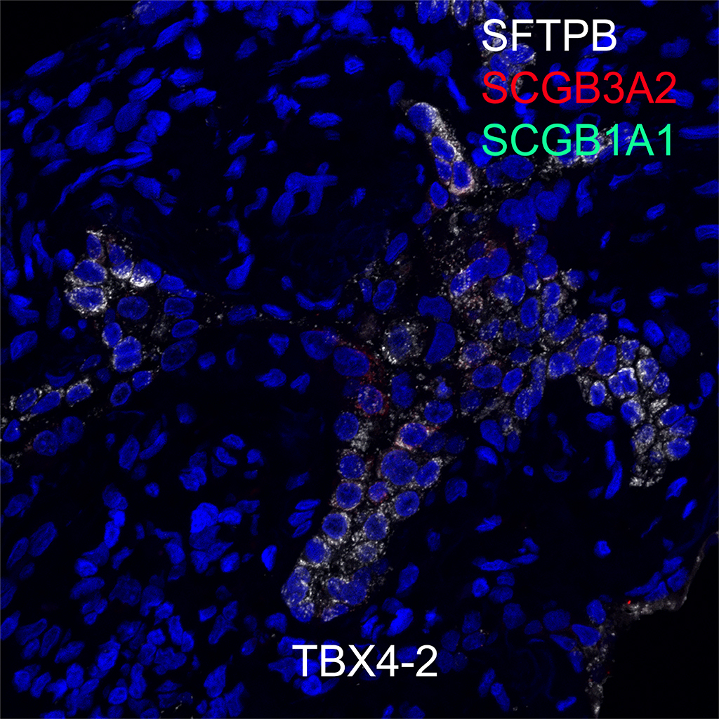
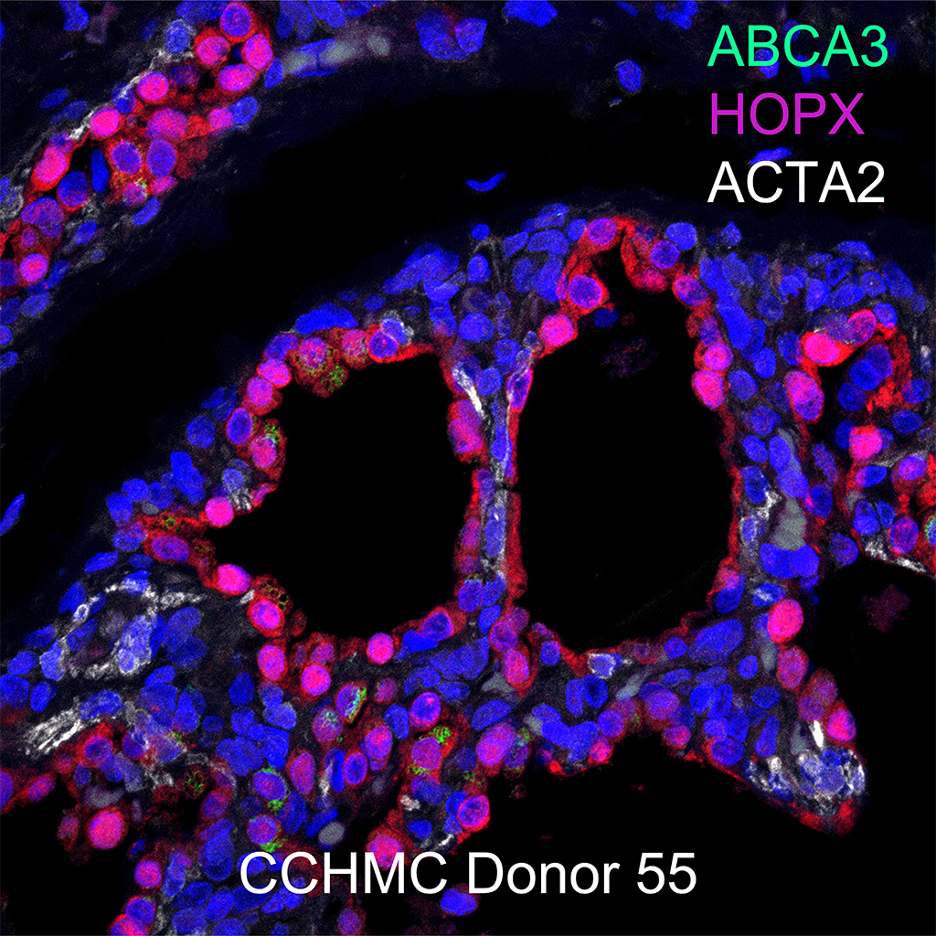
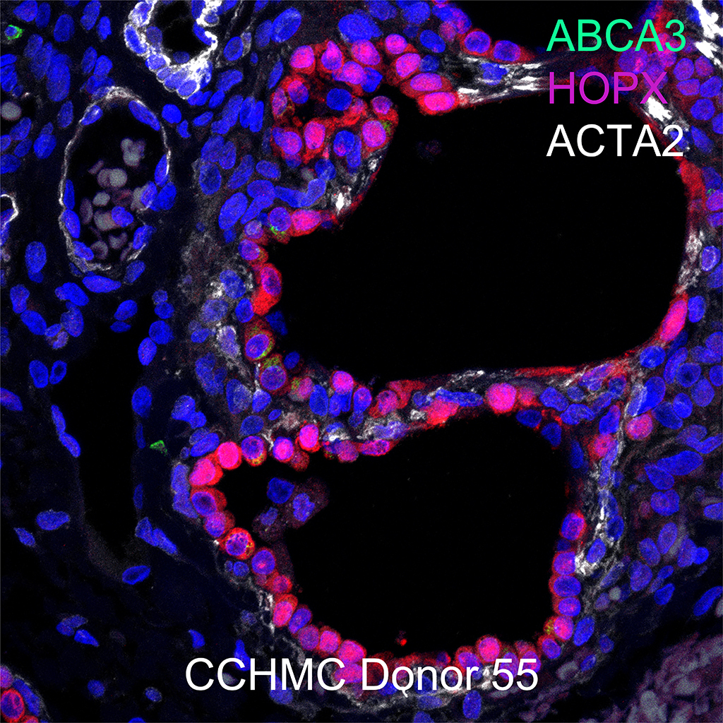
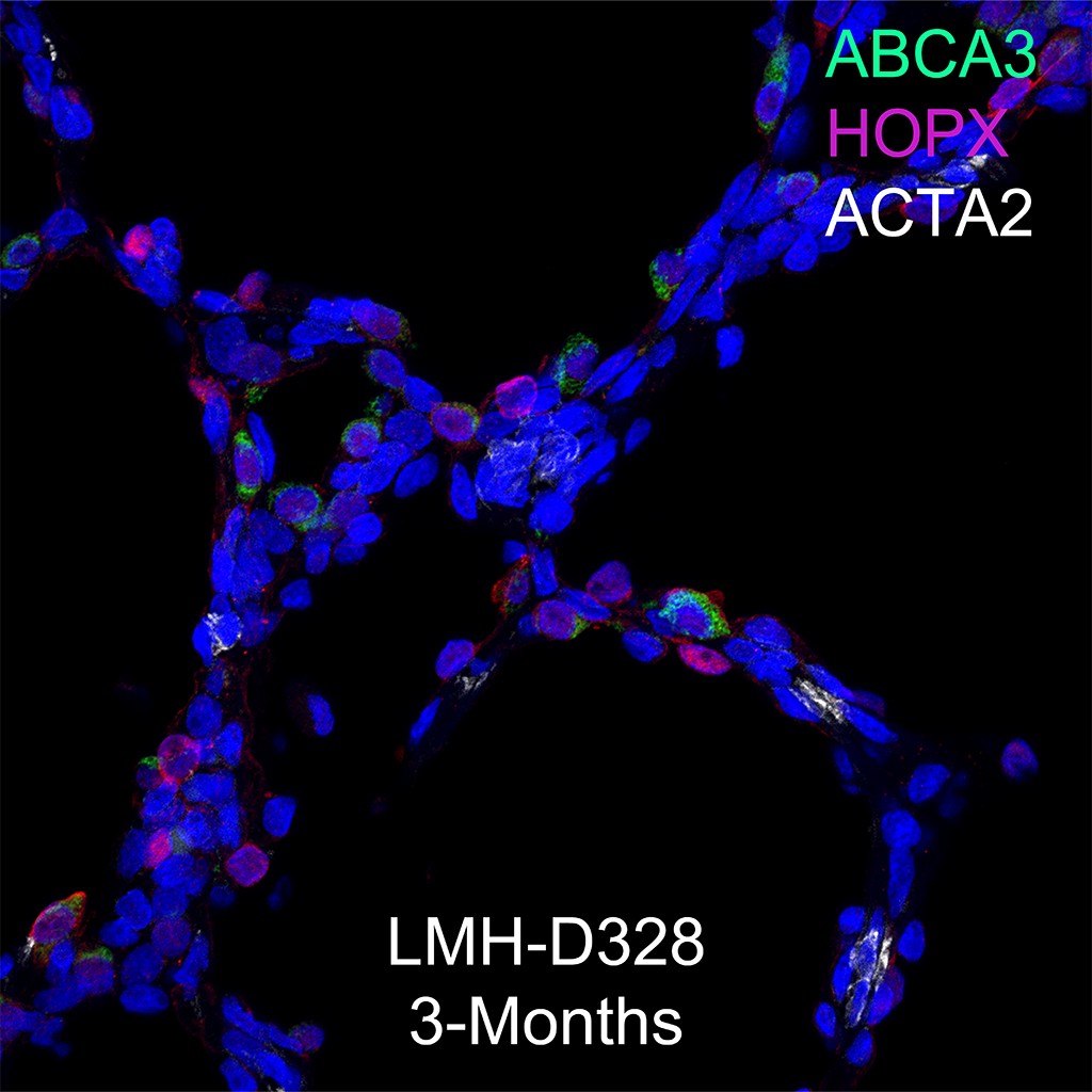
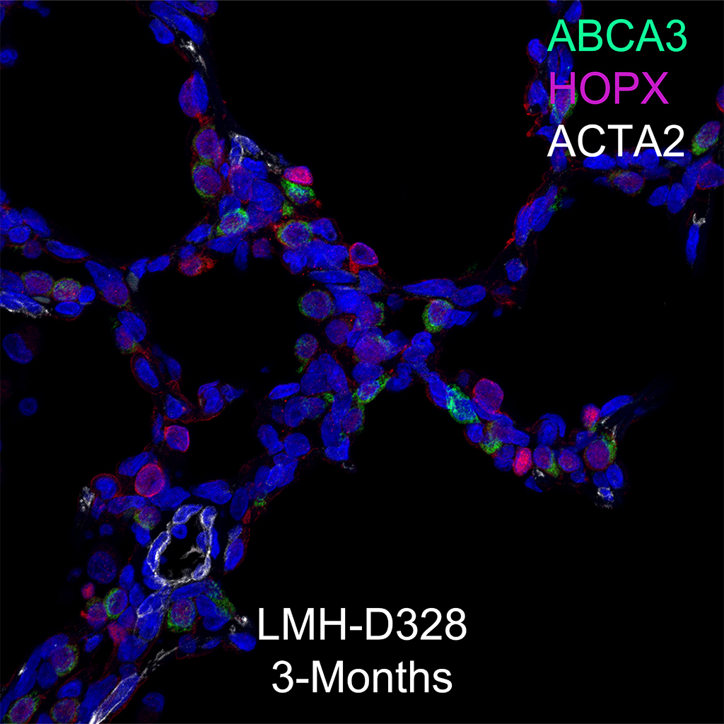
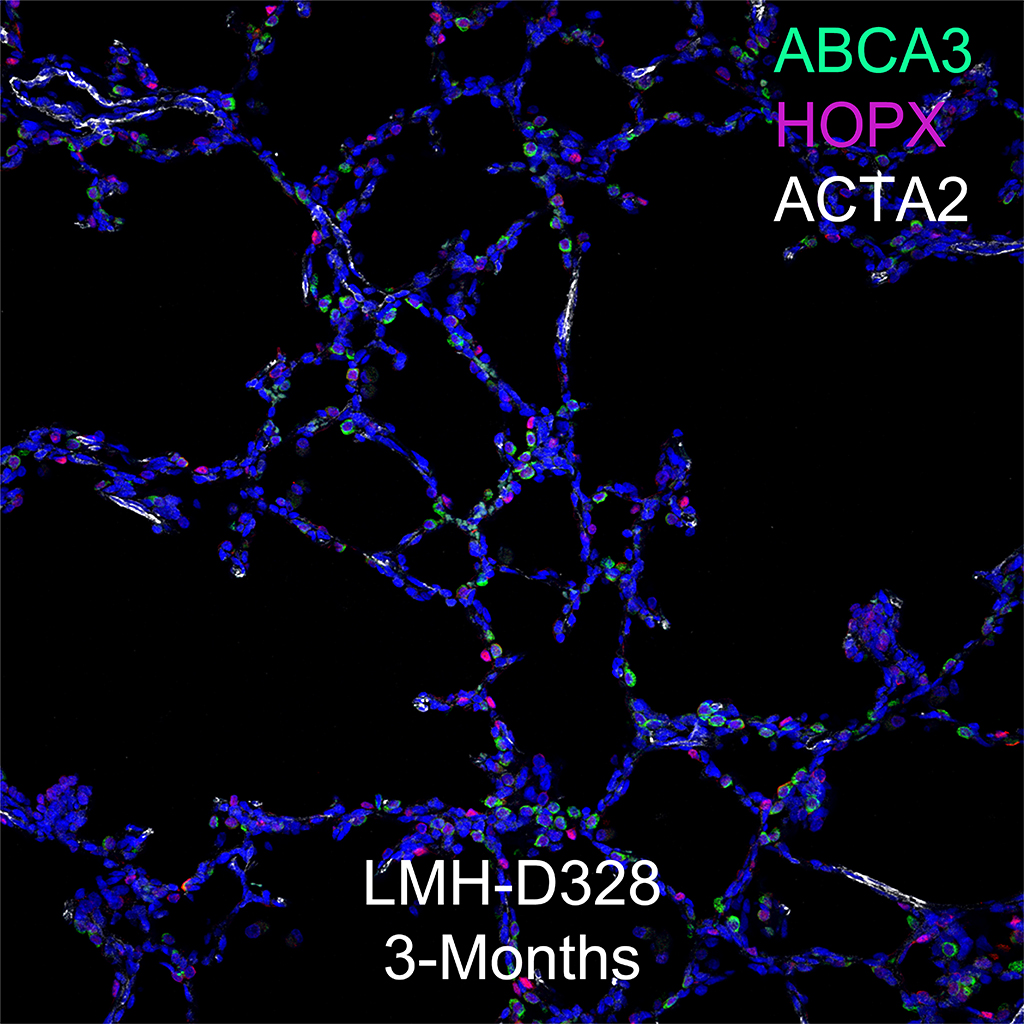
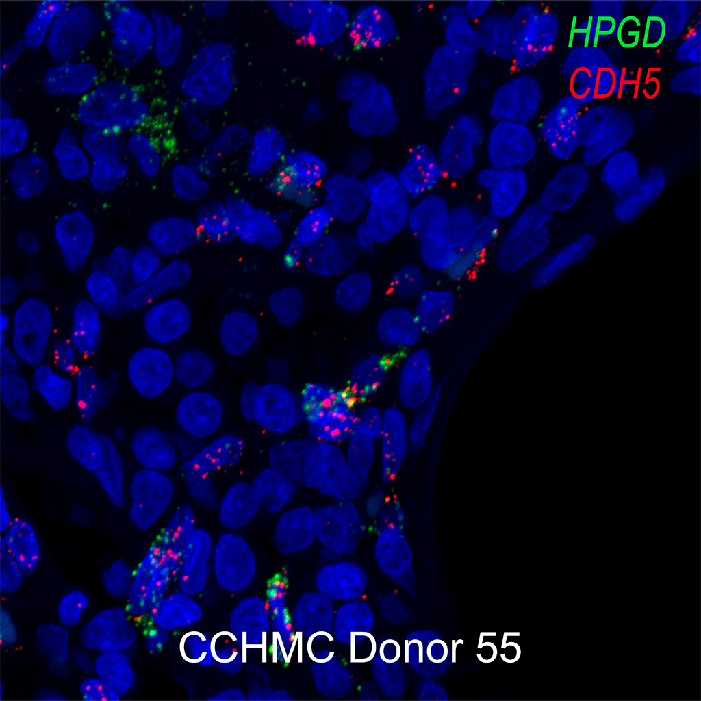
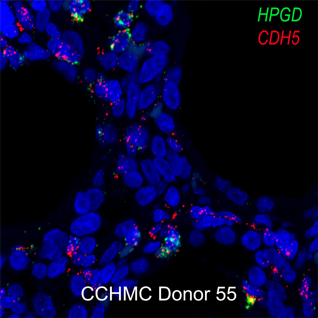
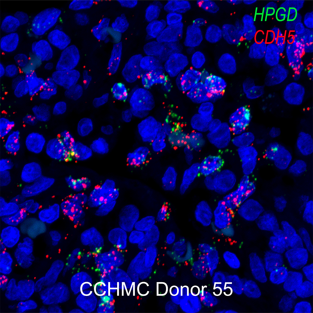
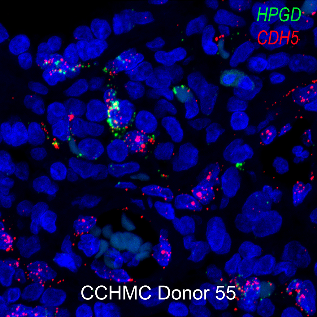
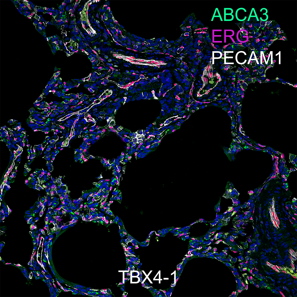
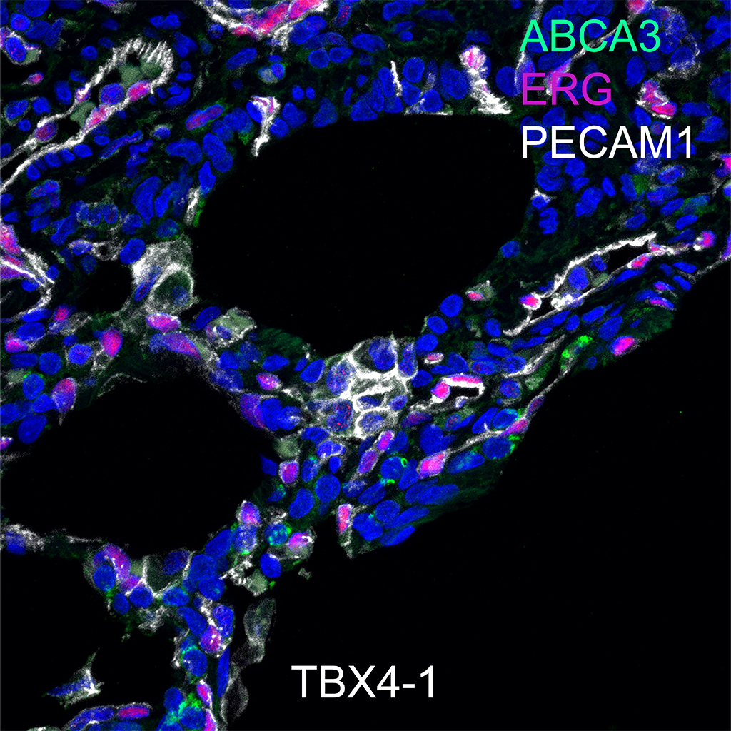
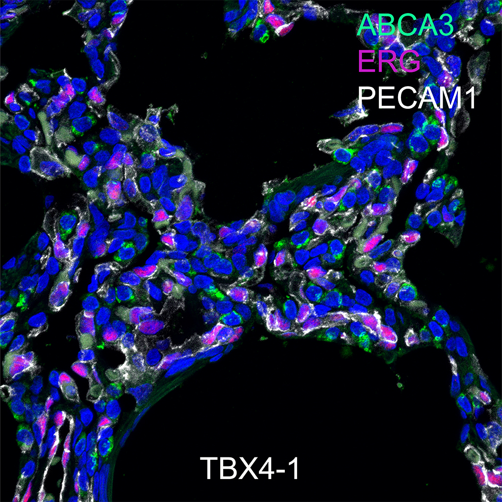
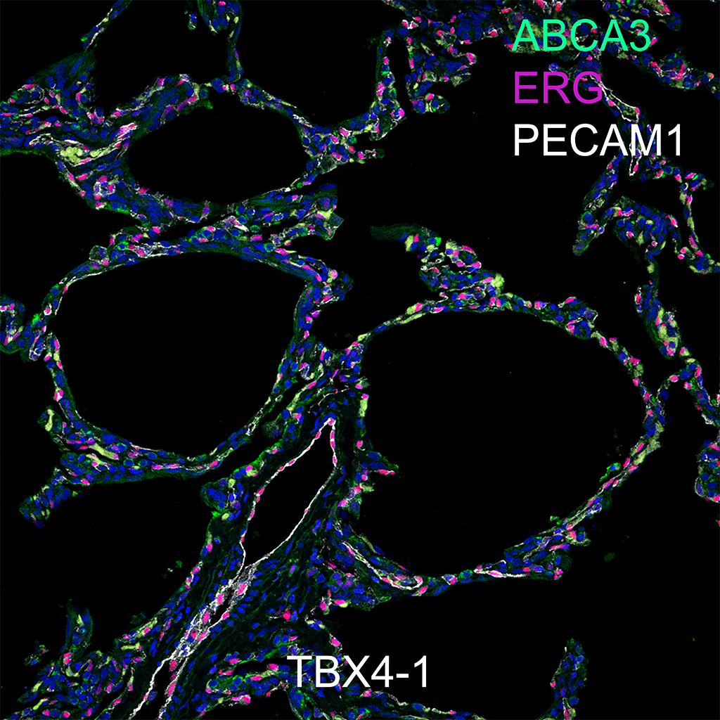
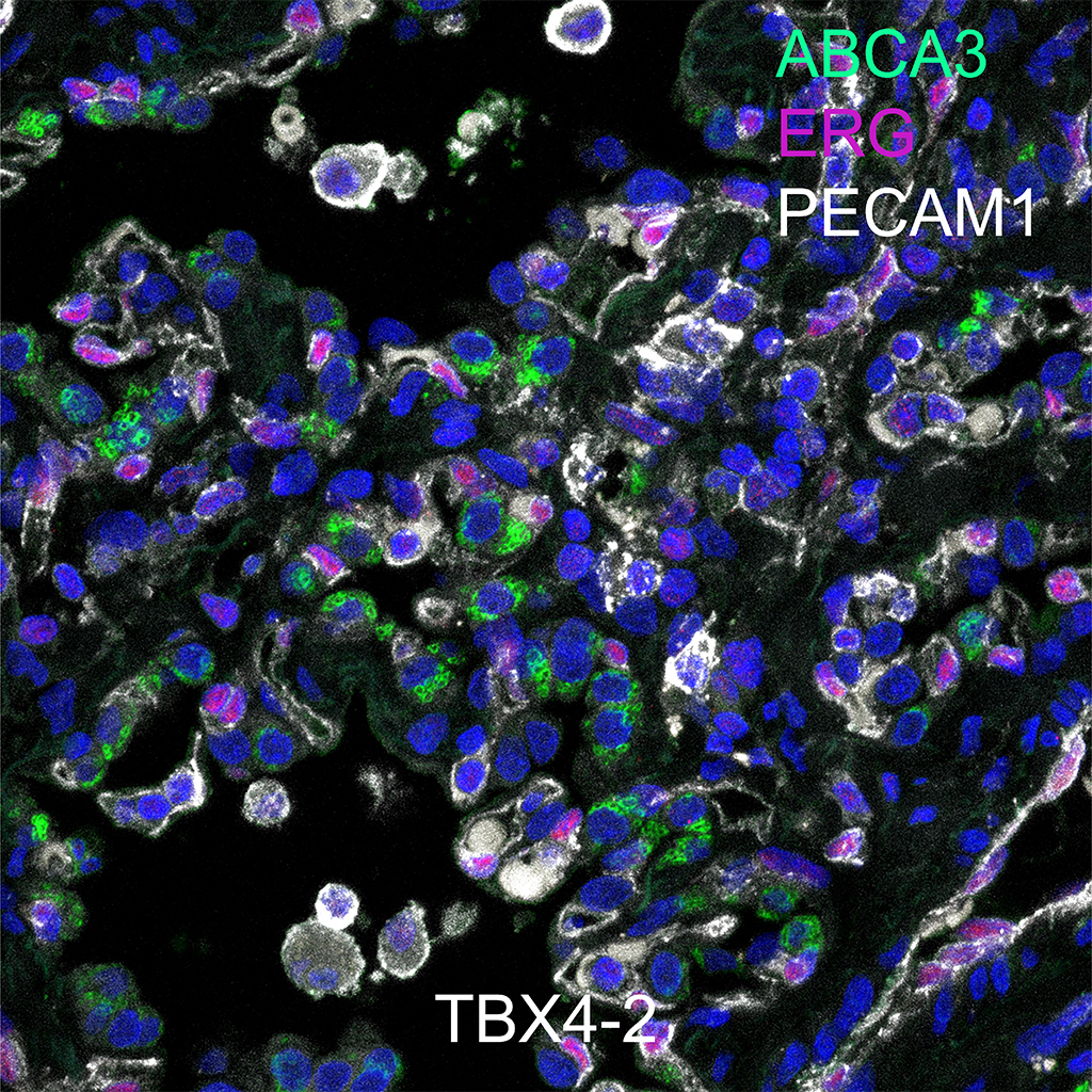
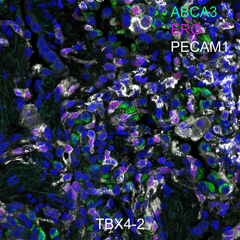
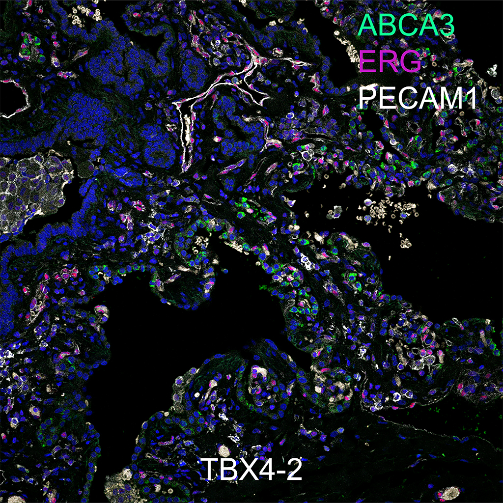
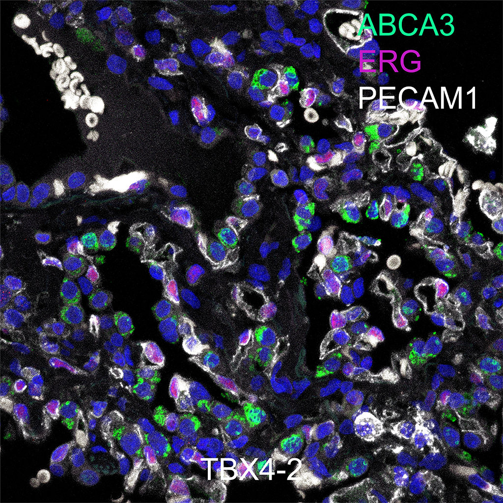
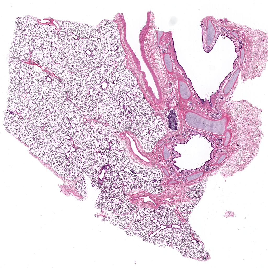
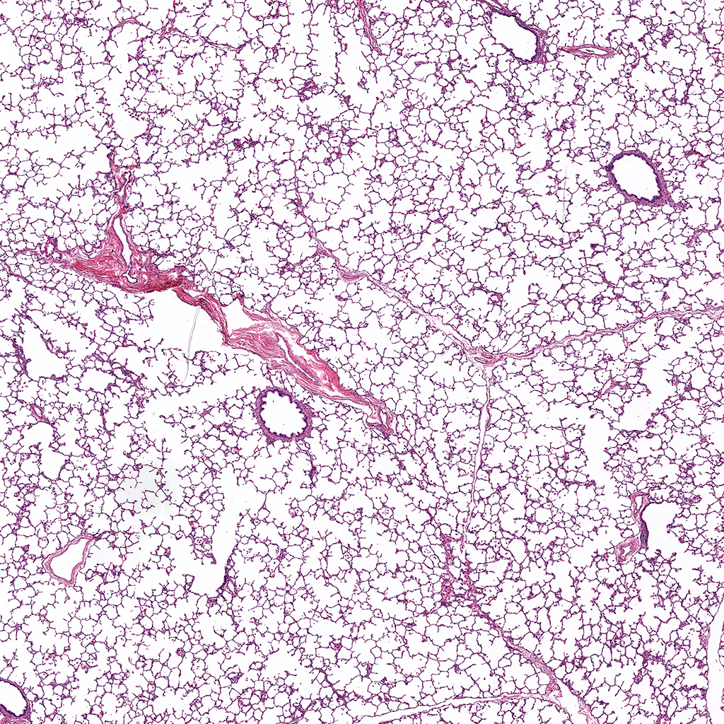
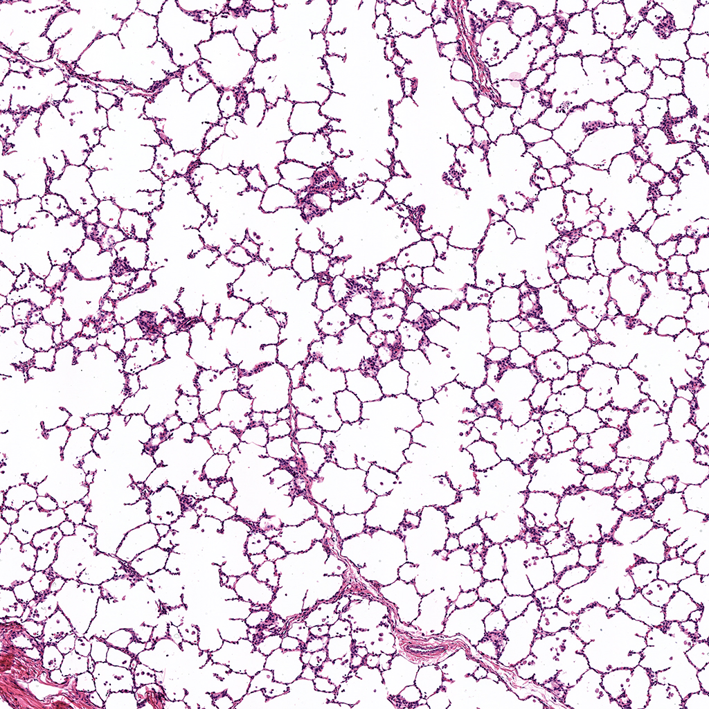
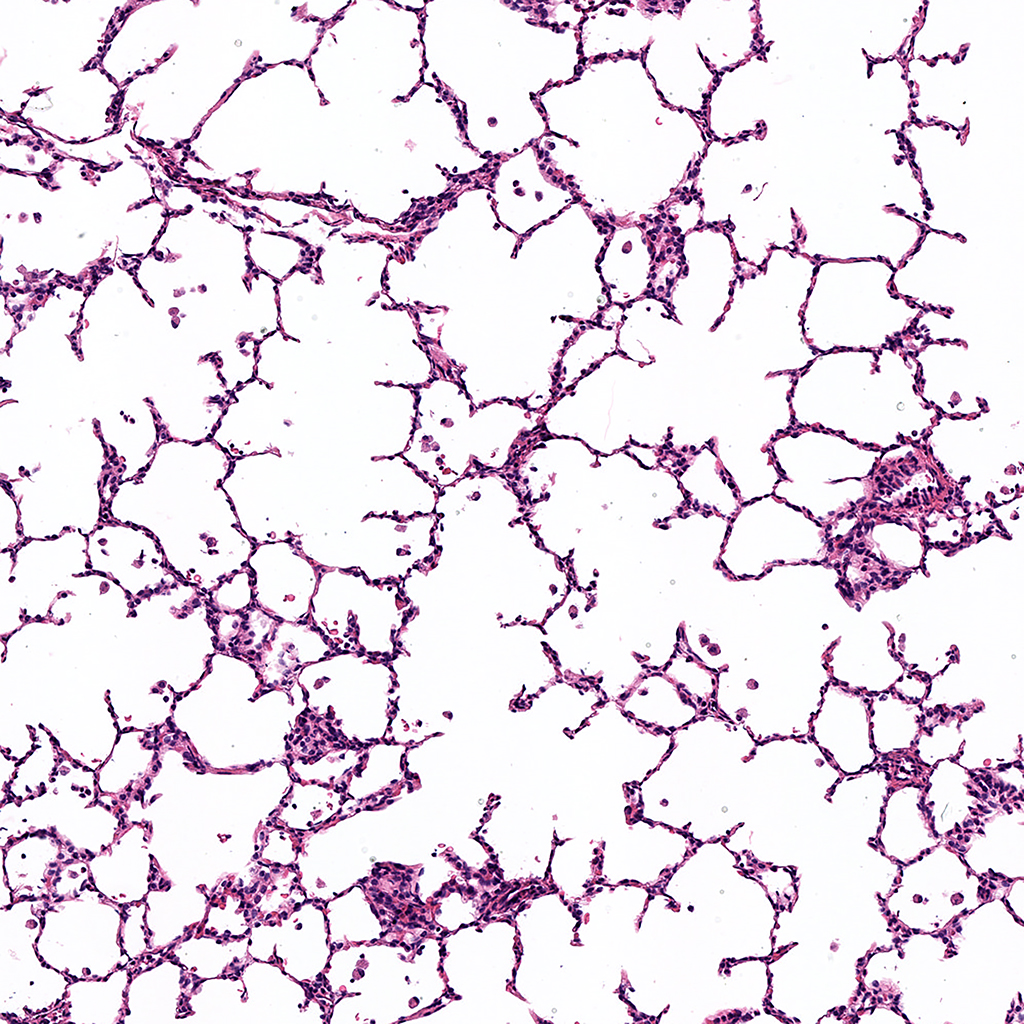
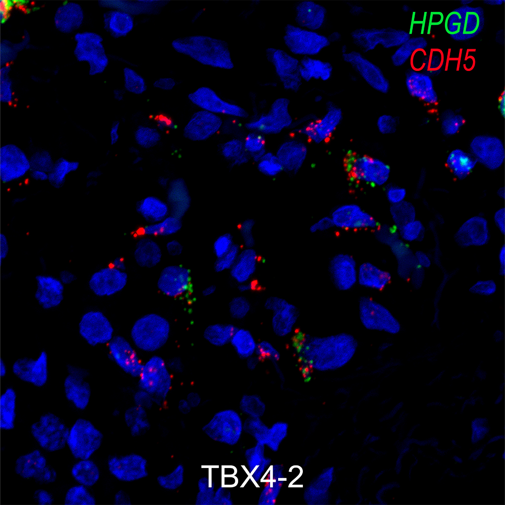
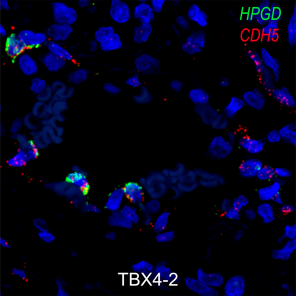
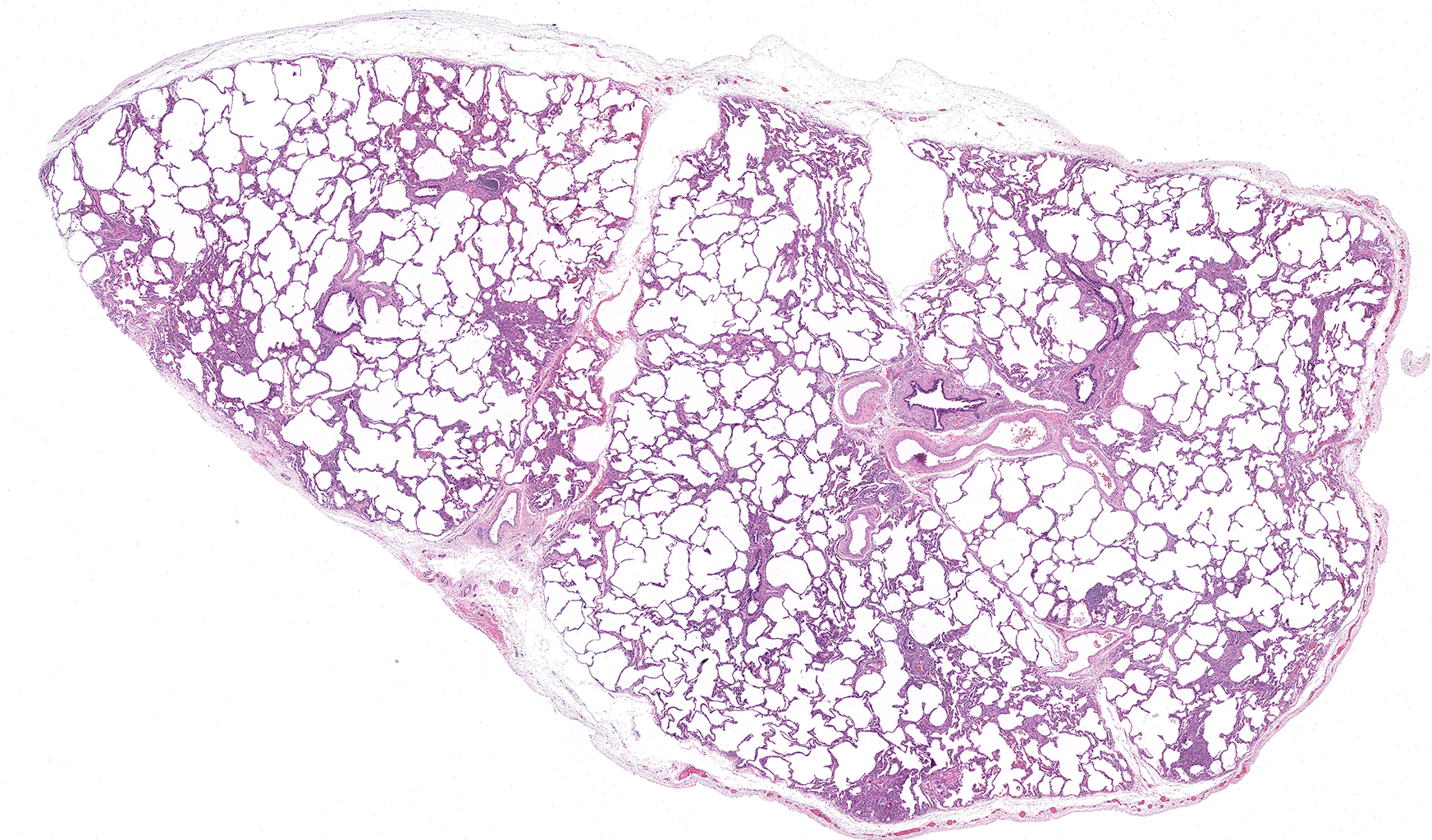
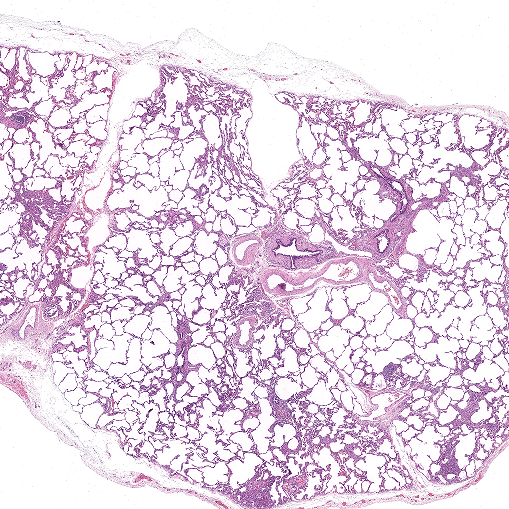
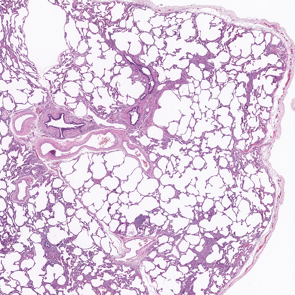
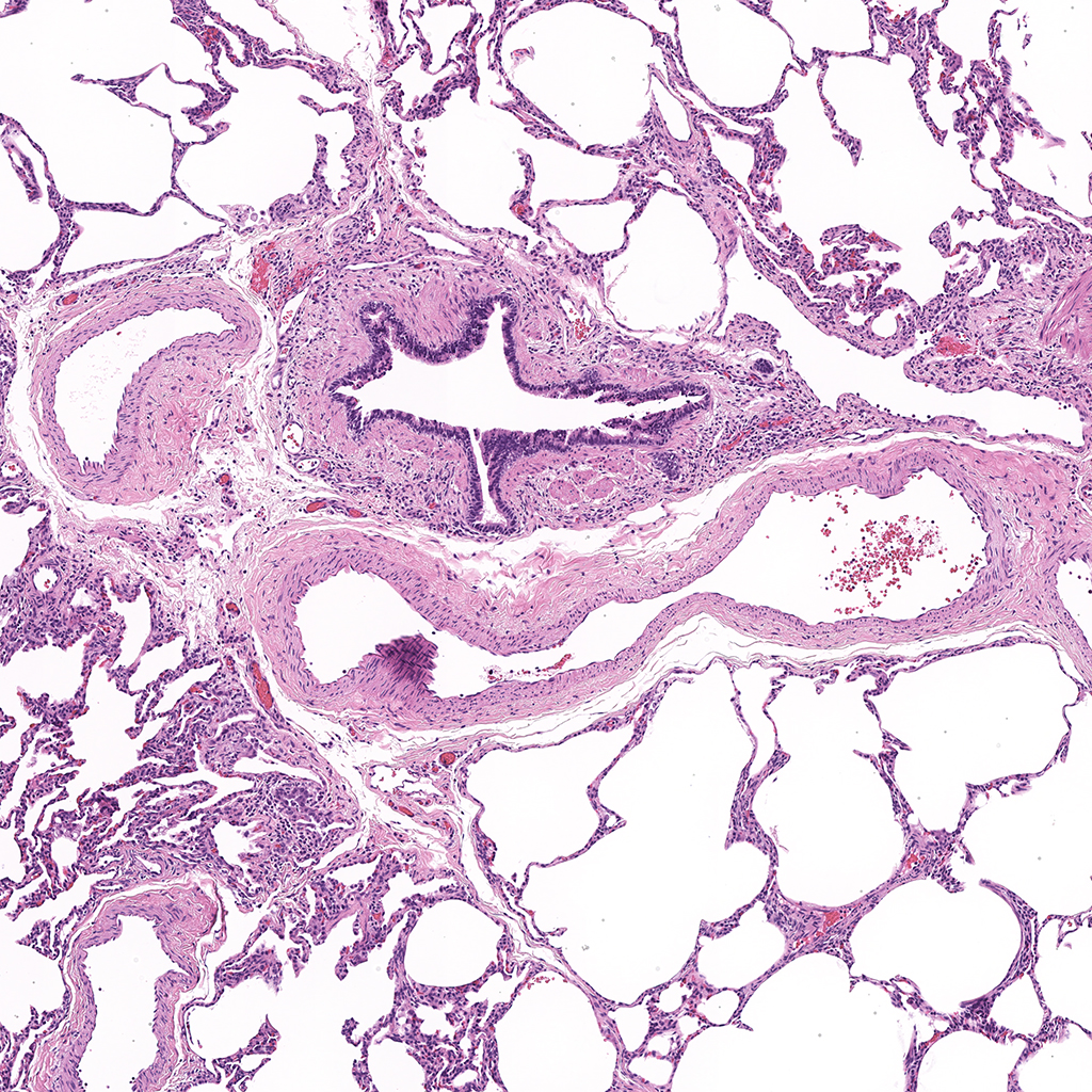
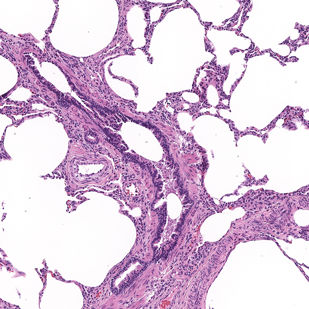
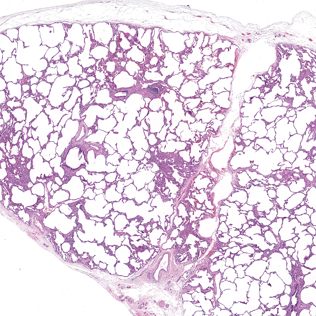
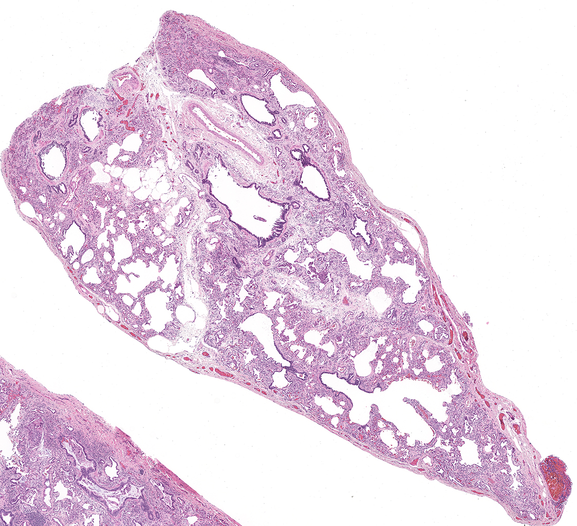
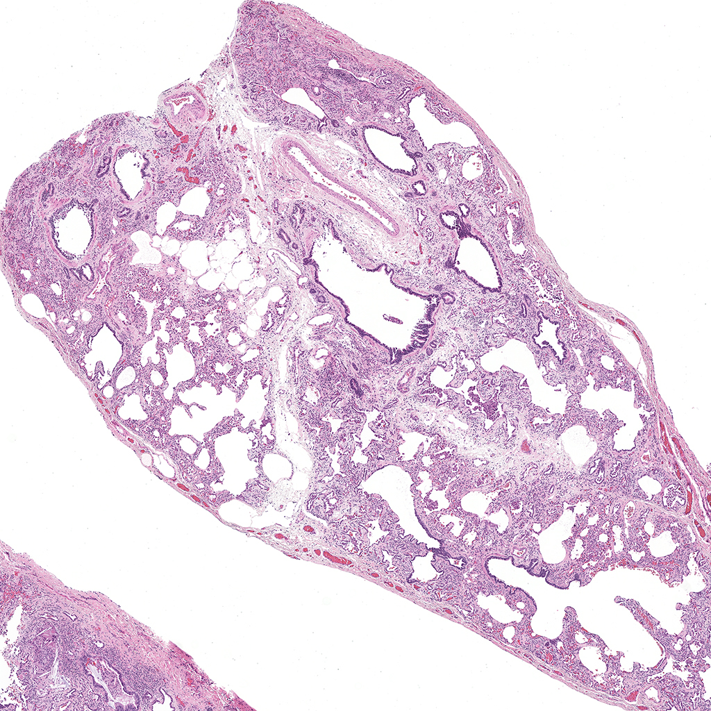
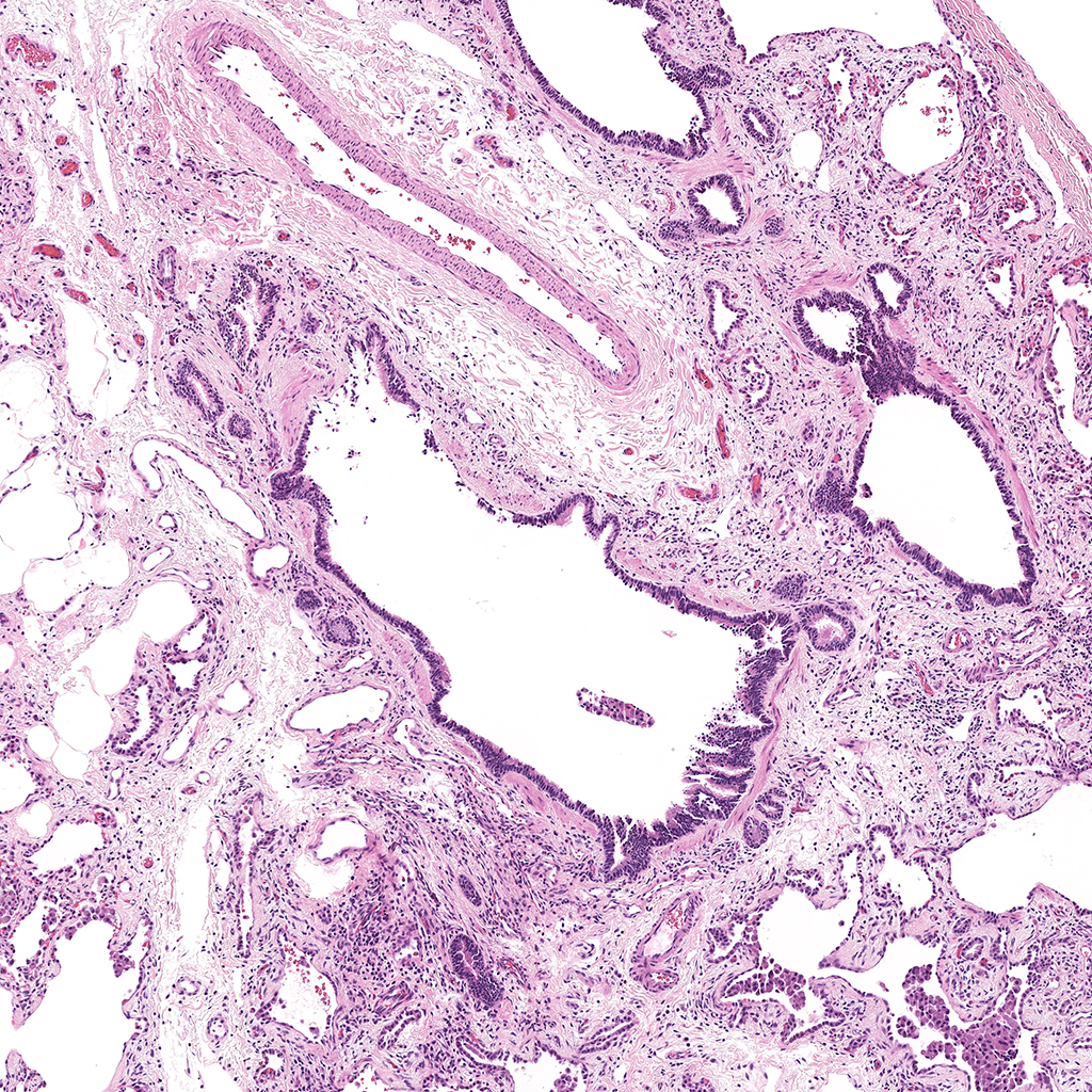
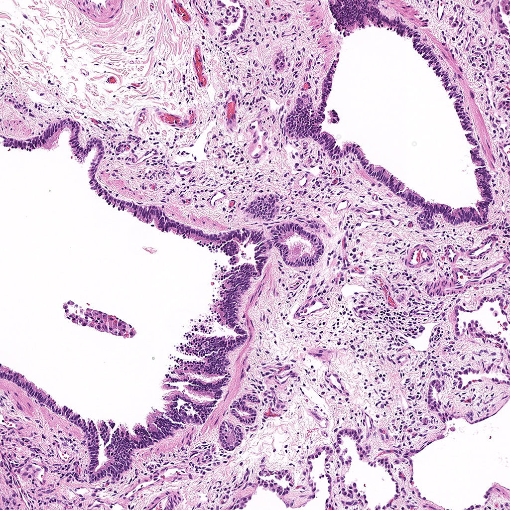
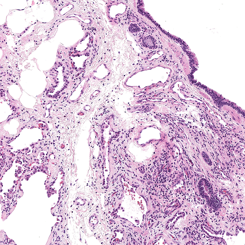
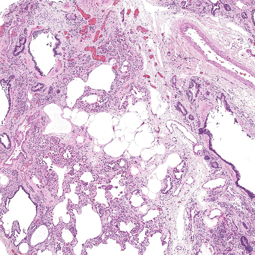
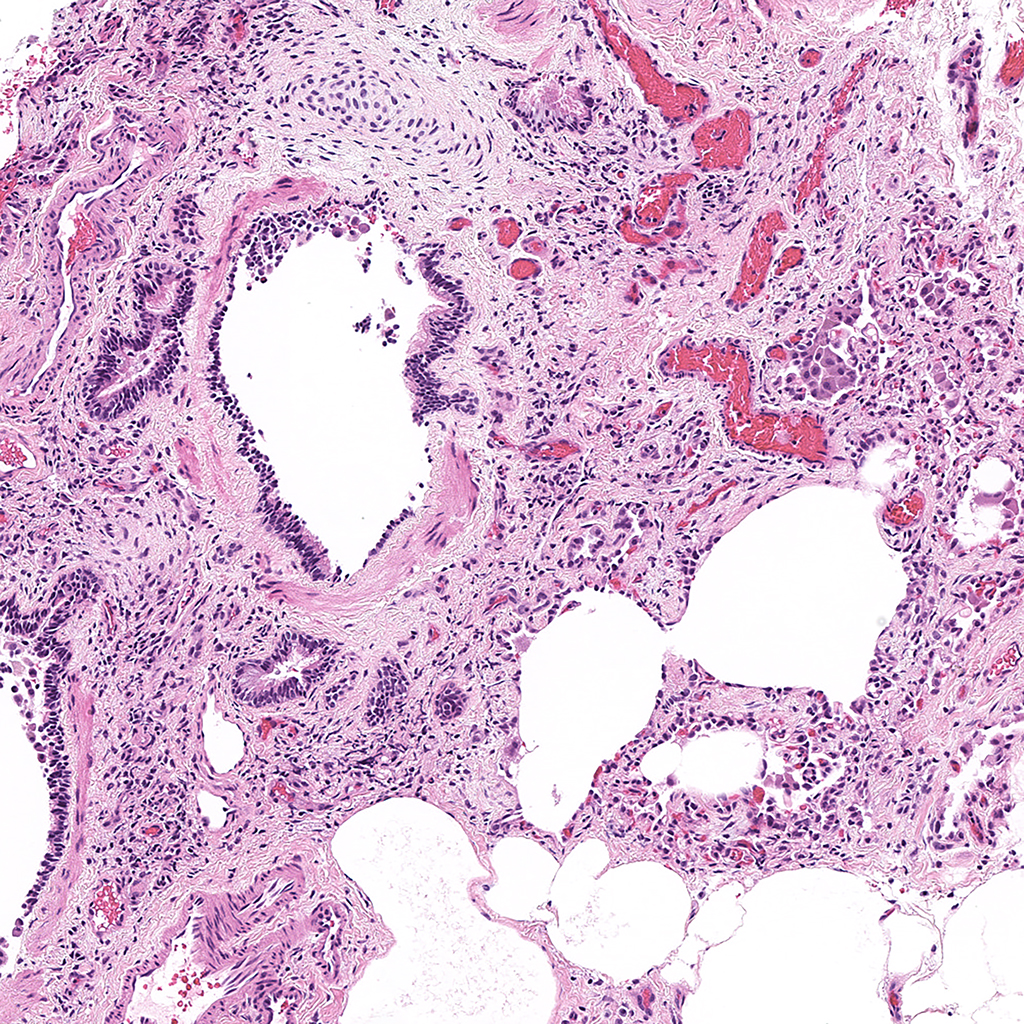
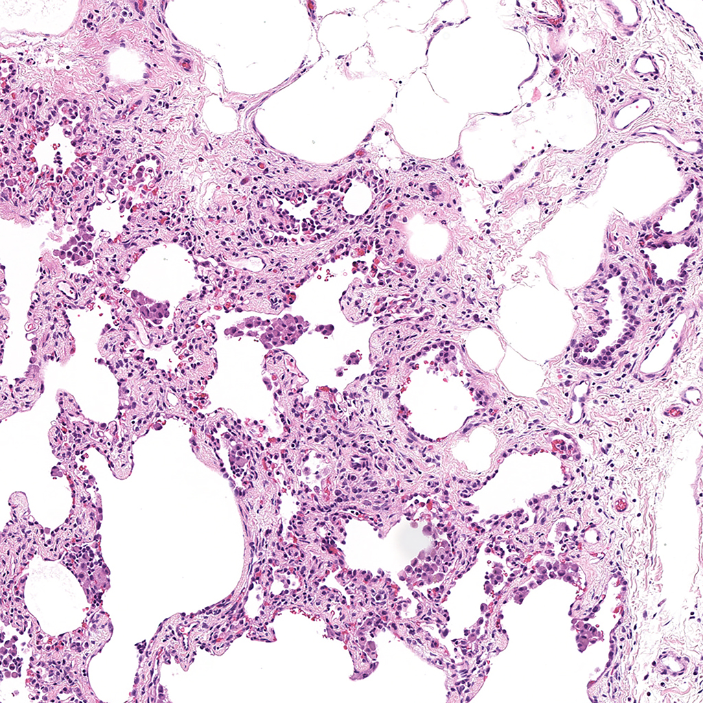
Donor Tissue Kindly Provided by Dr. Gloria Pryhuber from the University of Rochester Medical Center
![]()
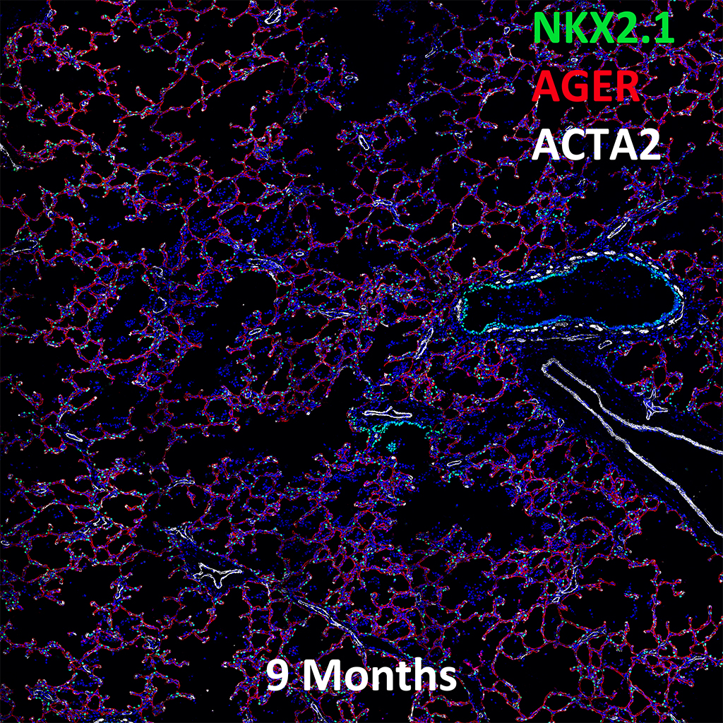
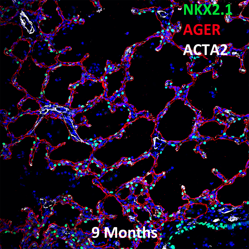
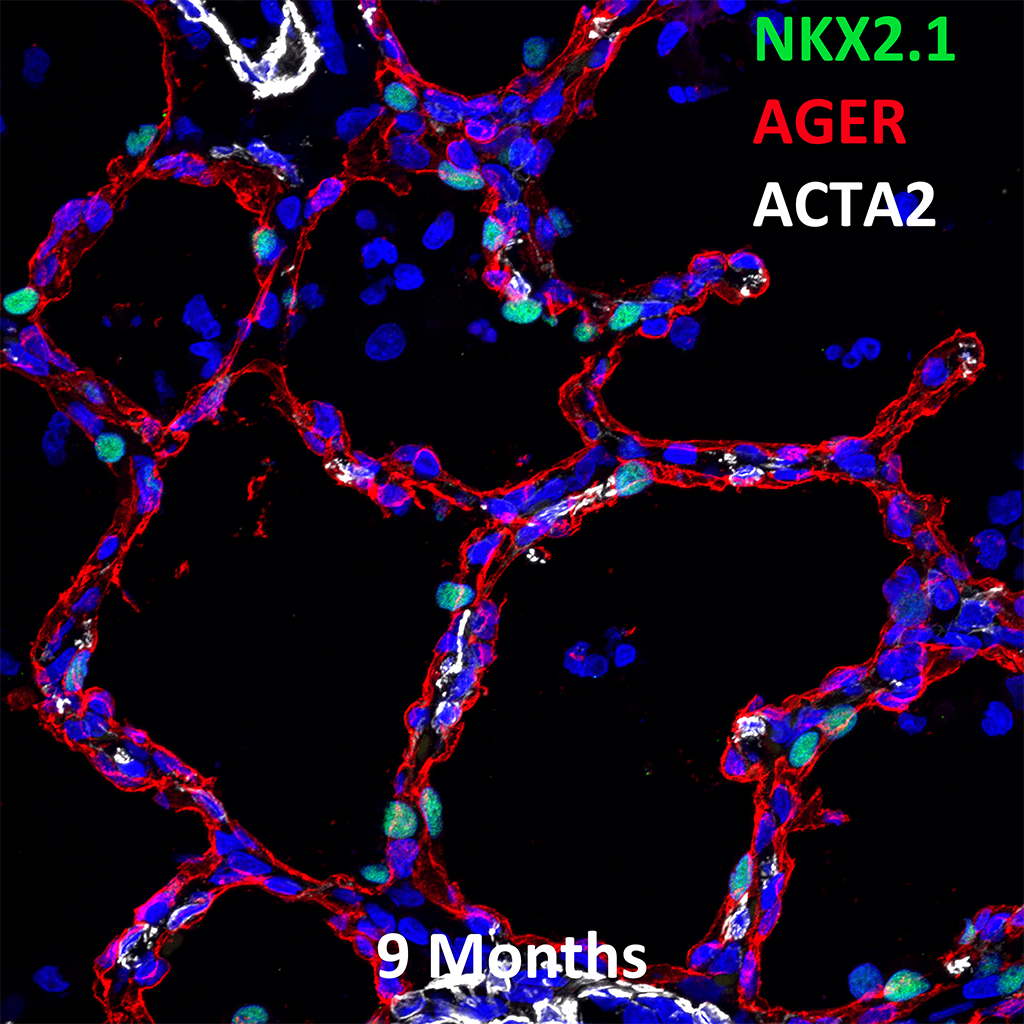
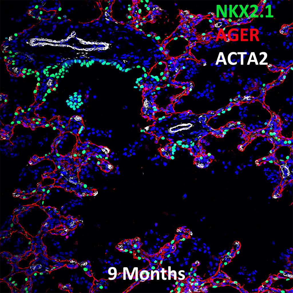
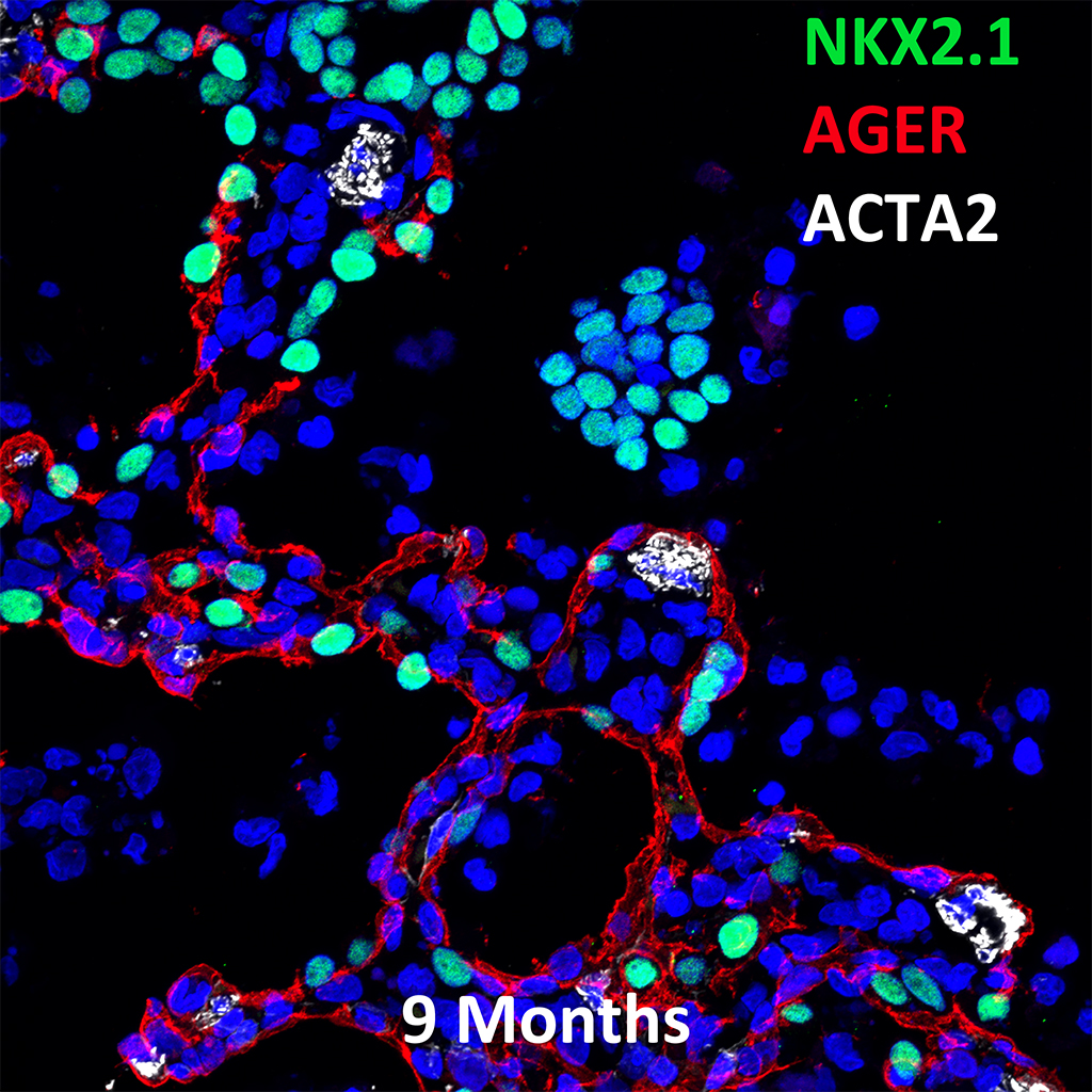
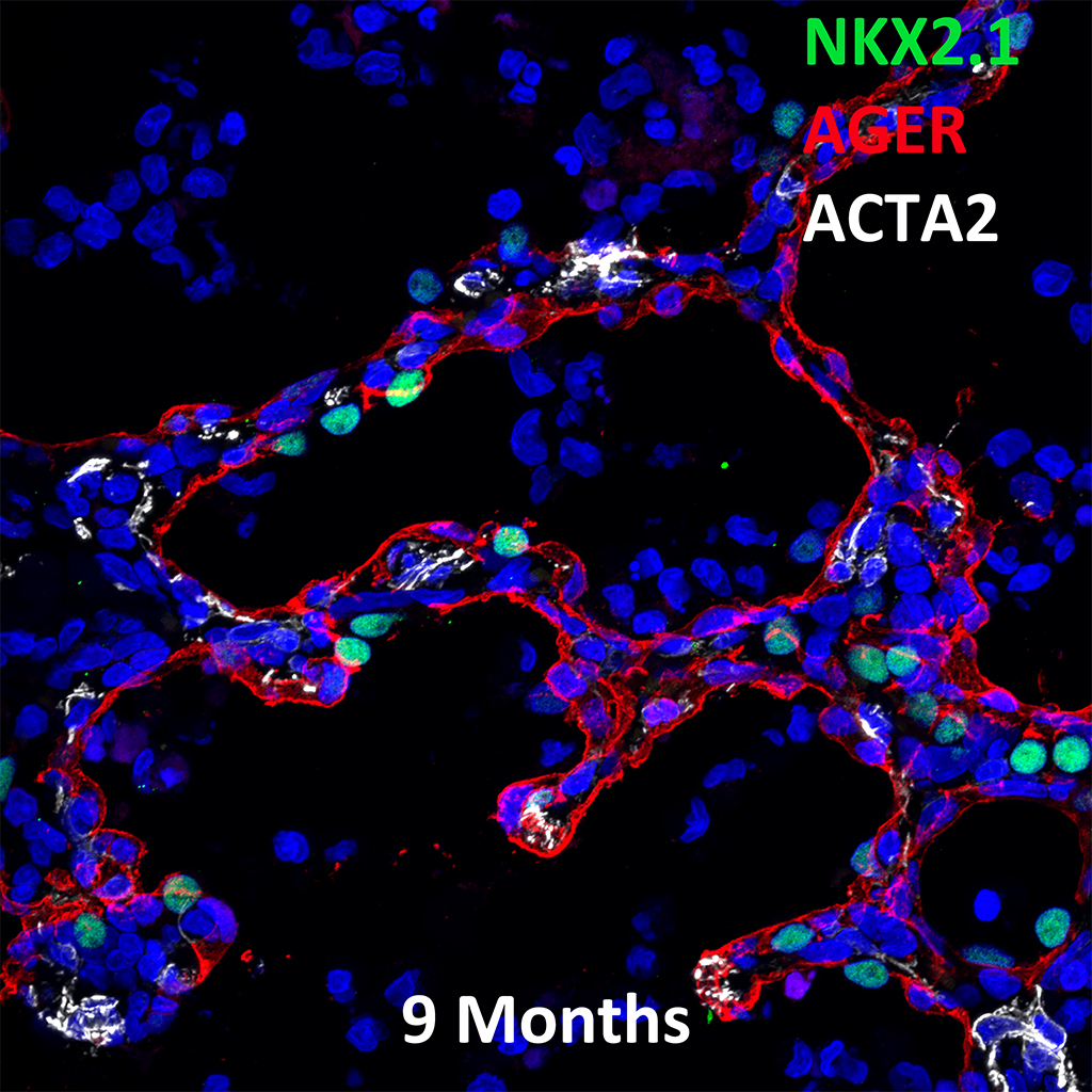
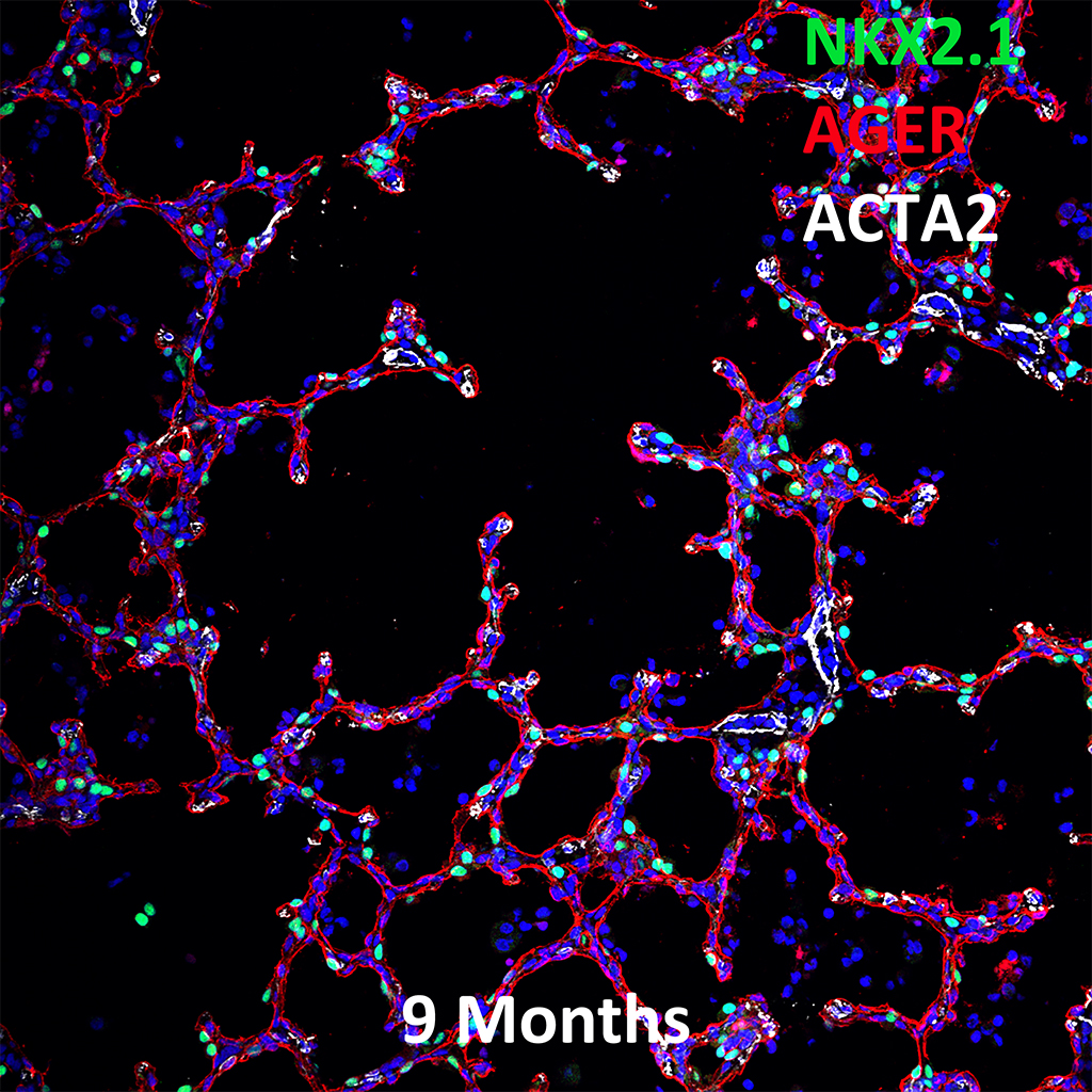
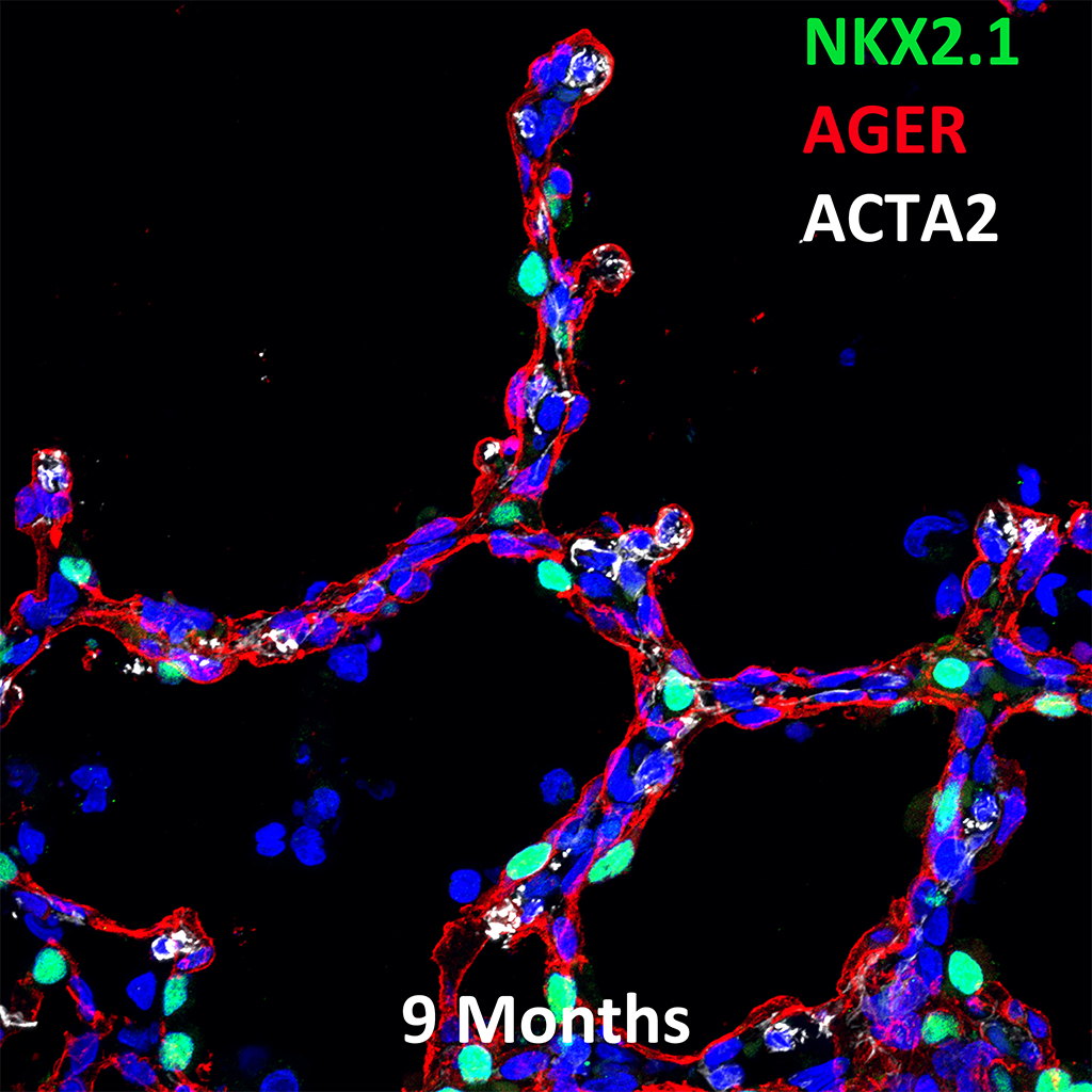
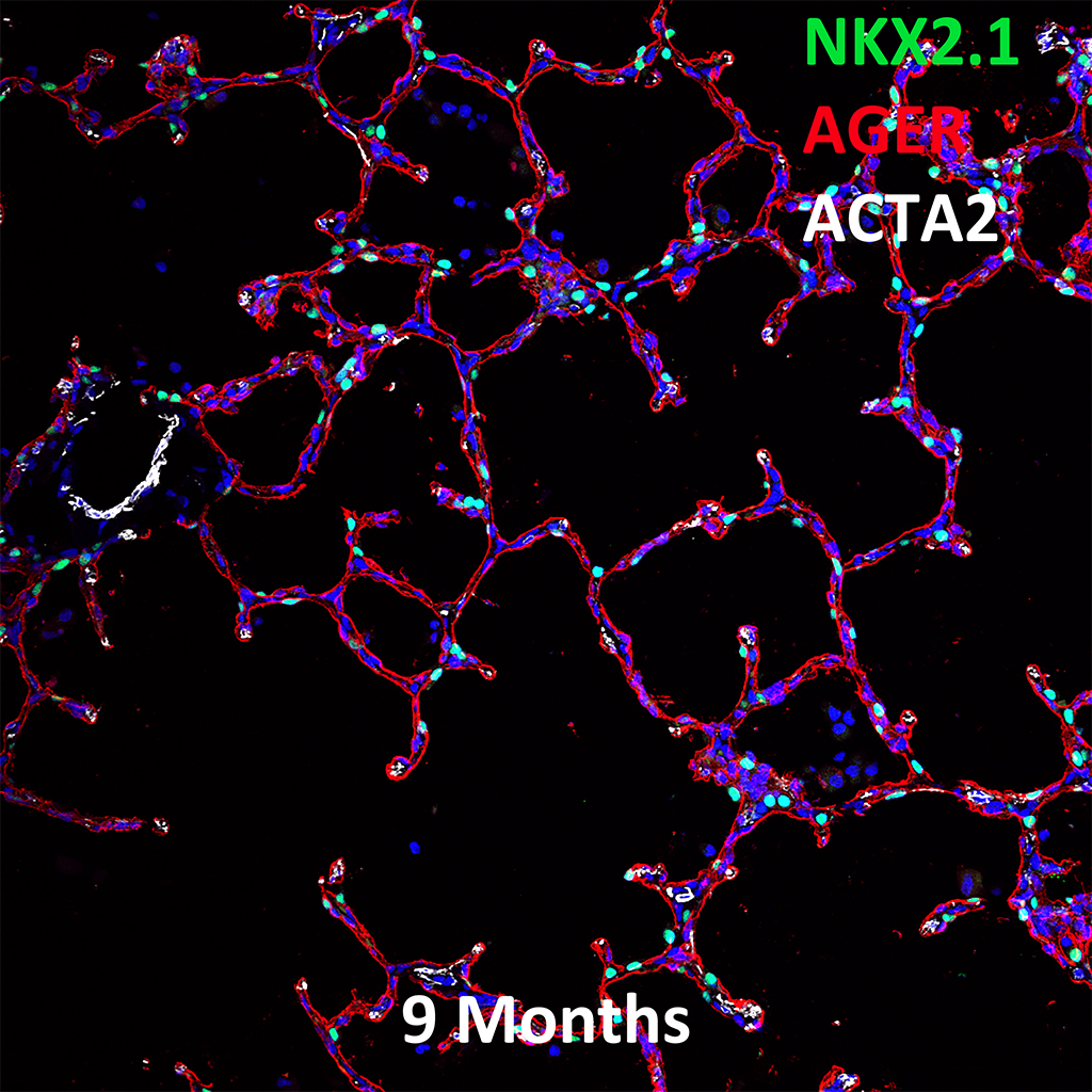
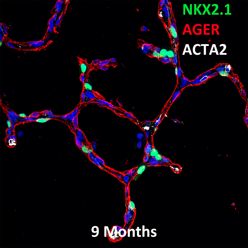
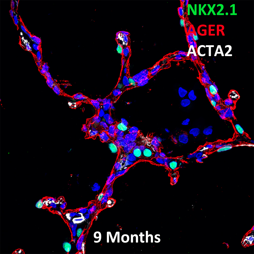
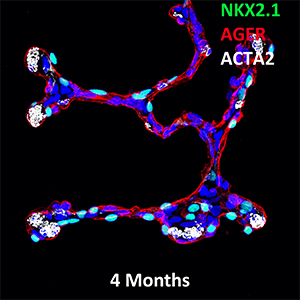
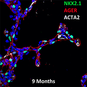
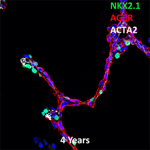
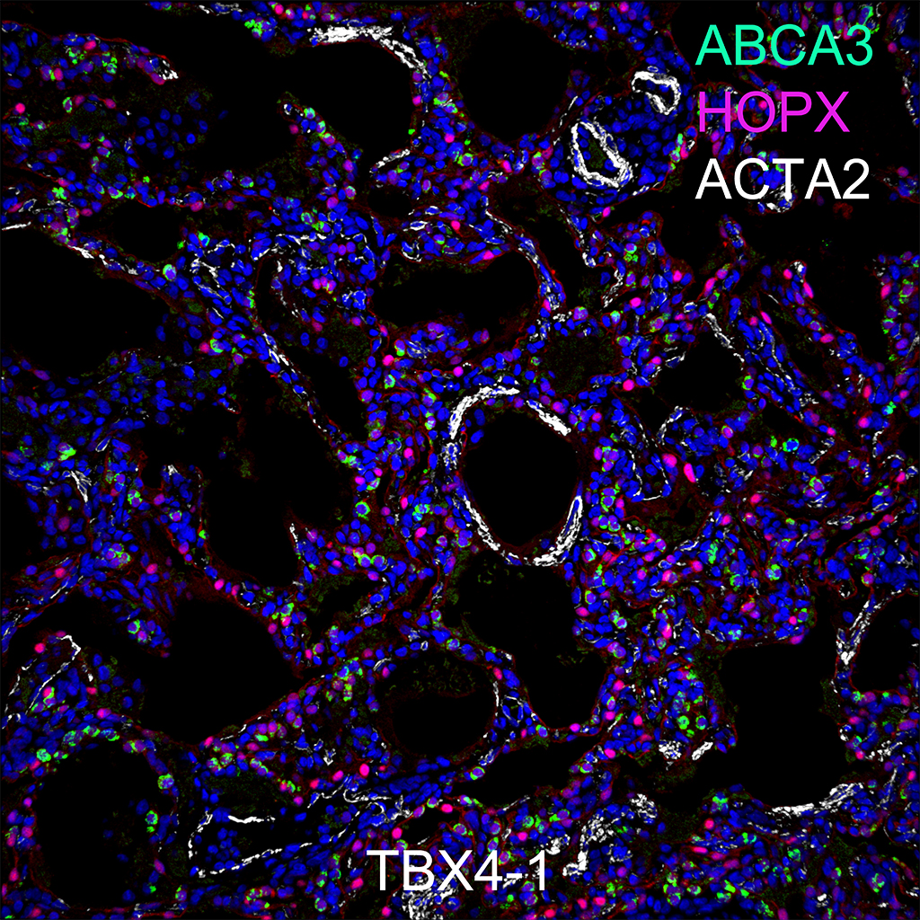
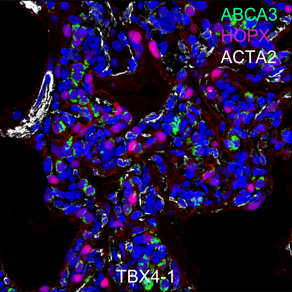
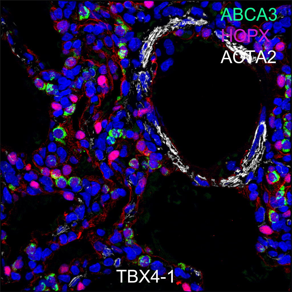
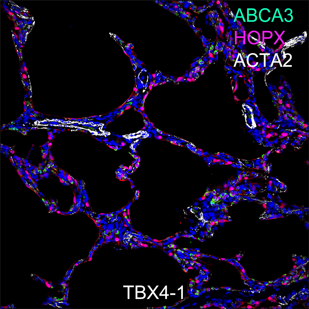
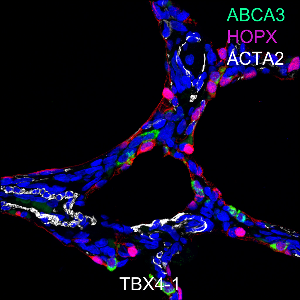
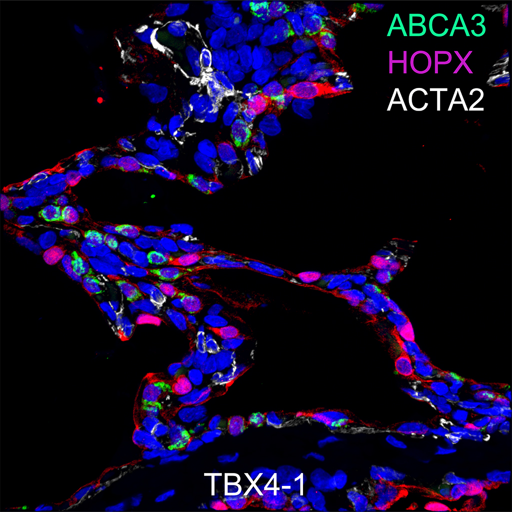
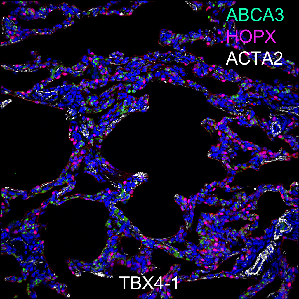
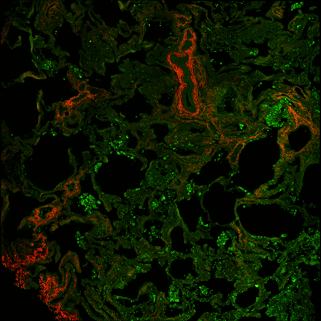
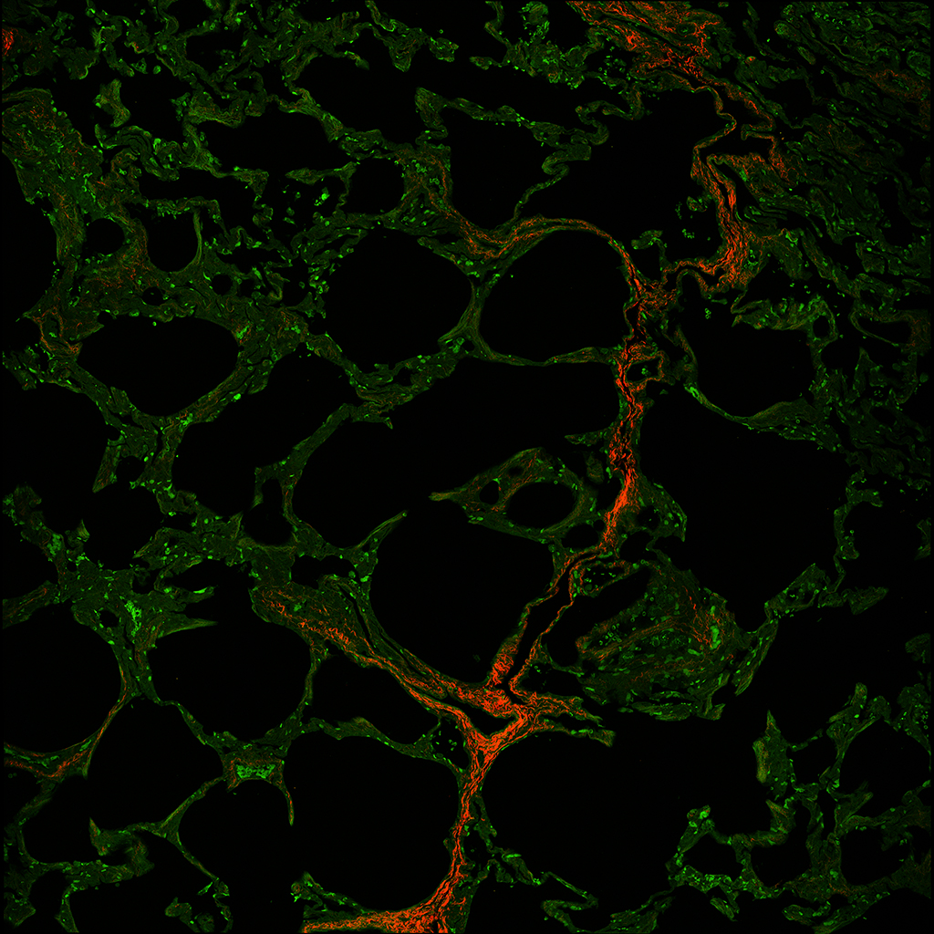
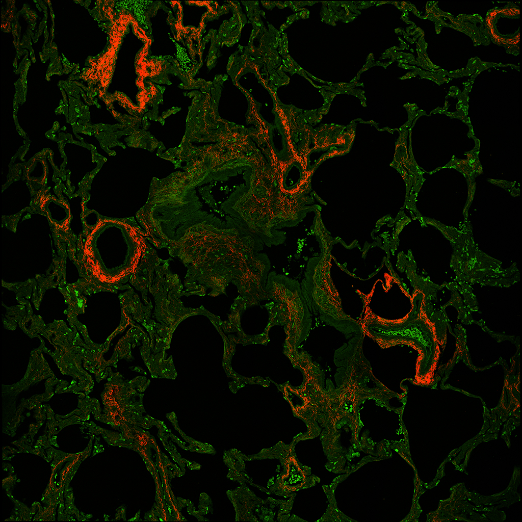
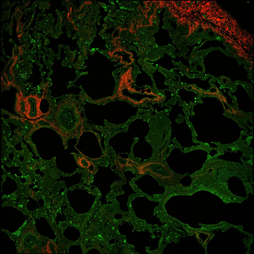
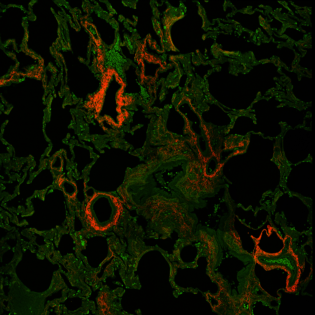
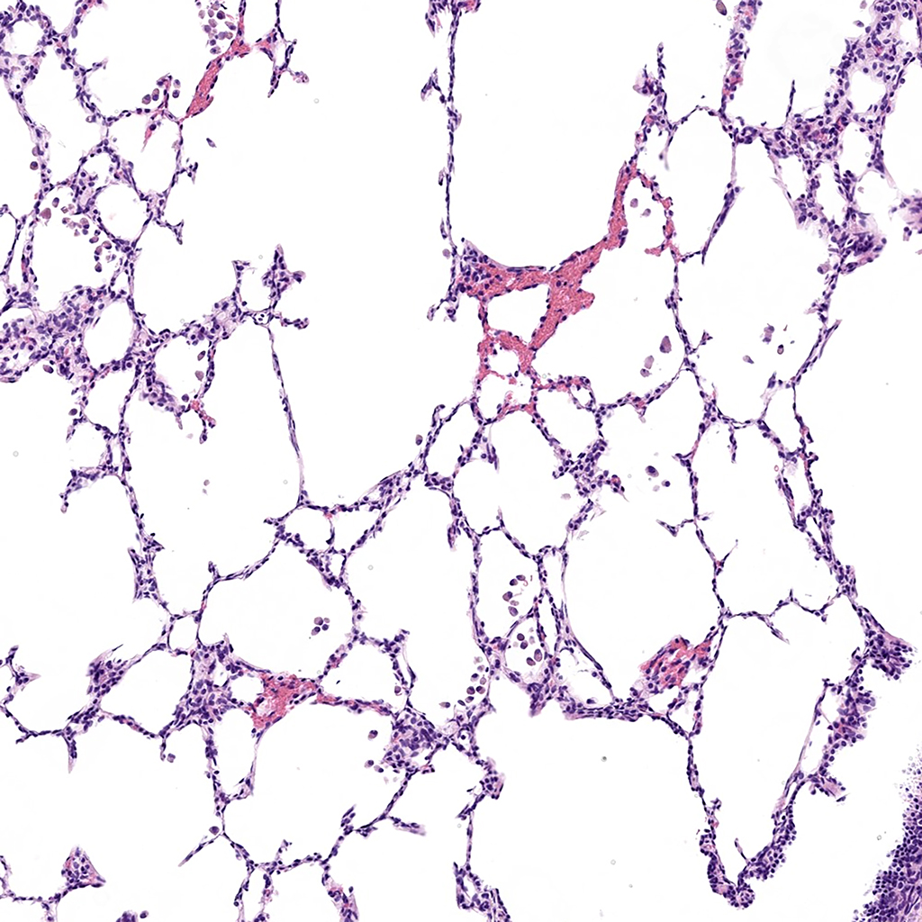
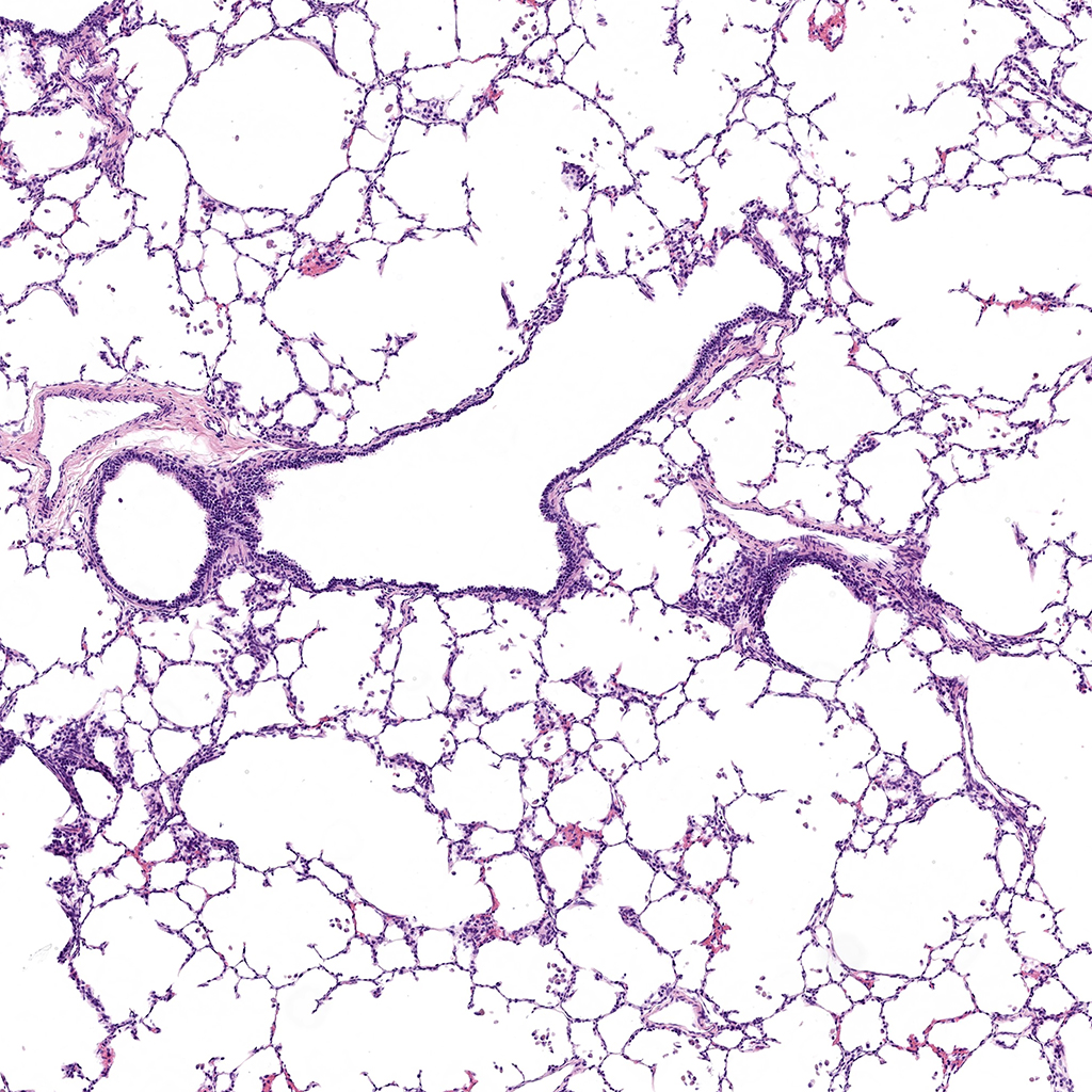
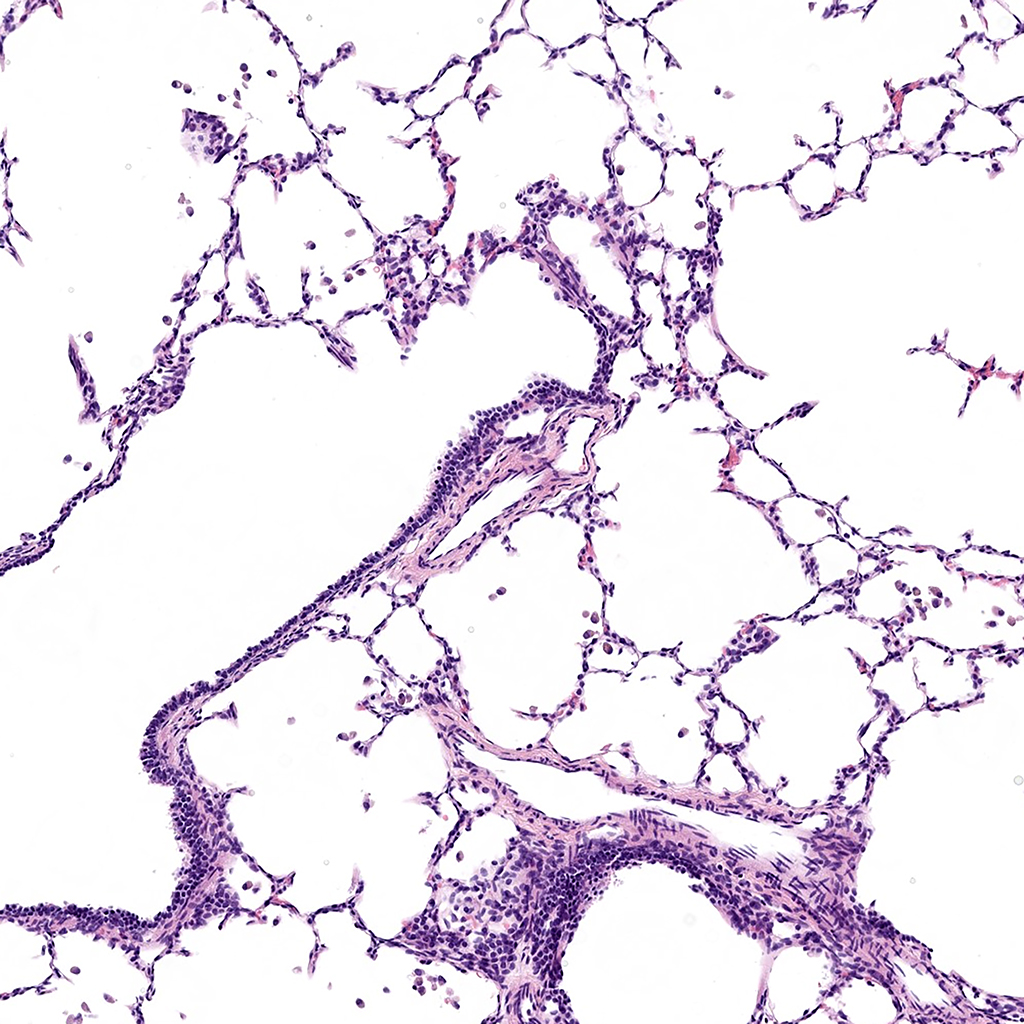
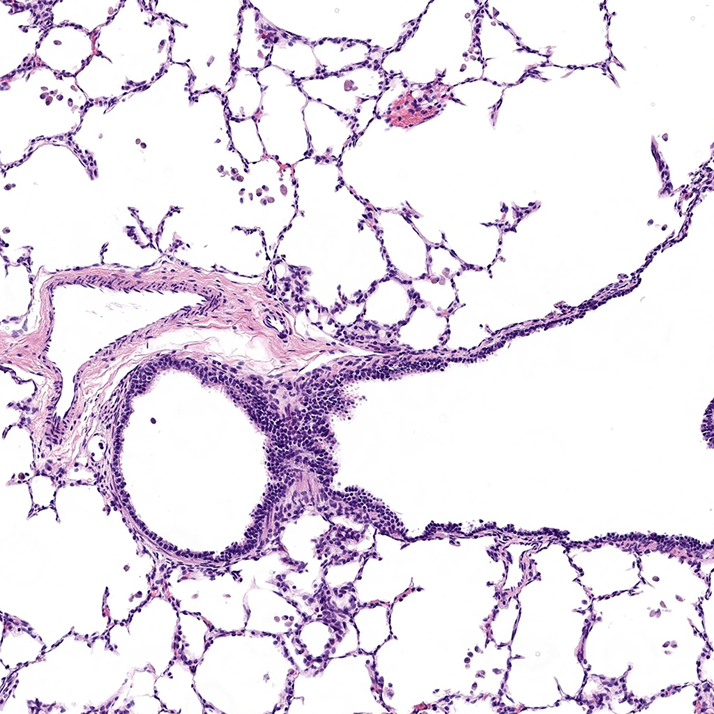
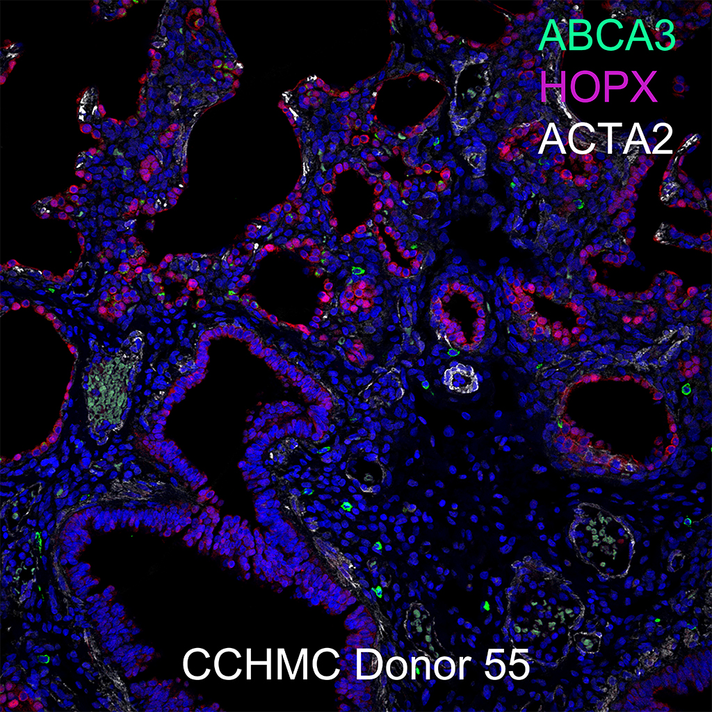
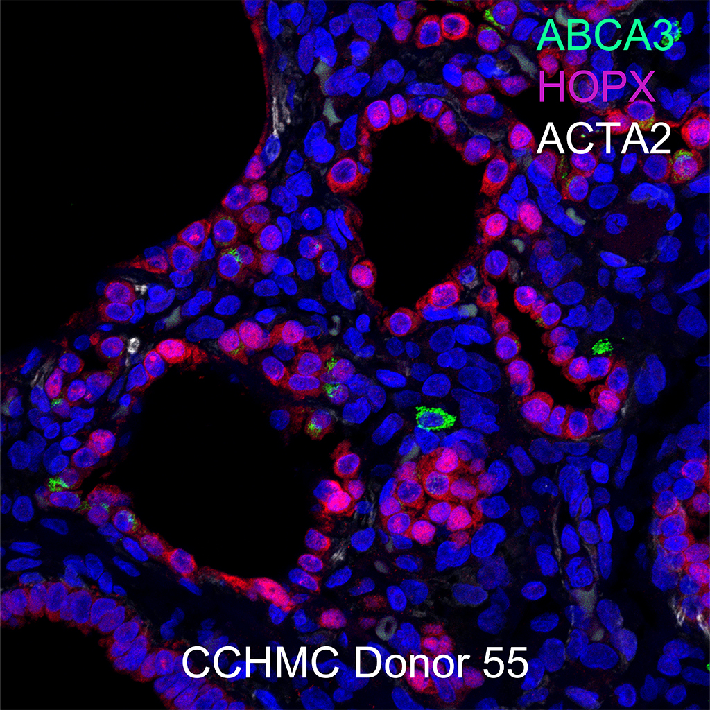
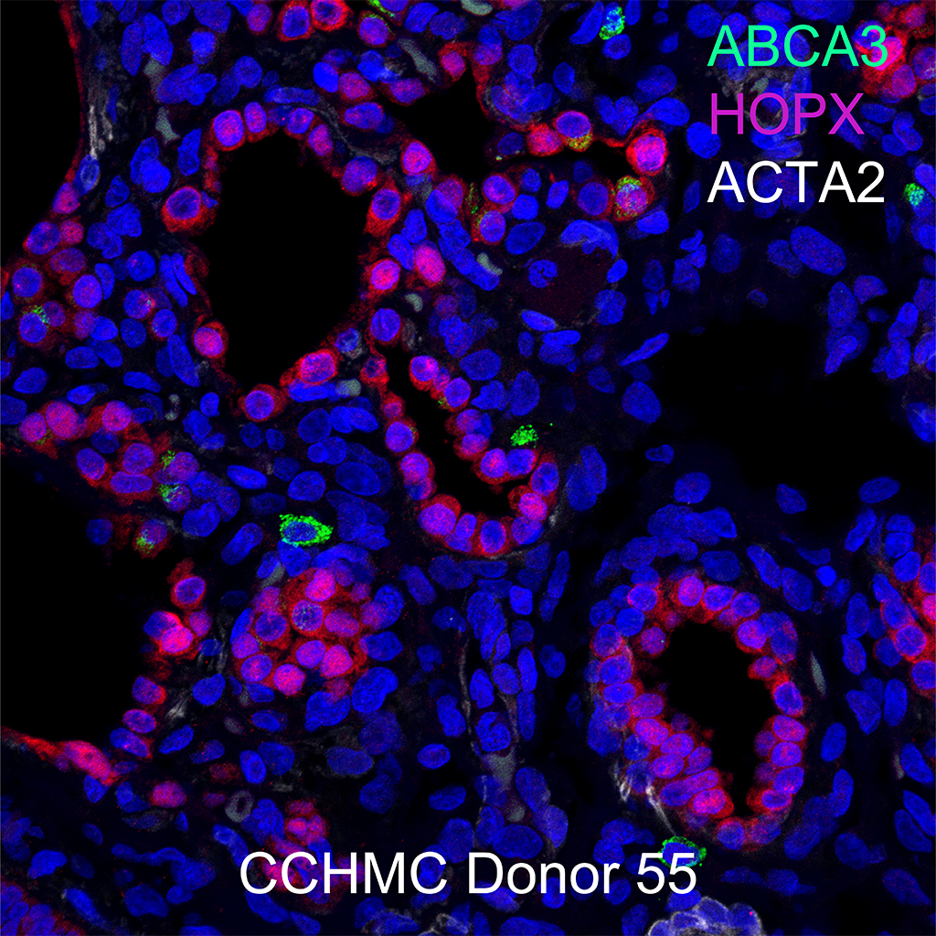
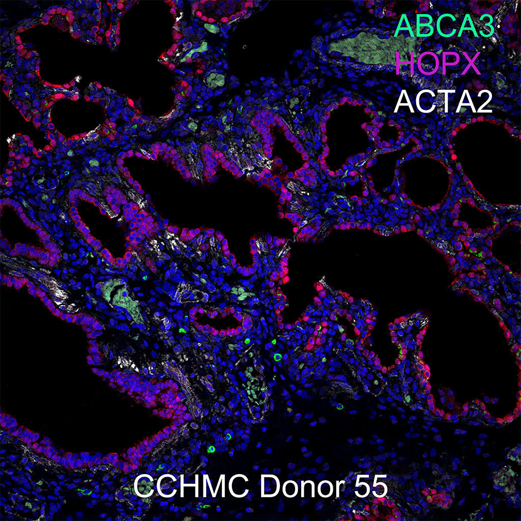
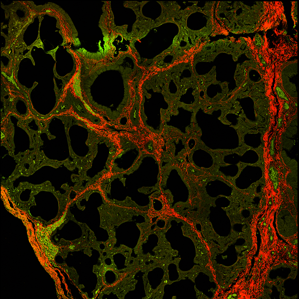
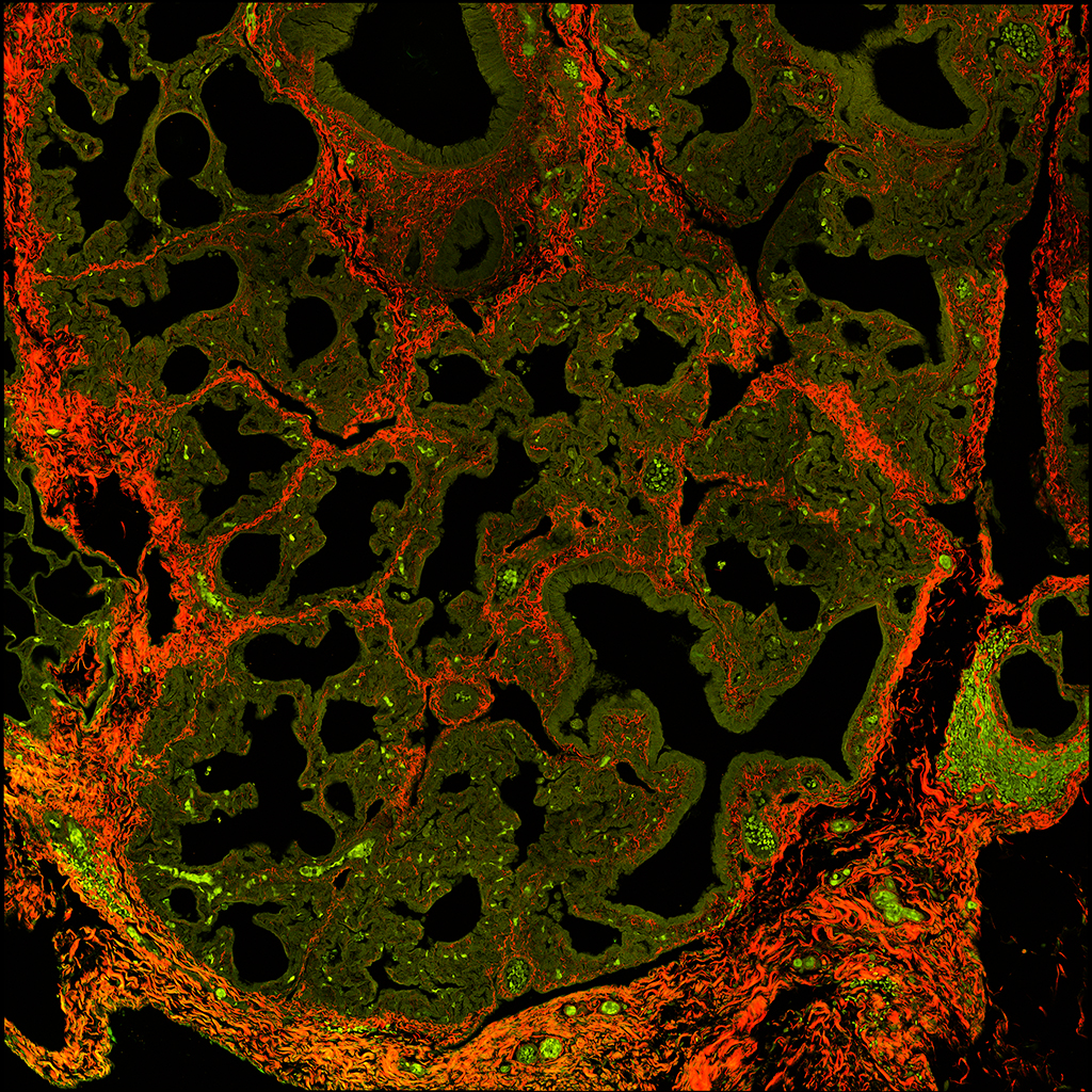
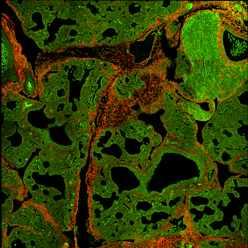
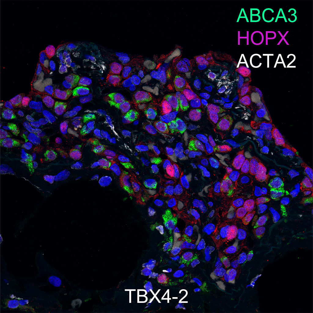
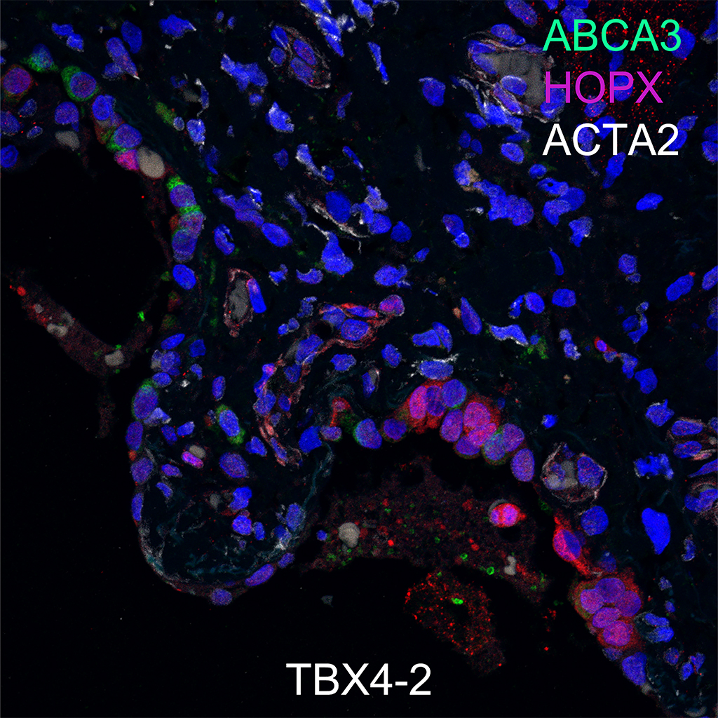
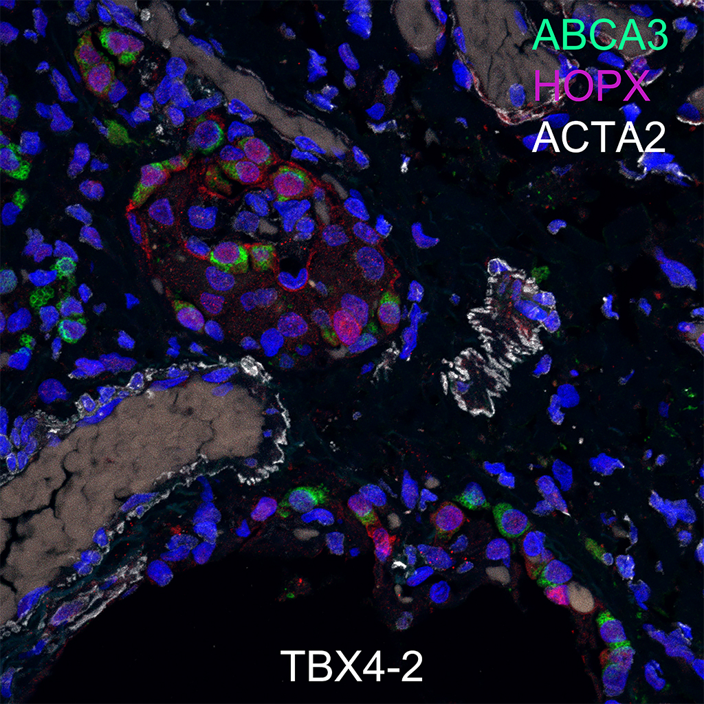
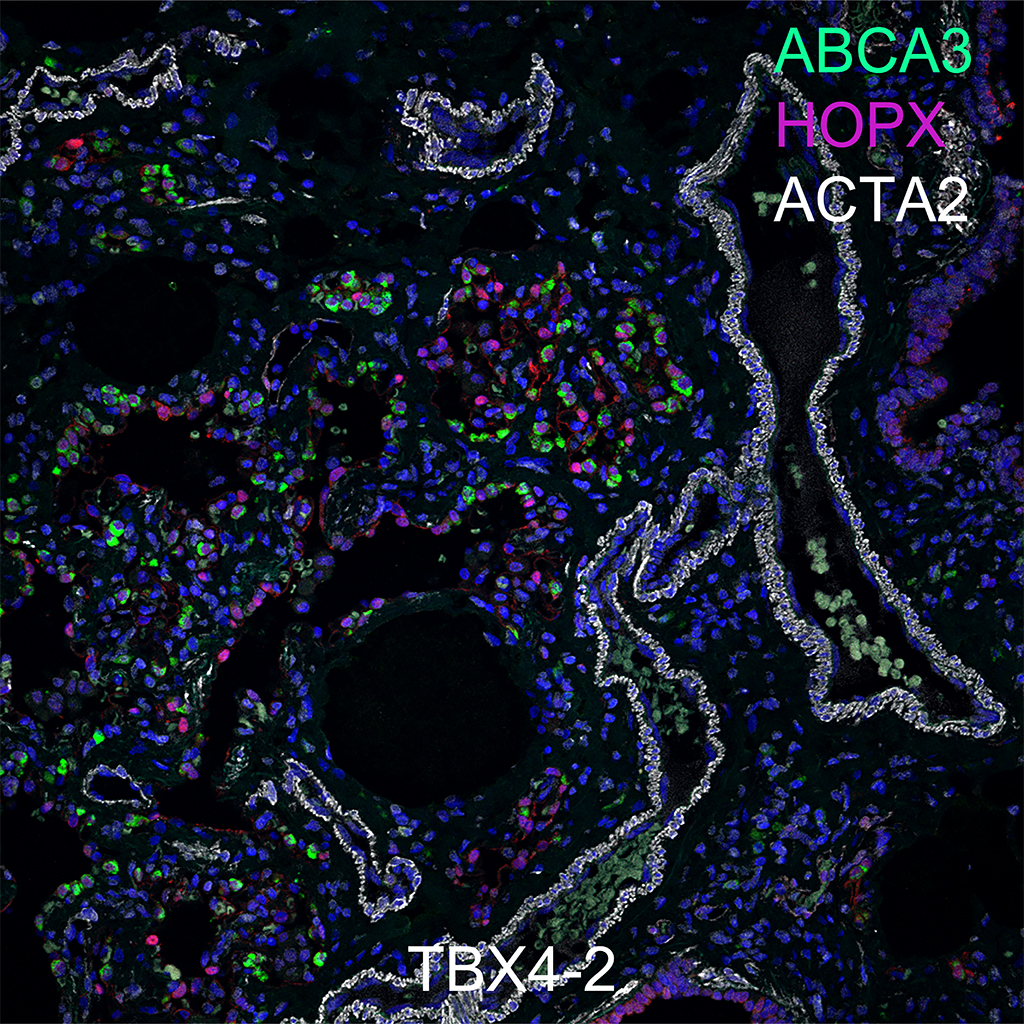
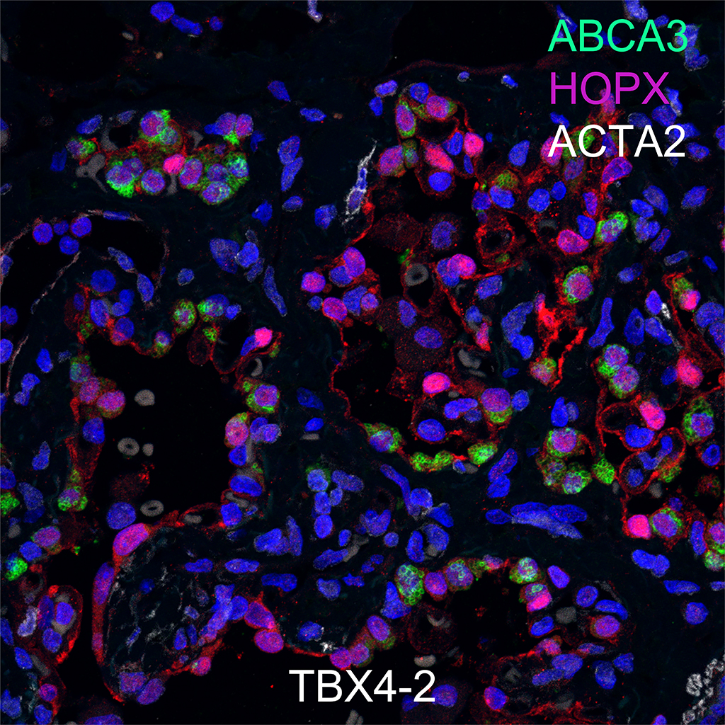
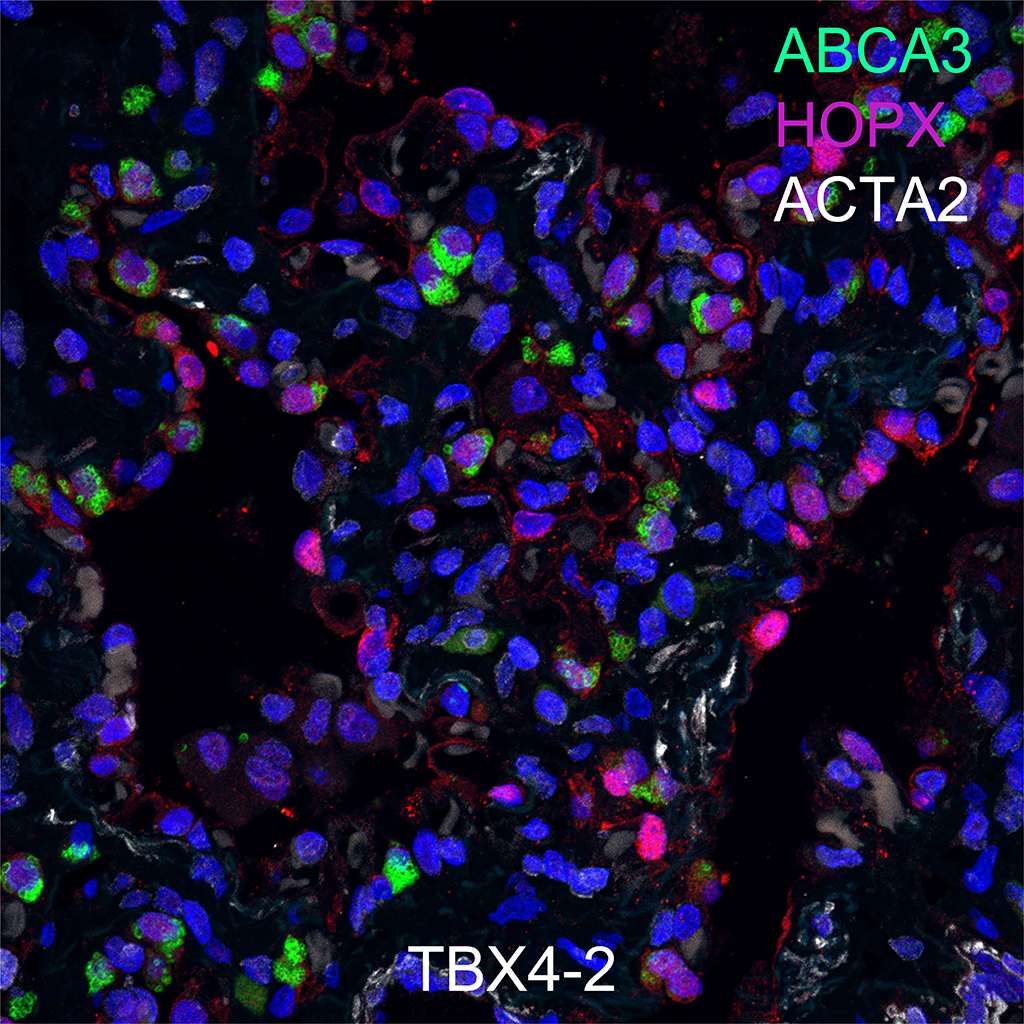
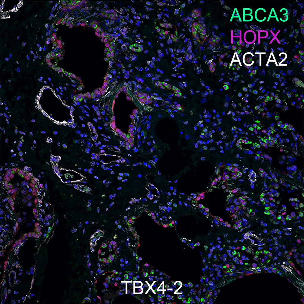
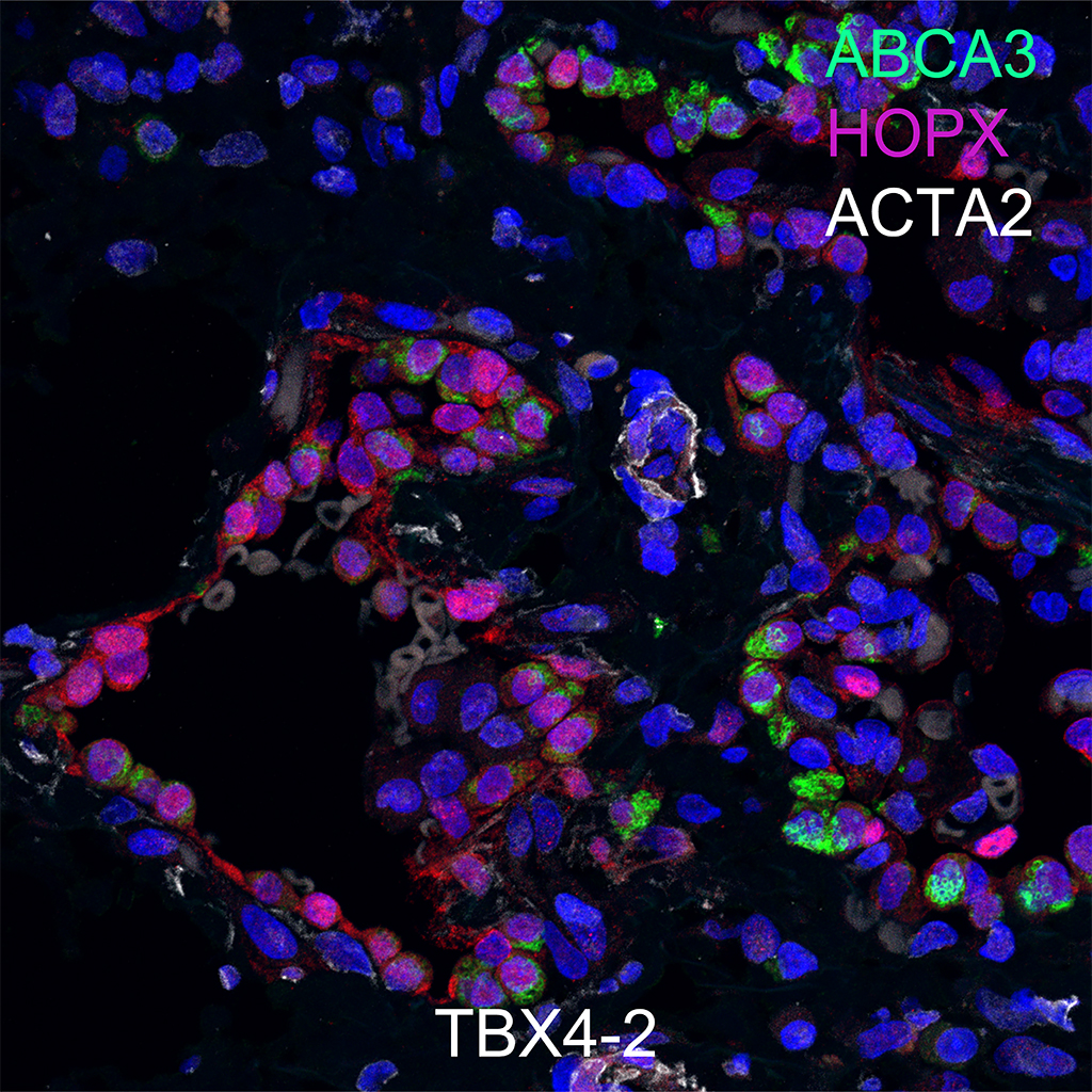
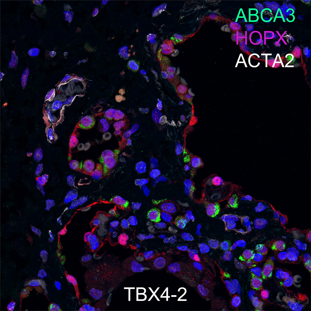
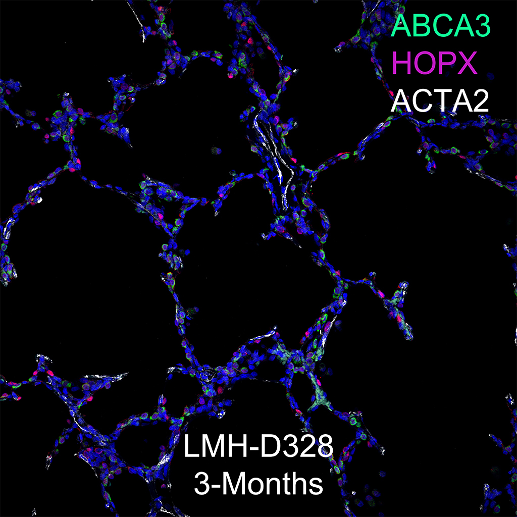
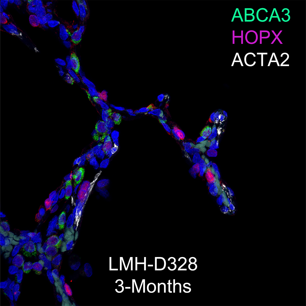
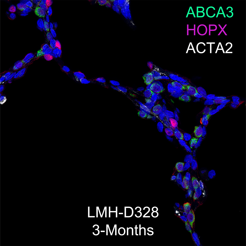
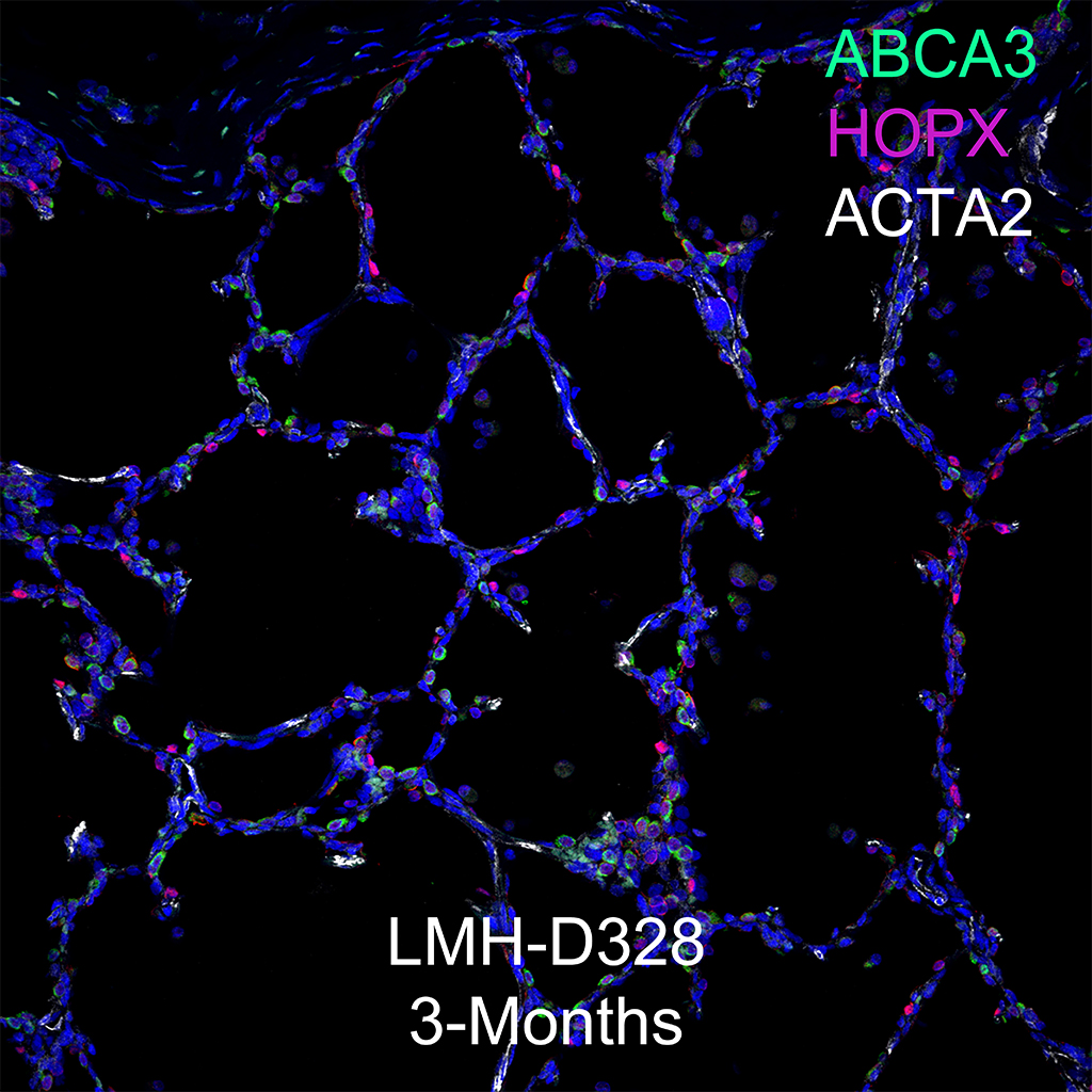
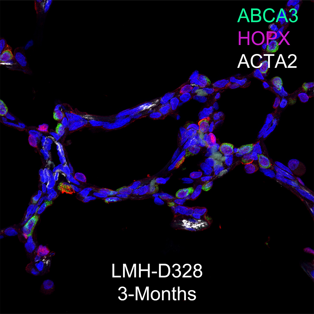
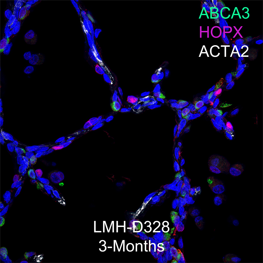
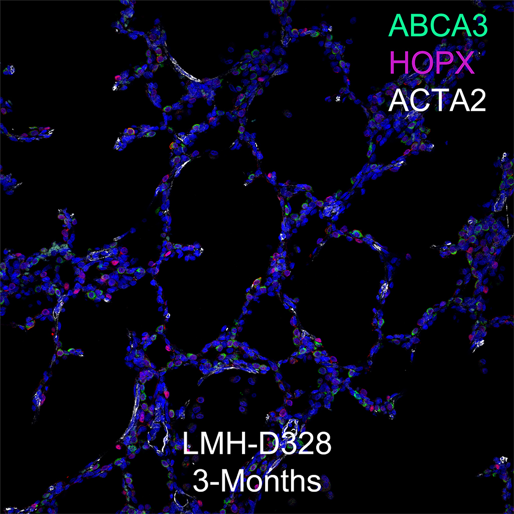
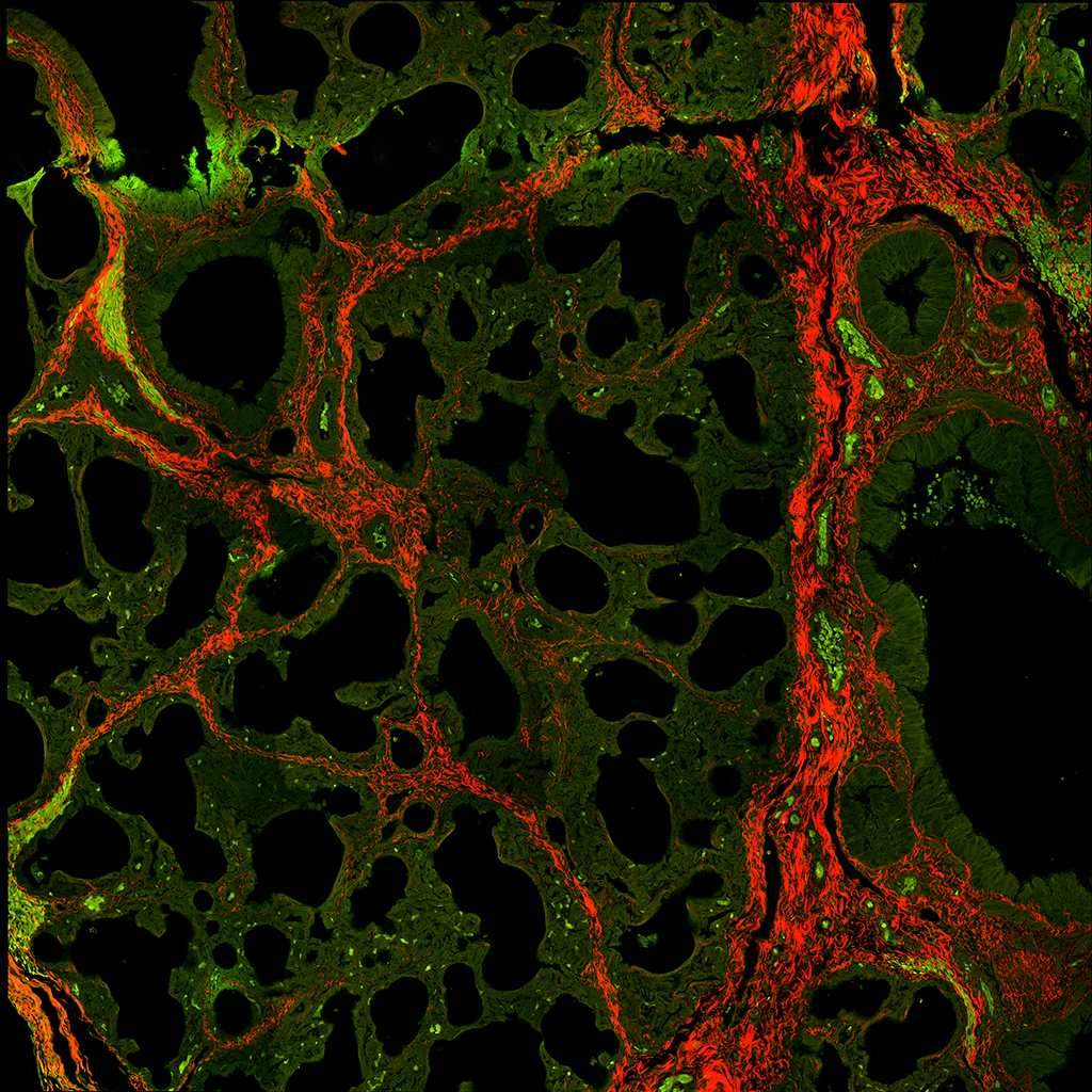
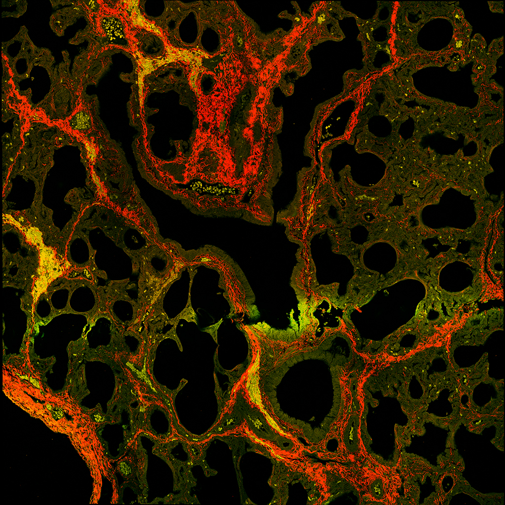
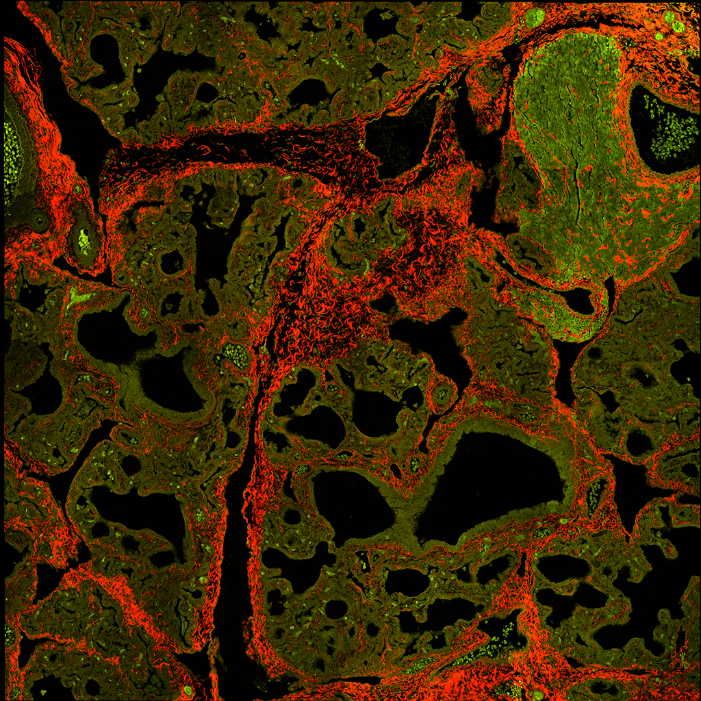
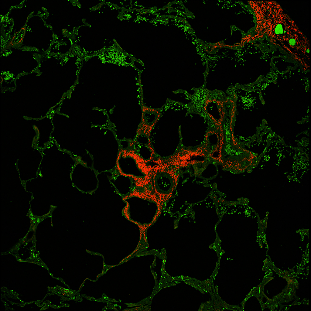
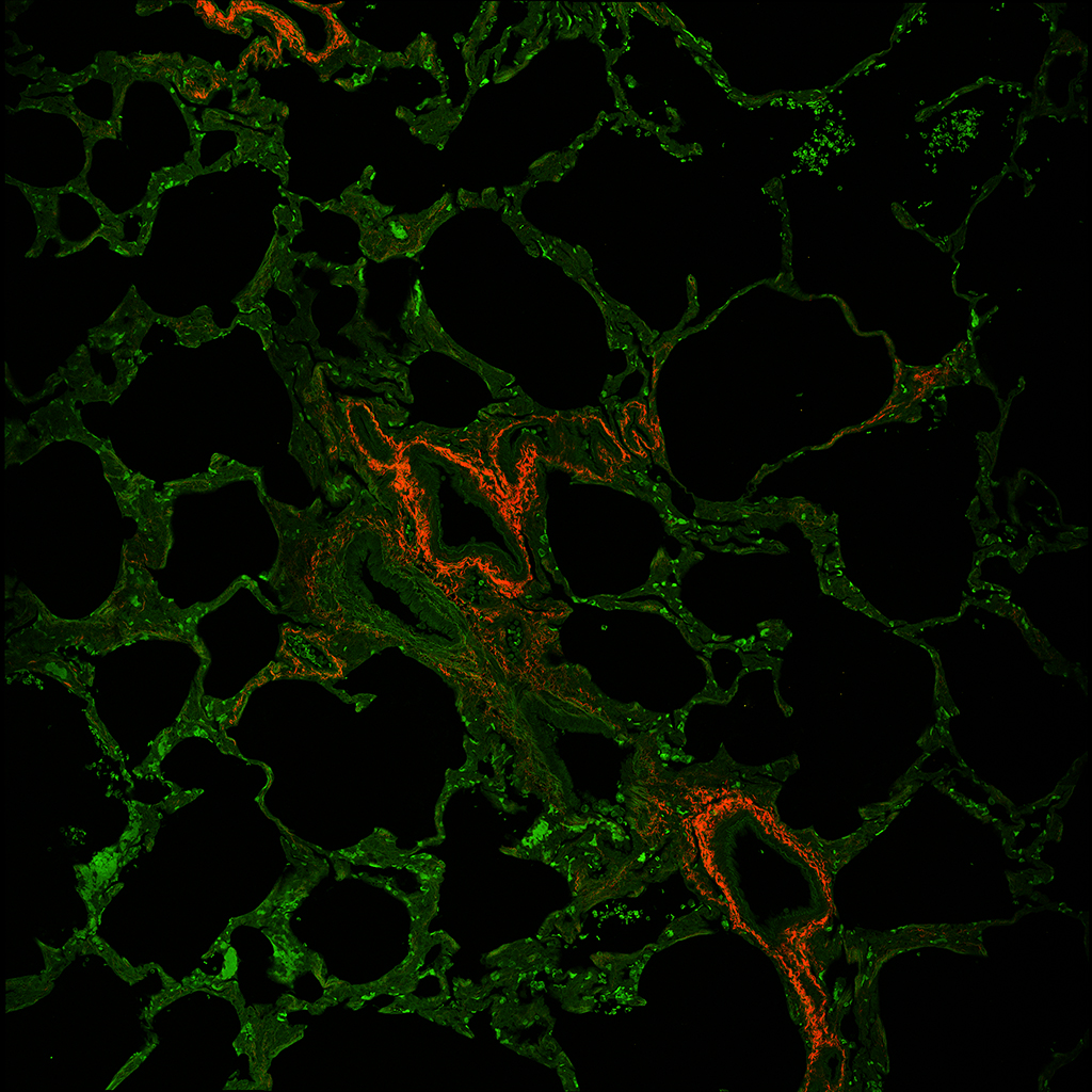
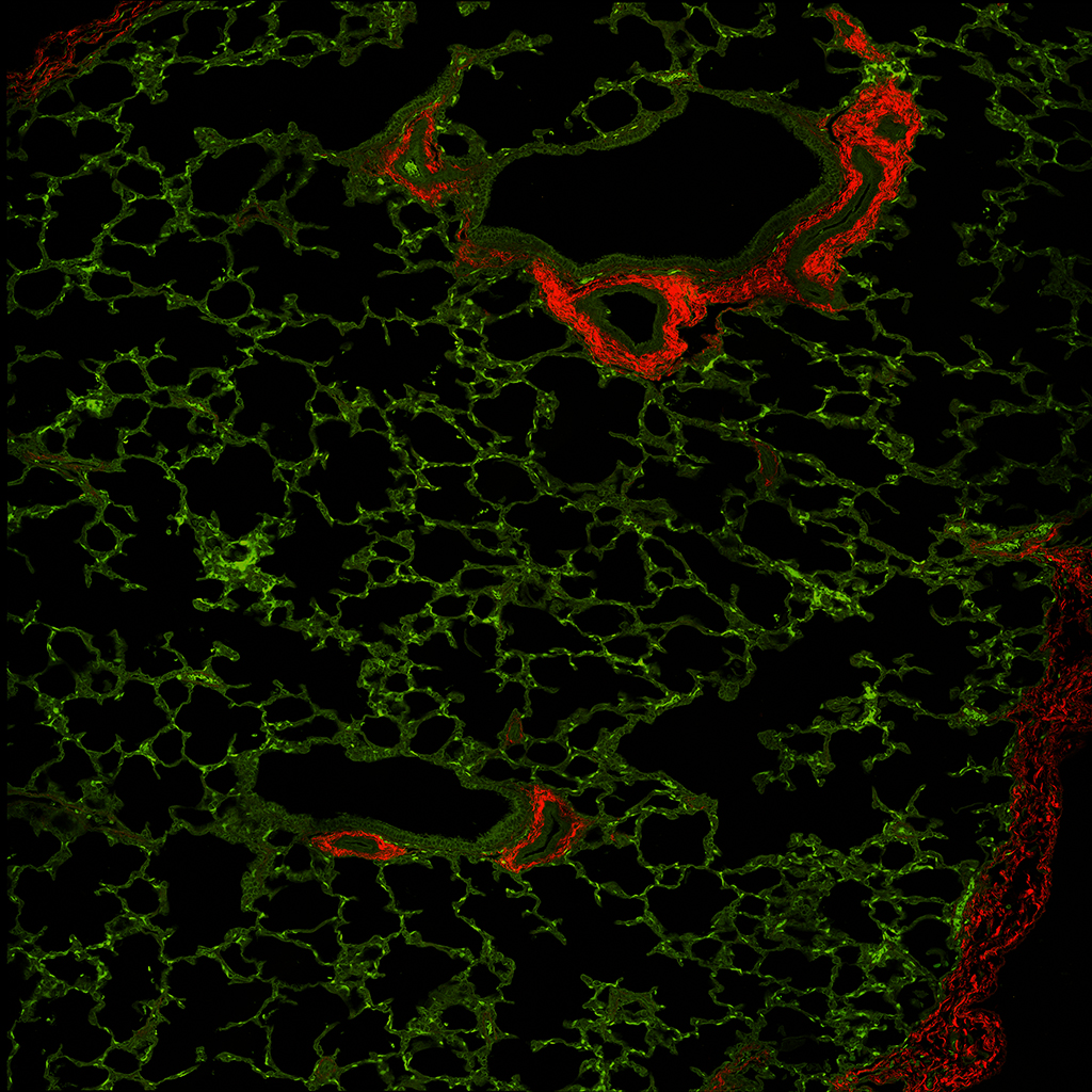
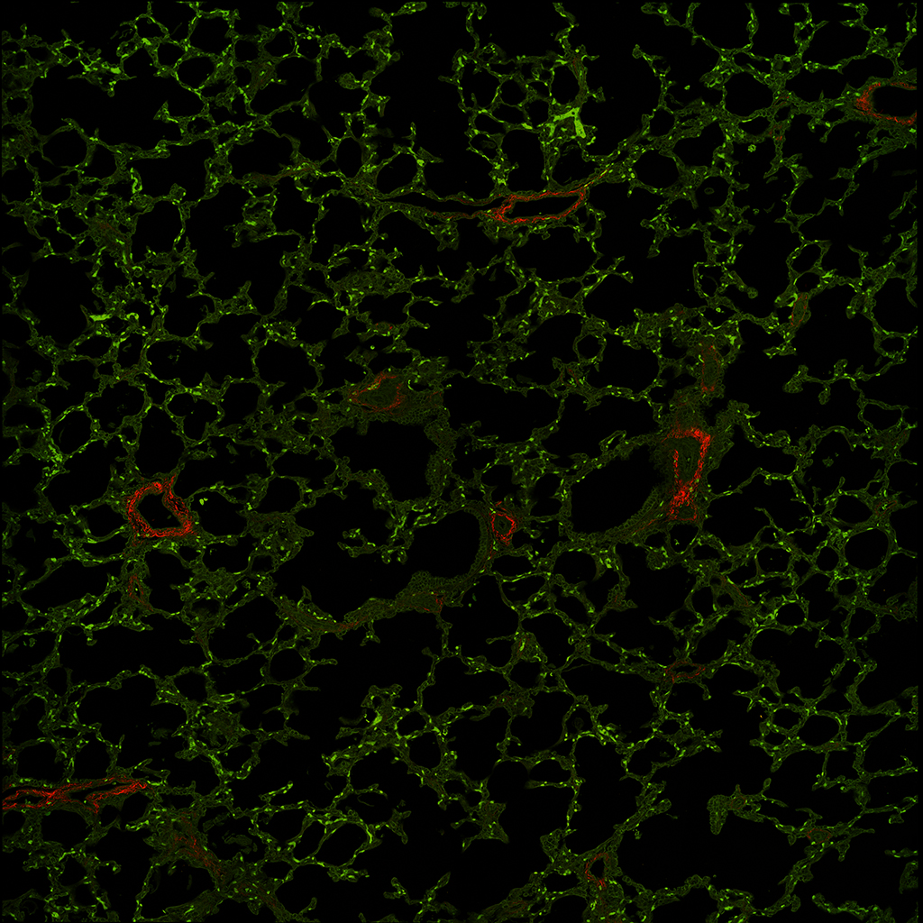
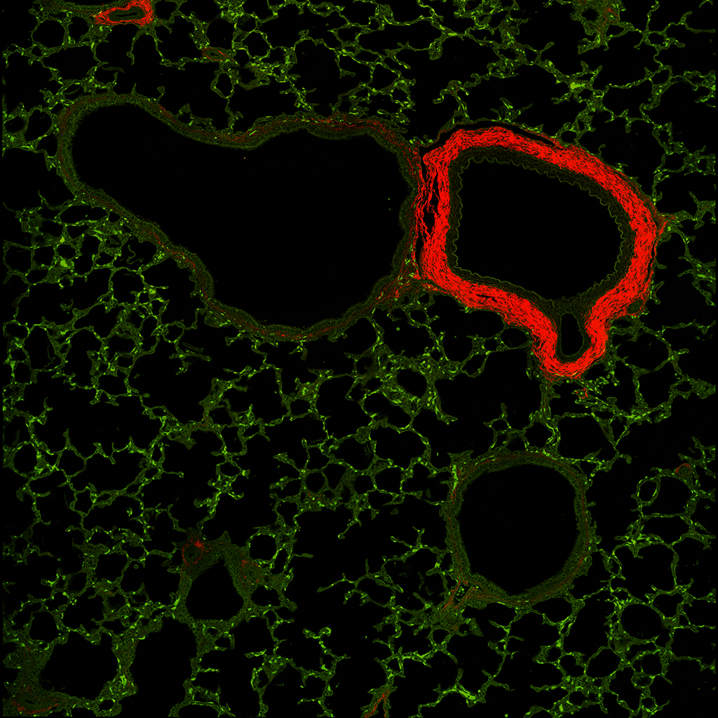
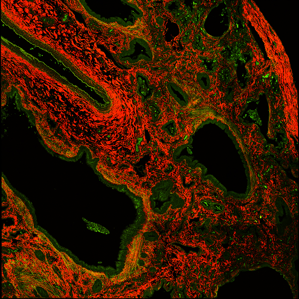
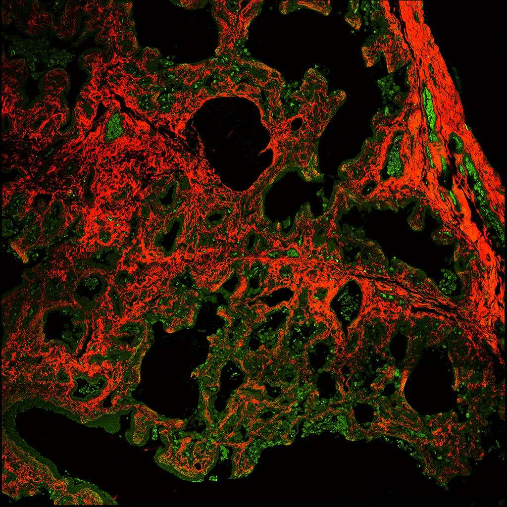
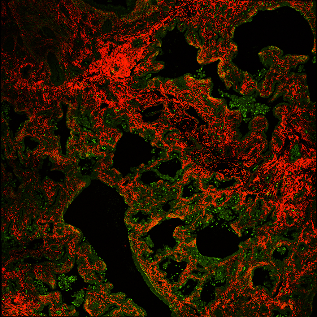
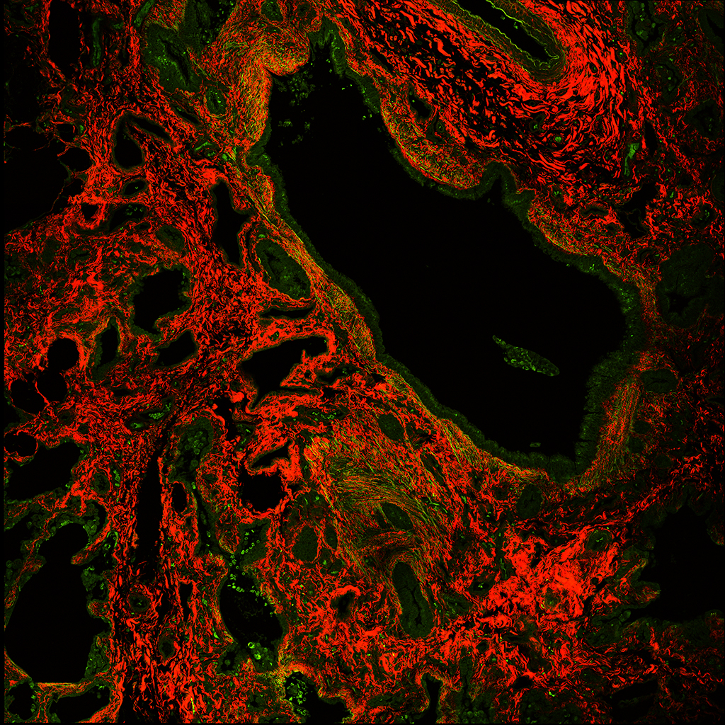
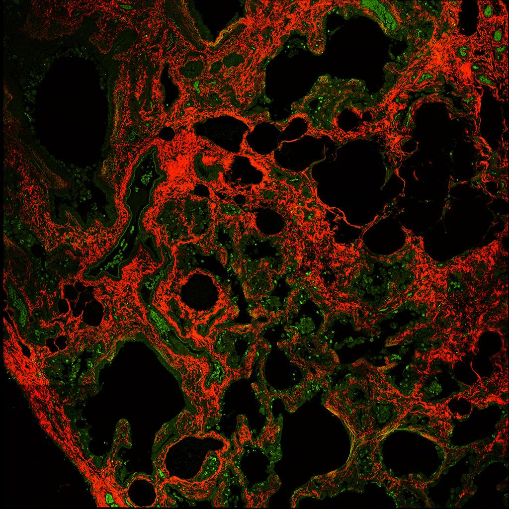
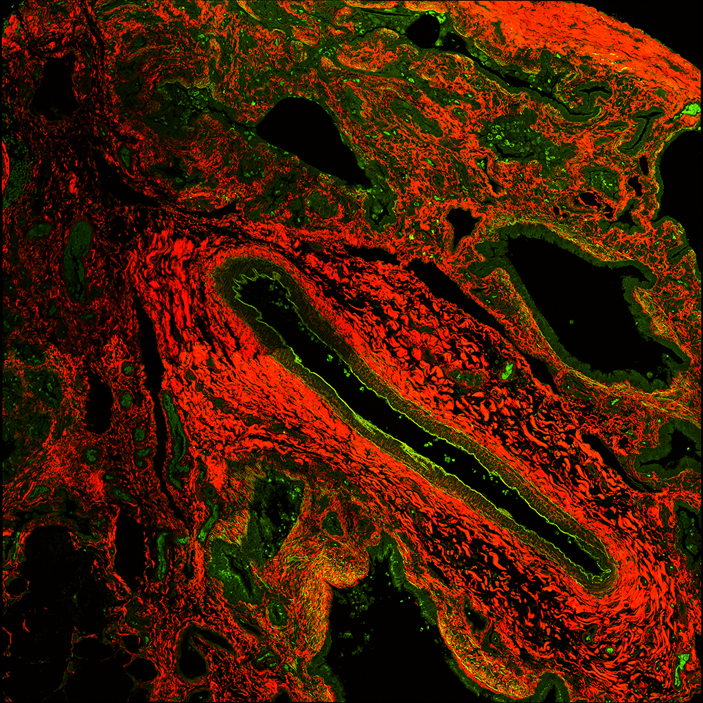
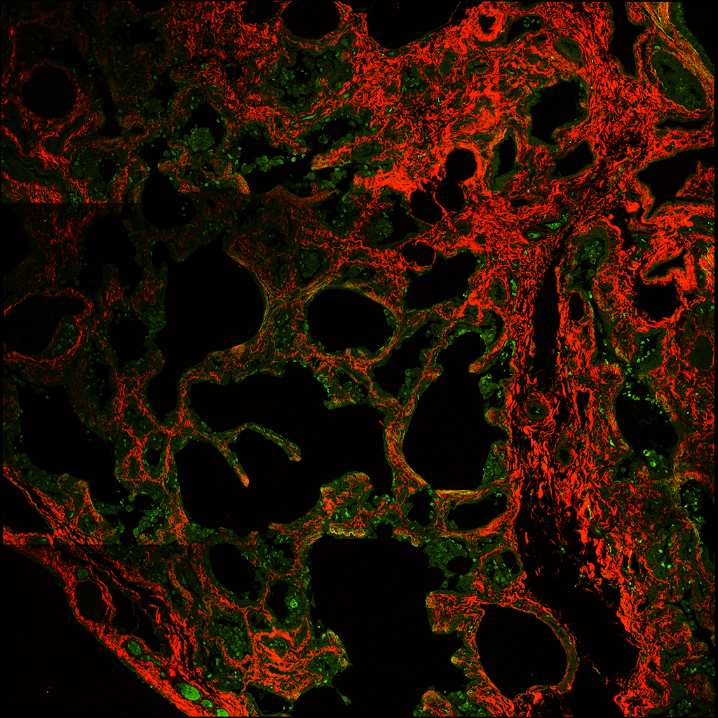
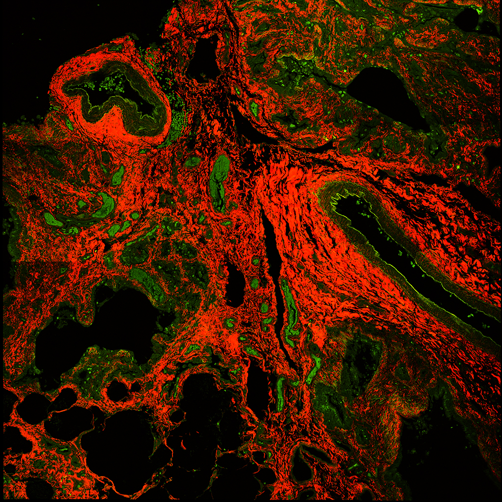
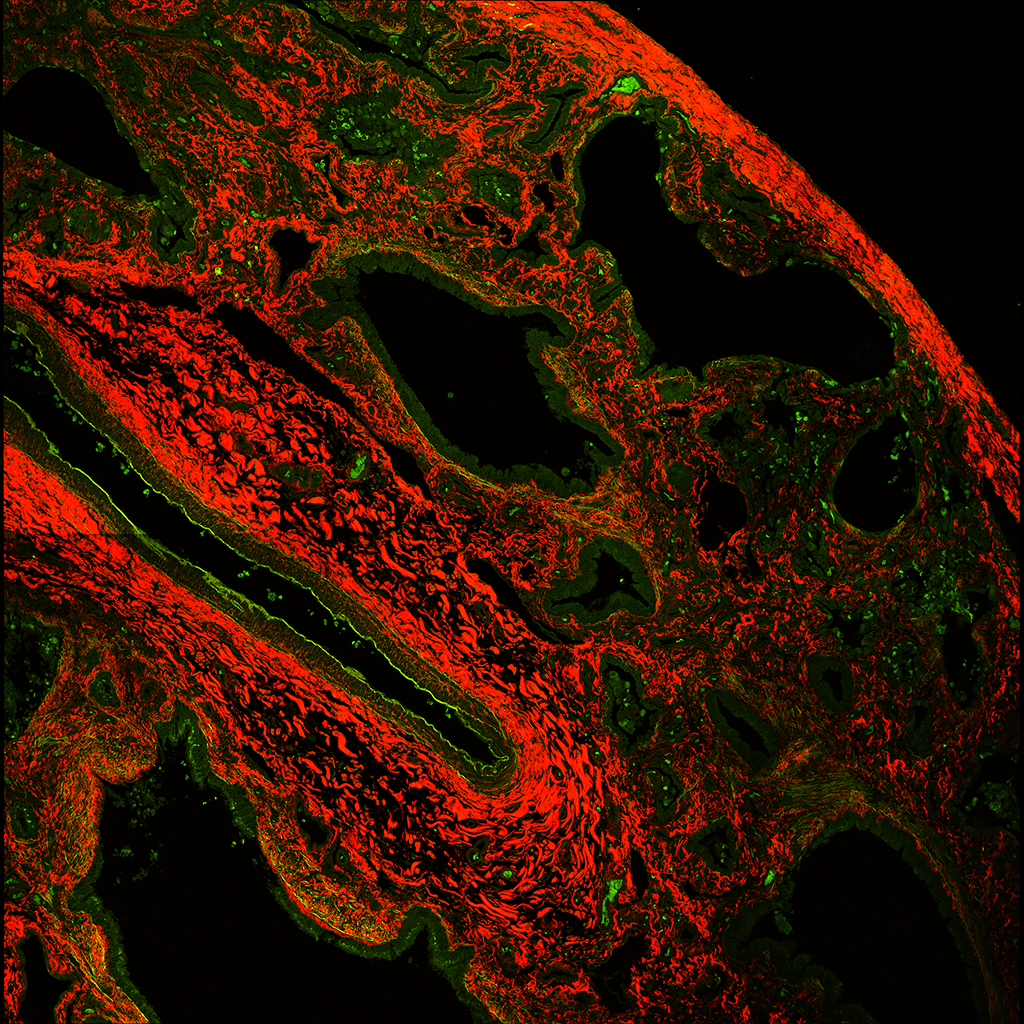
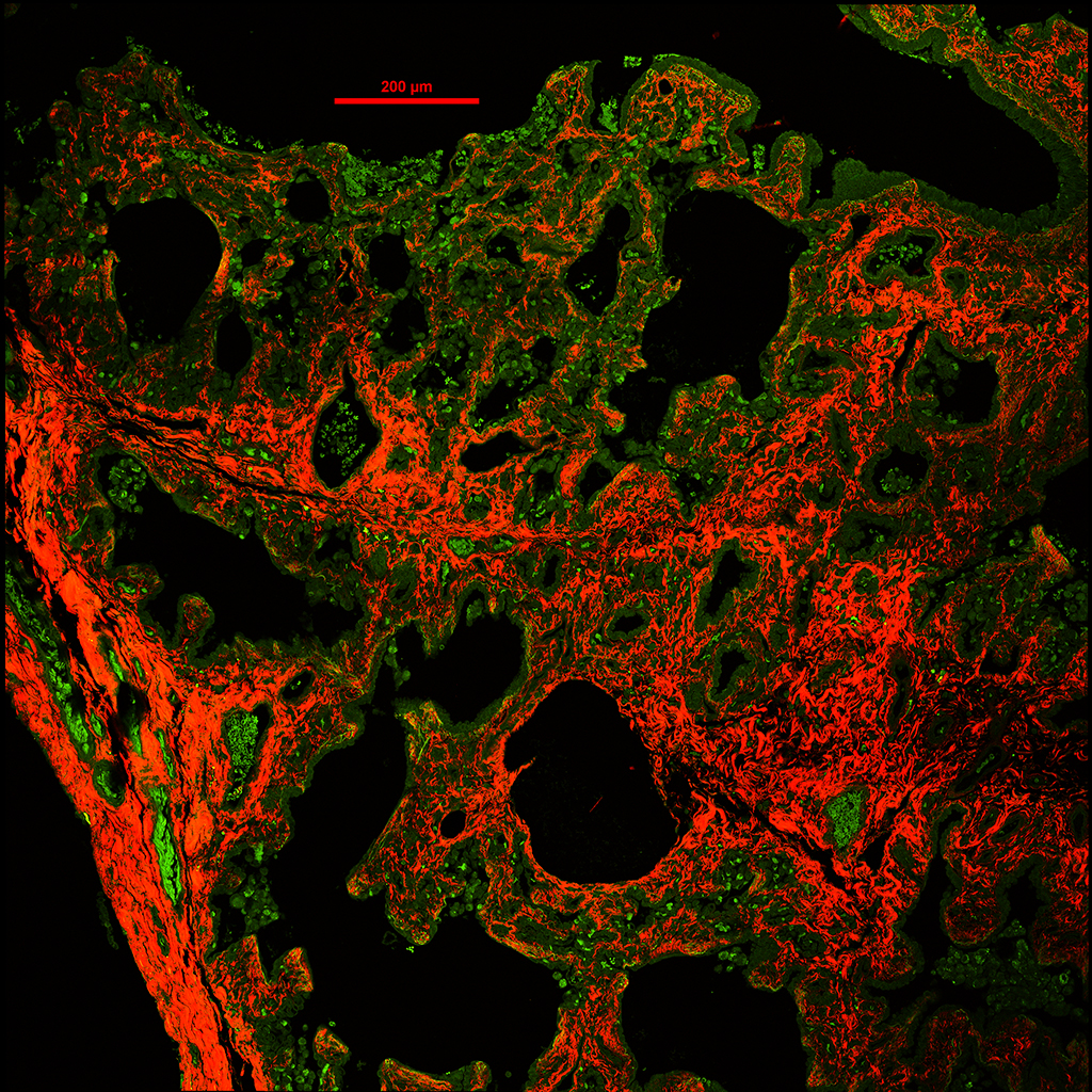
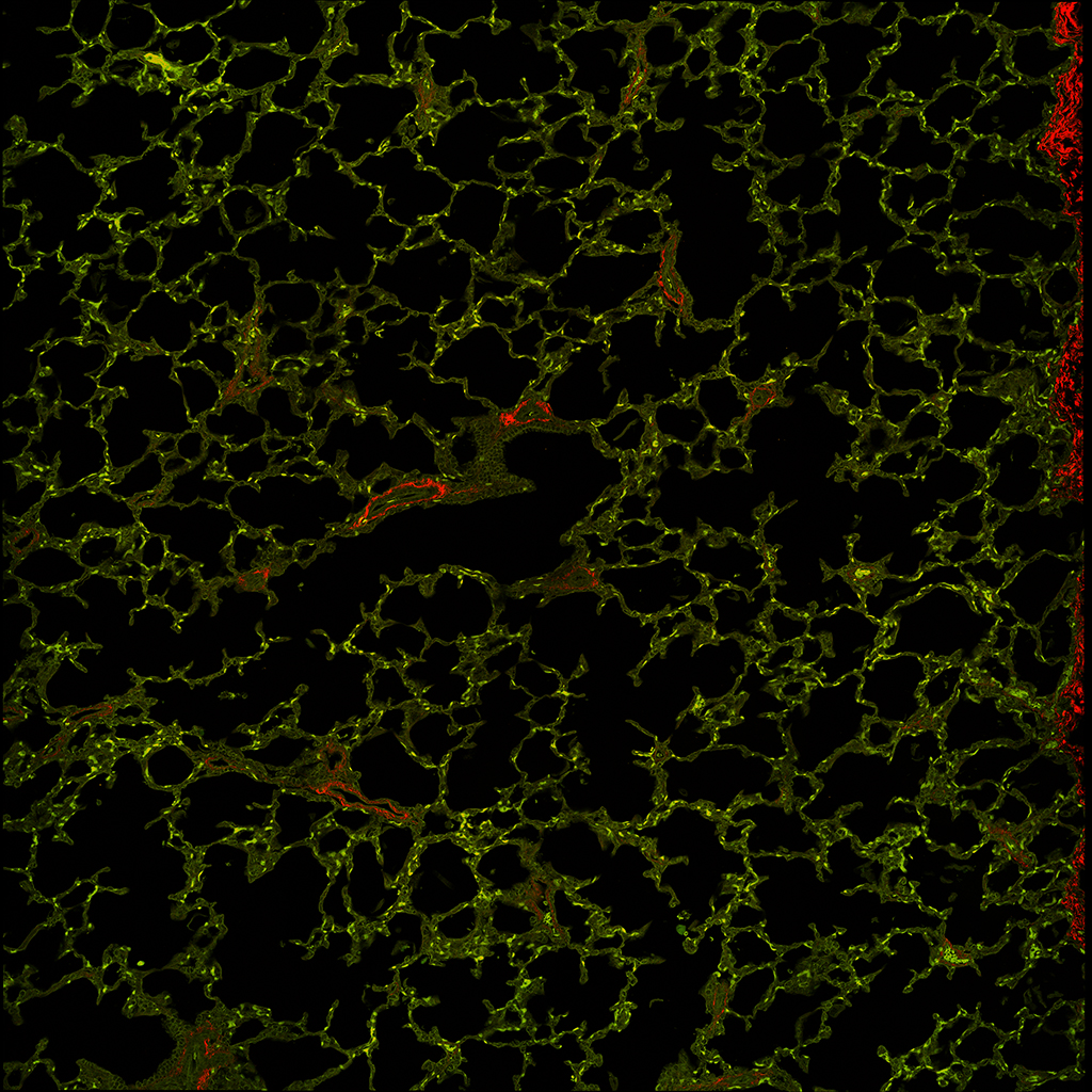
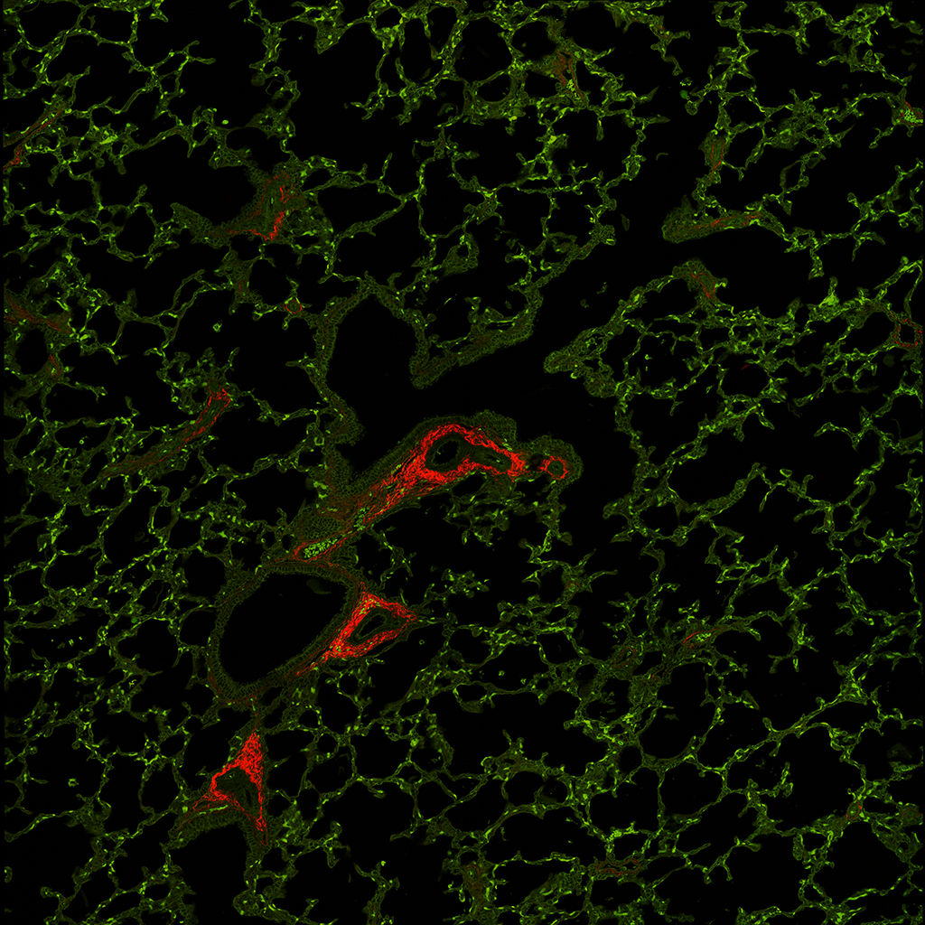
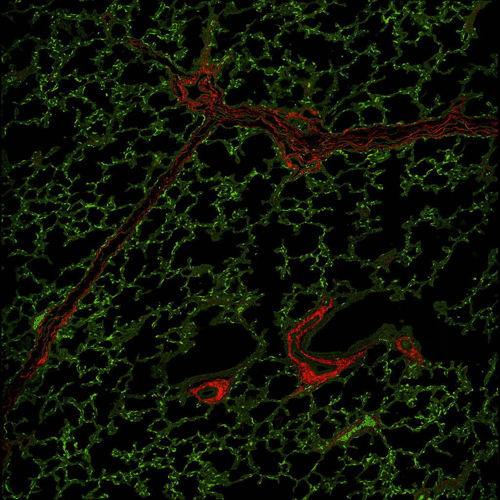
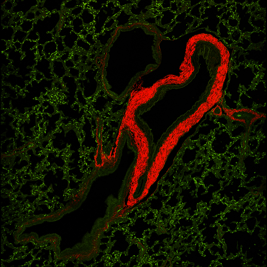
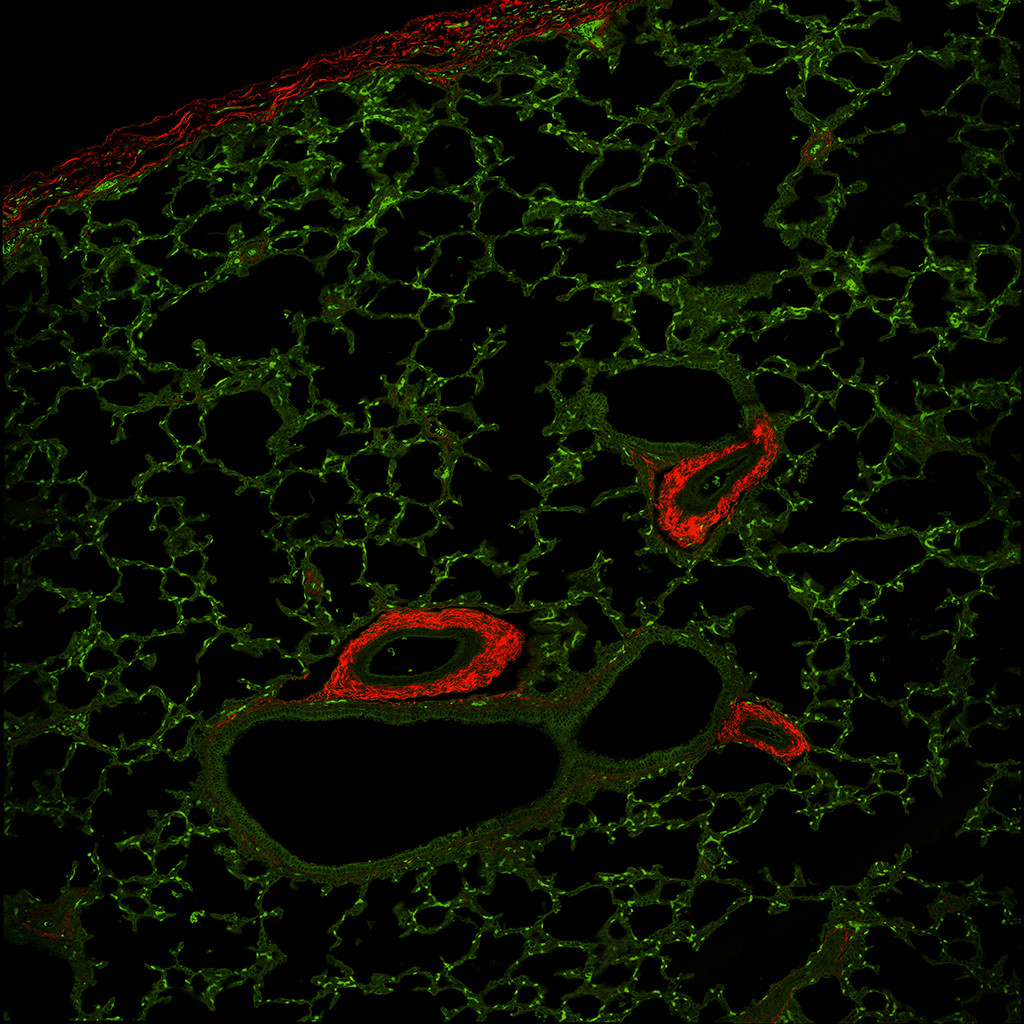
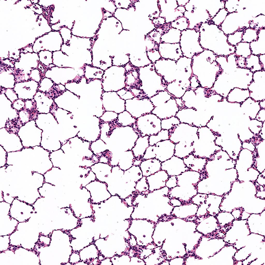
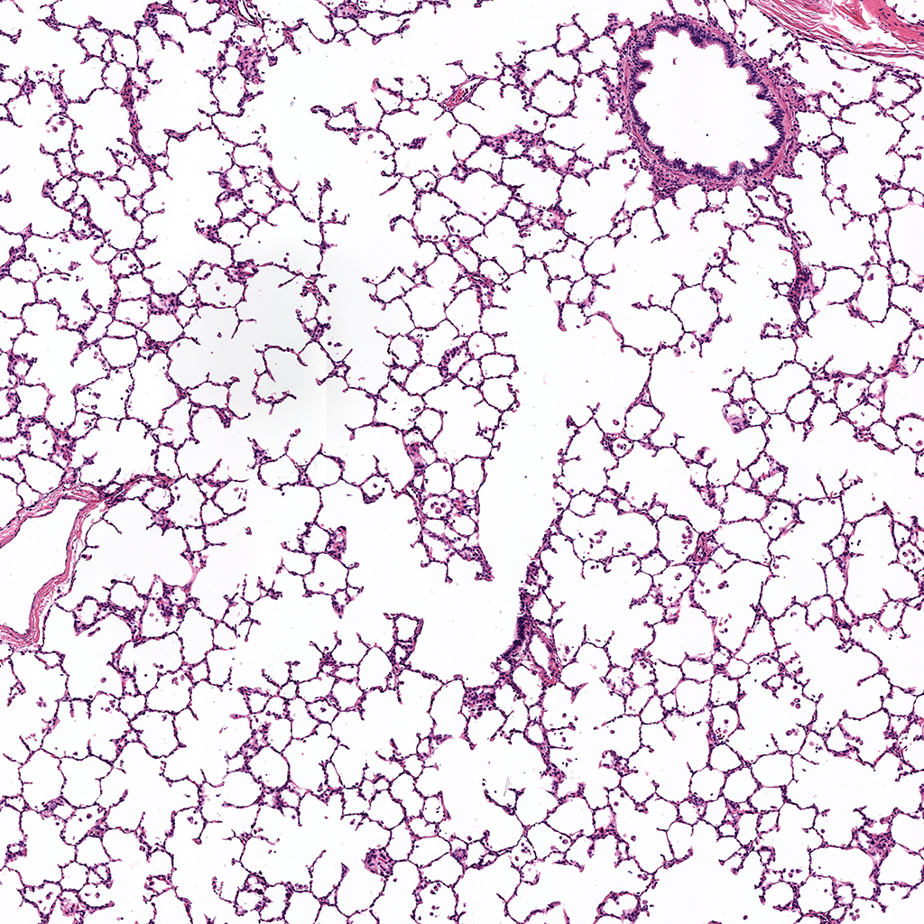
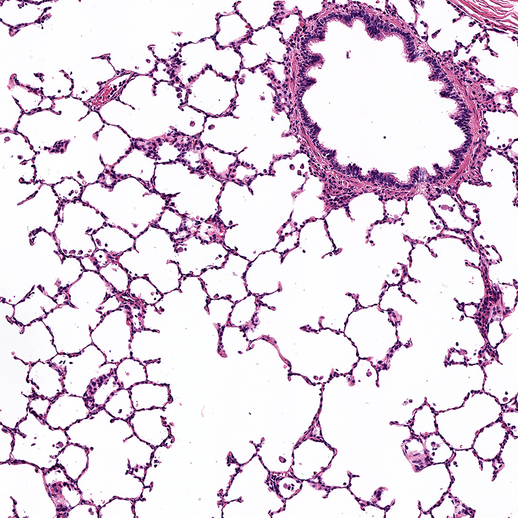
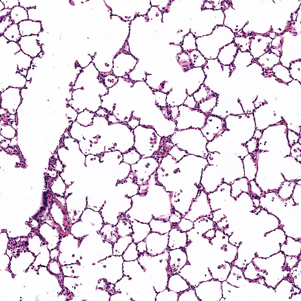
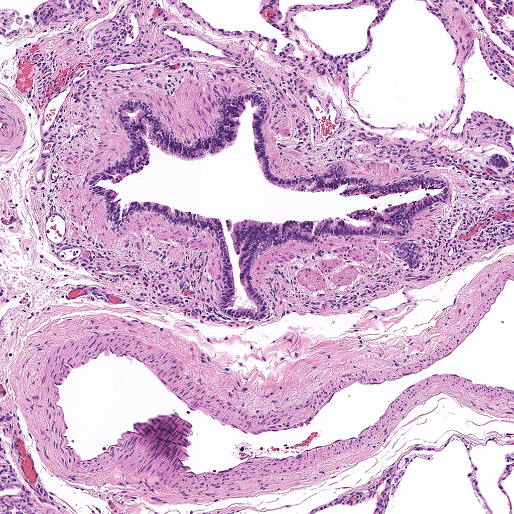
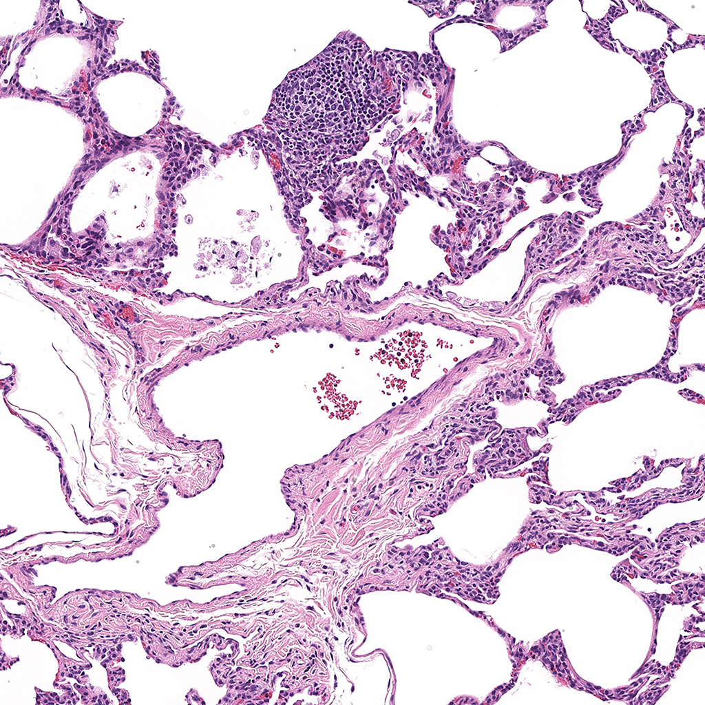
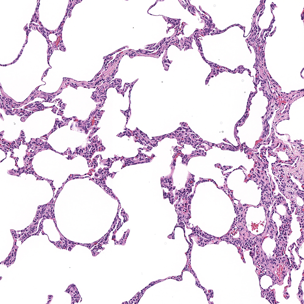
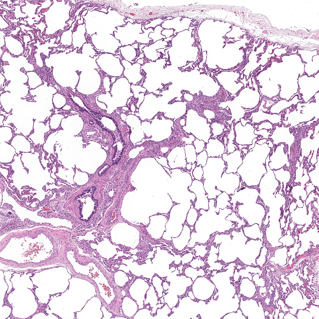
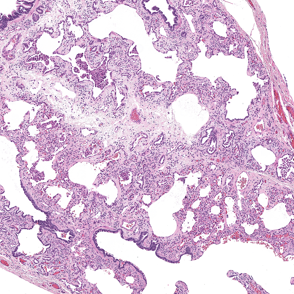
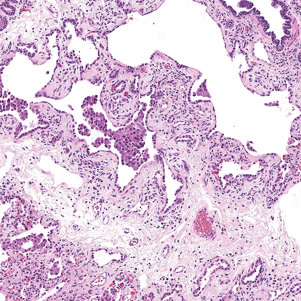
Immunofluorescence- NKX2.1, AGER, and ACTA2
Purpose: Stain slides of 7µm frozen sections of 9 month-old lung provided by the HTC for NKX2.1, AGER, and ACTA2 with antigen retrieval.
Day 1
Day 2
Tissue Used:
LMH-16-D003-LLL-9B3.5
Gender: Male
Age: 9 Months
Appendix
Antigen Retrieval Solution
18ml Solution A- 0.1M Citric Acid (Sigma, C1909) pH 2.5
82ml Reagent B- 0.1M Sodium Citrate (Sigma, S4641 ) pH 8.2
1L dH2O
pH 6.0
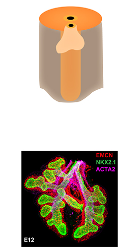
Embryonic
3-7 Weeks PC Human
E9.5-12 Mouse
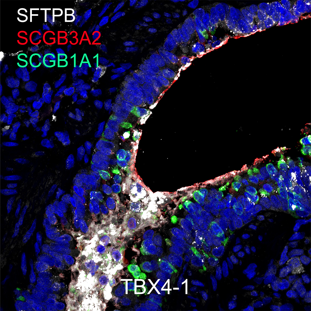
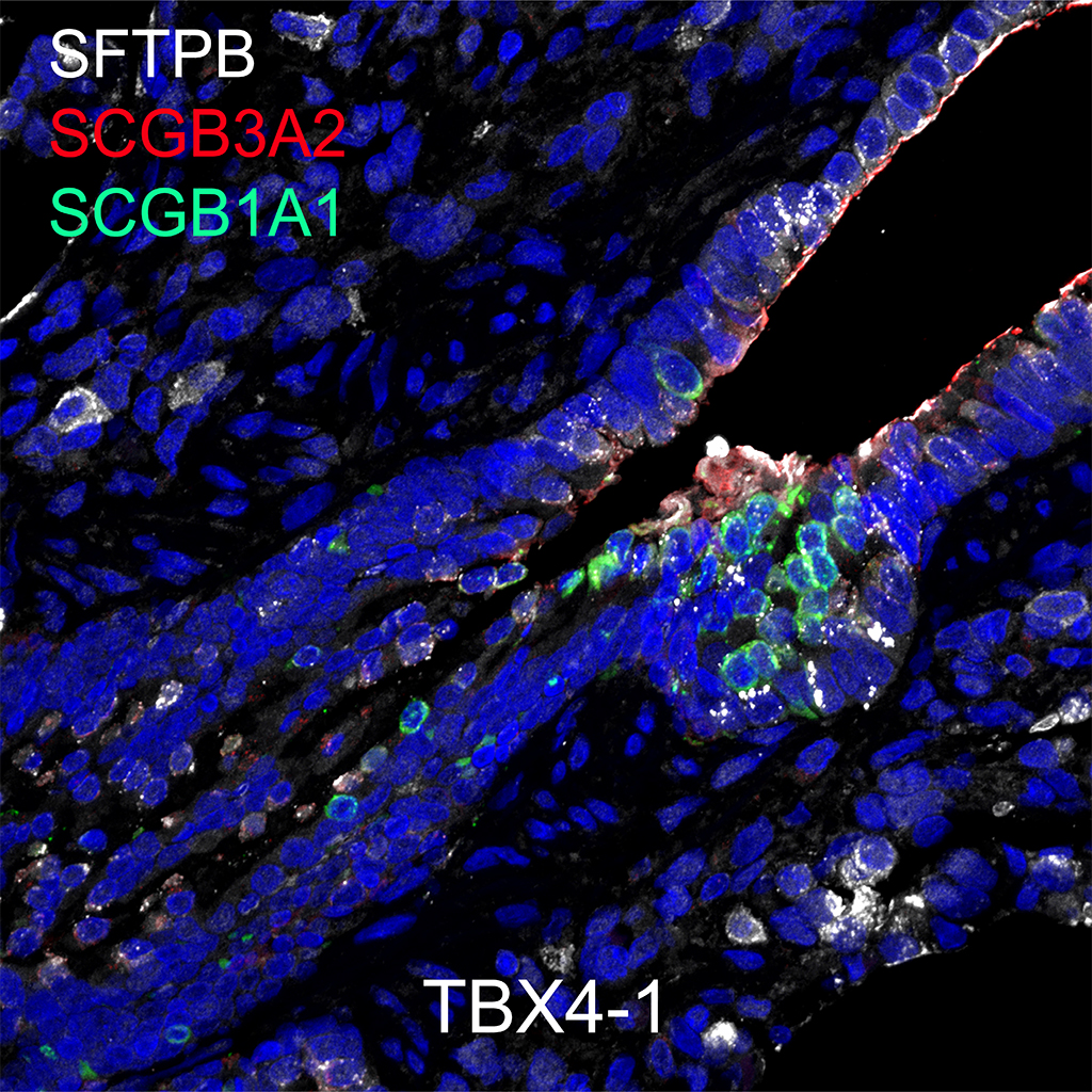
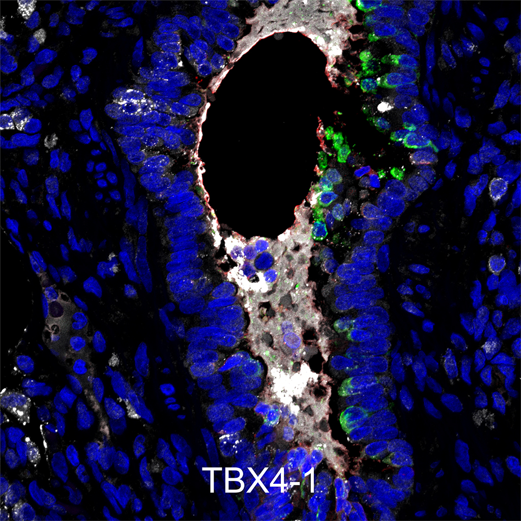
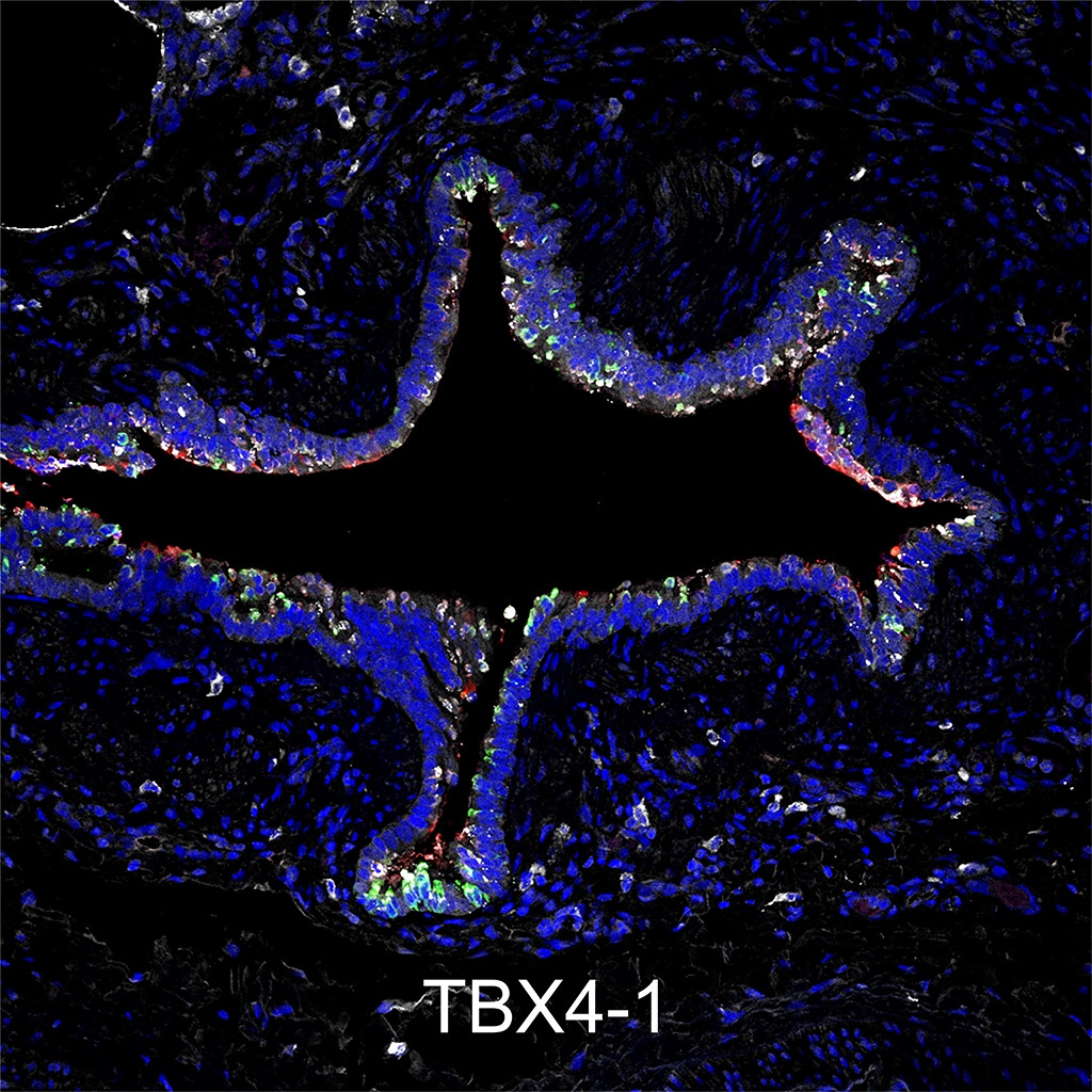
The Embryonic stage of lung development occurs from 4-7 weeks PC in human and embryonic (E) day 9.5-E12 in mouse (Ref). During the embryonic period, trachea, main stem, lobar and segmental bronchi are formed and the trachea and esophagus separate.
.
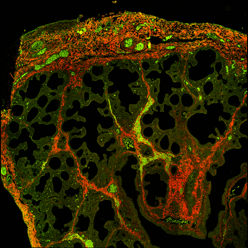
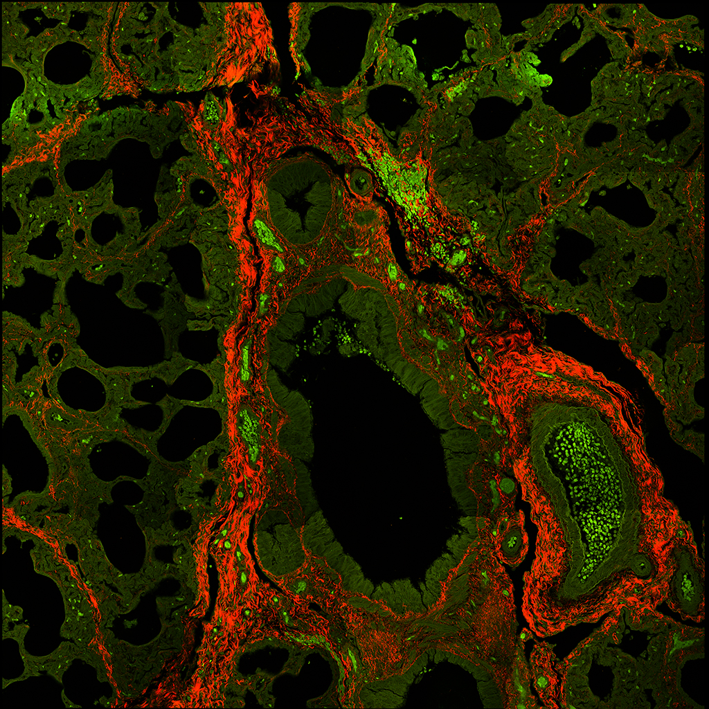
| Gene | Cell Type(s) | Subcellular Localization | LungGENS | Mouse LungImage | Ensembl | Genecards | NCBI | Human Protein Atlas | Wikipedia |
|---|---|---|---|---|---|---|---|---|---|
| Acta2 | smooth muscle cell | cytoskeleton / microfilaments | Acta2 E16.5 | E16.5 | Ensembl | Genecards | NCBI | The Human Protein Atlas | Wikipedia |
| alpha-actin-2 | (conducting airways; trachea, bronchi, bronchioles) | Acta2 E18.5 | E18.5 | ||||||
| syn: a-SMA, alpha-smooth muscle actin | (pulmonary blood vessels) | Acta2 P01 | |||||||
| *cytoskeletal protein | myofibroblast | Acta2 P03 | |||||||
| (pre-alveoar and alveolar parenchyma) | Acta2 P07 | ||||||||
| Acta2 P10 | |||||||||
| Acta2 P14 | |||||||||
| Acta2 P28 |
.
.
.
.
.
.
.
.
.
.
.
.
.
.
.
.
.
.
.
.
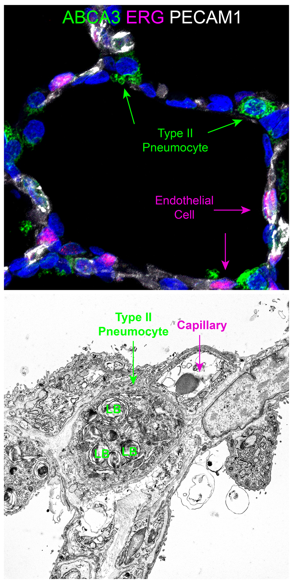
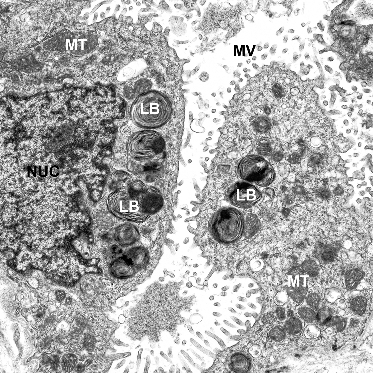
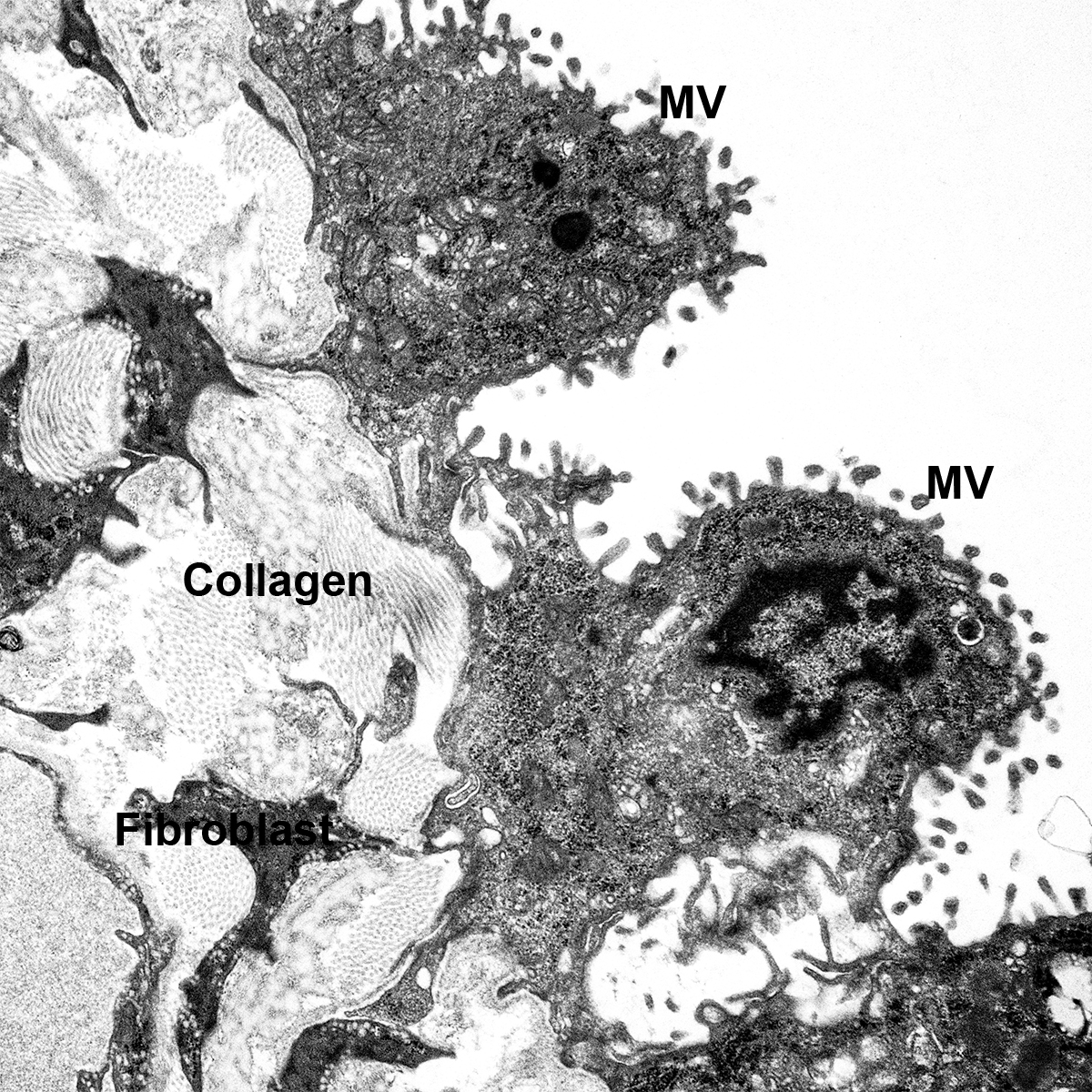
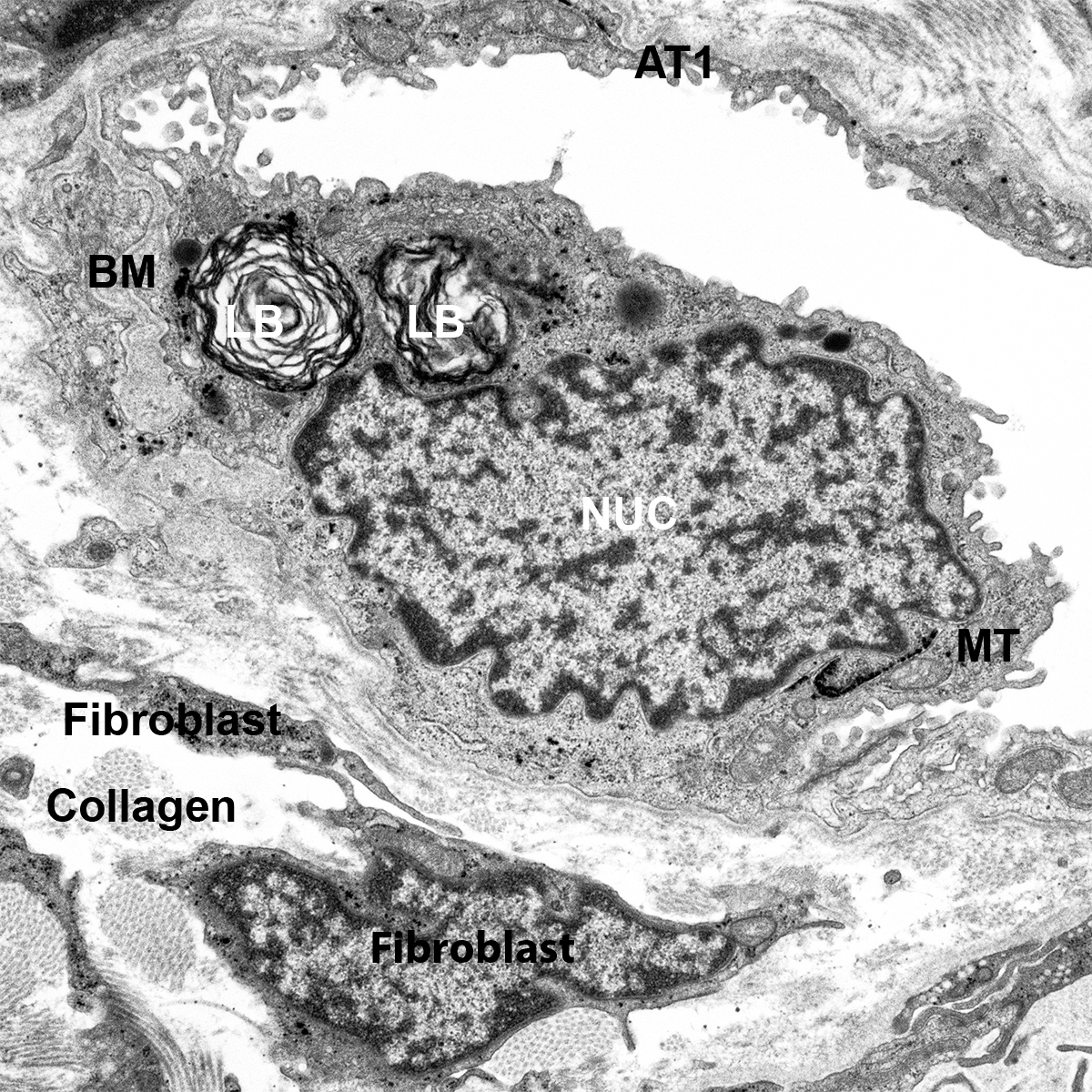
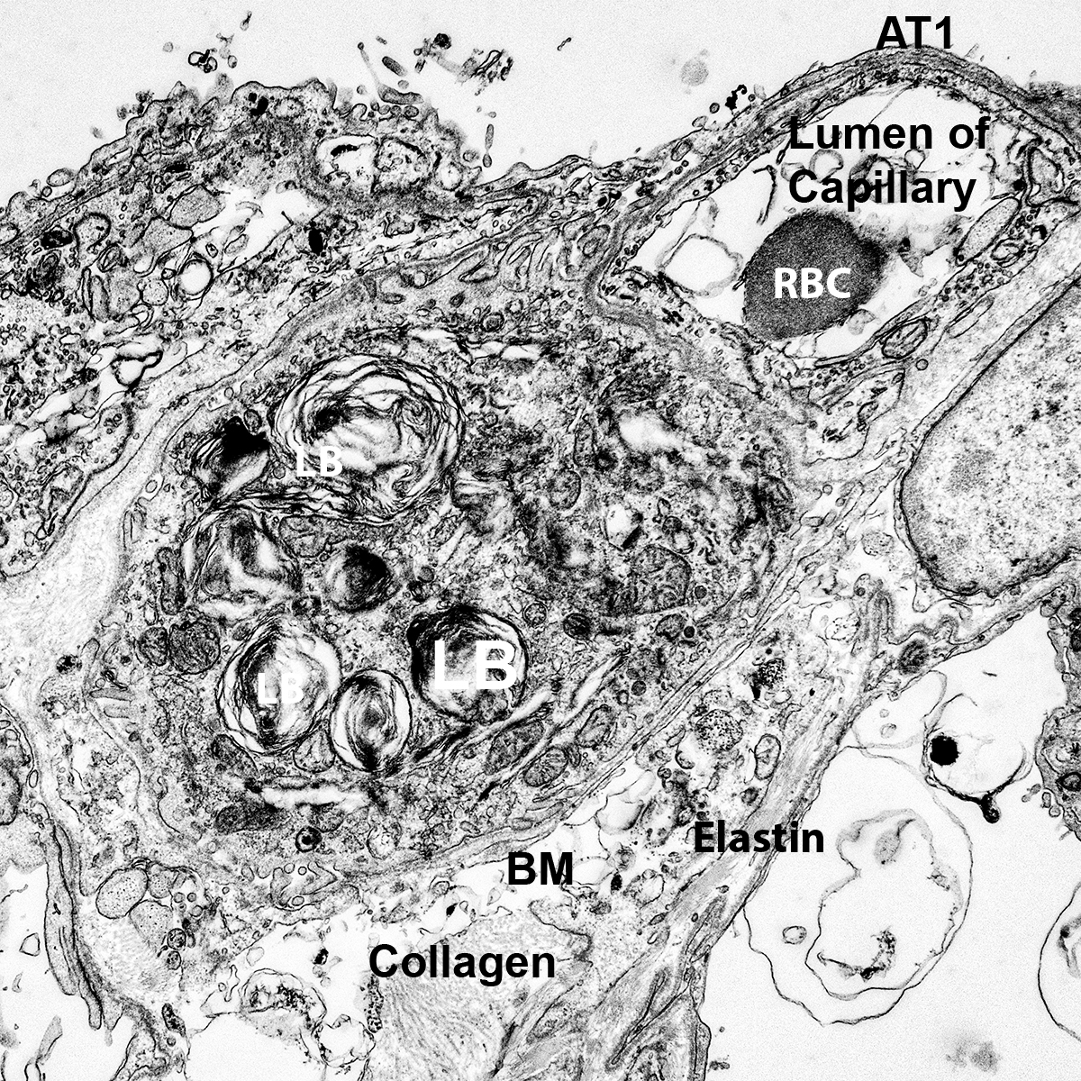
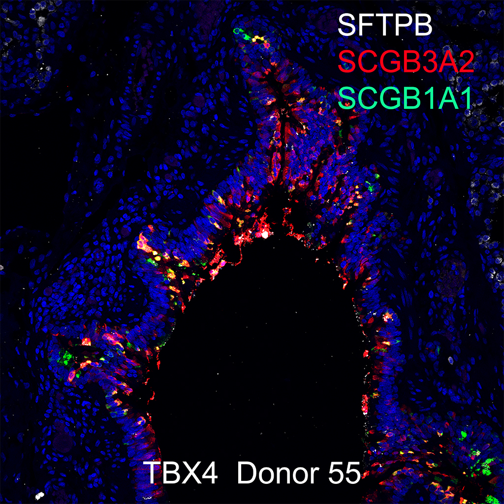
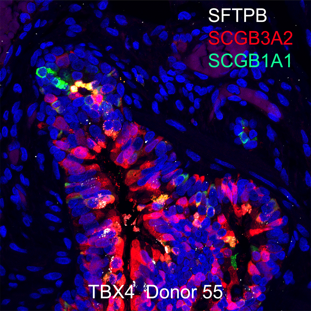
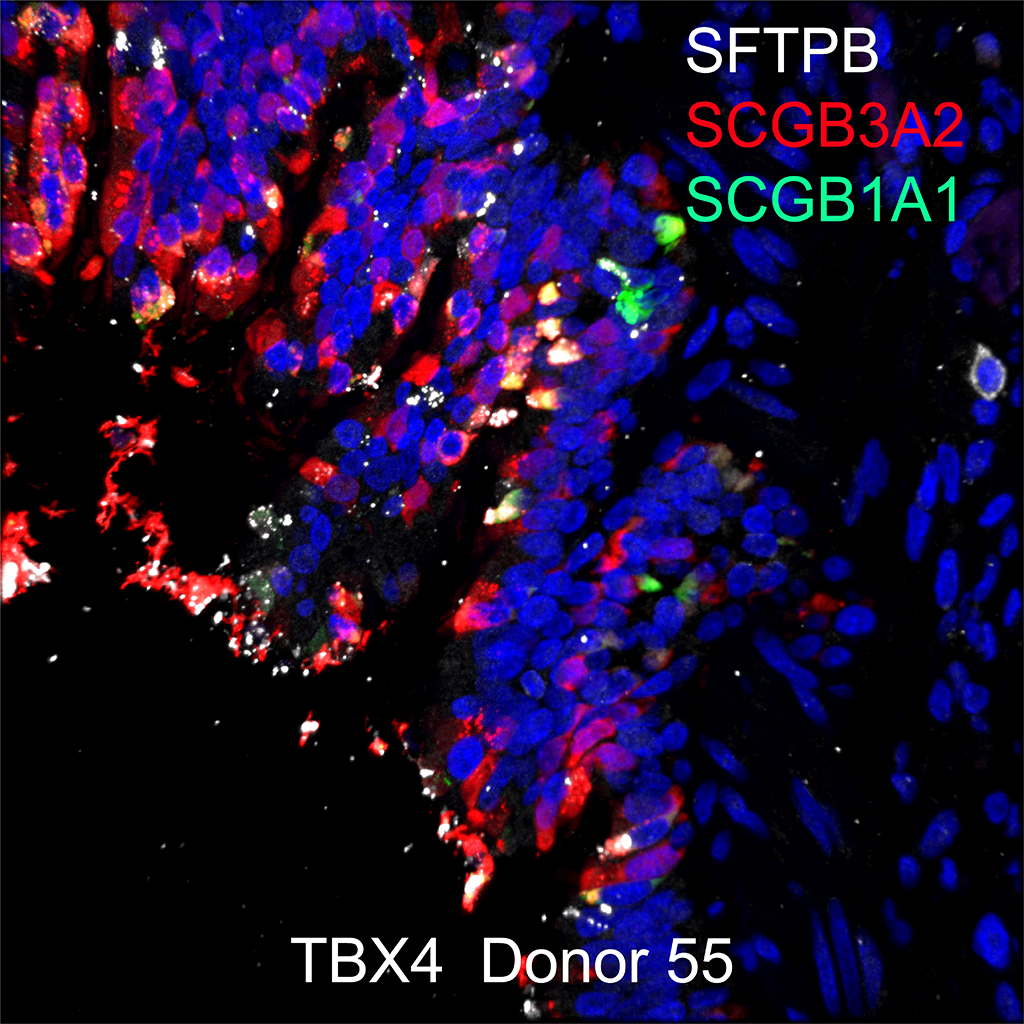
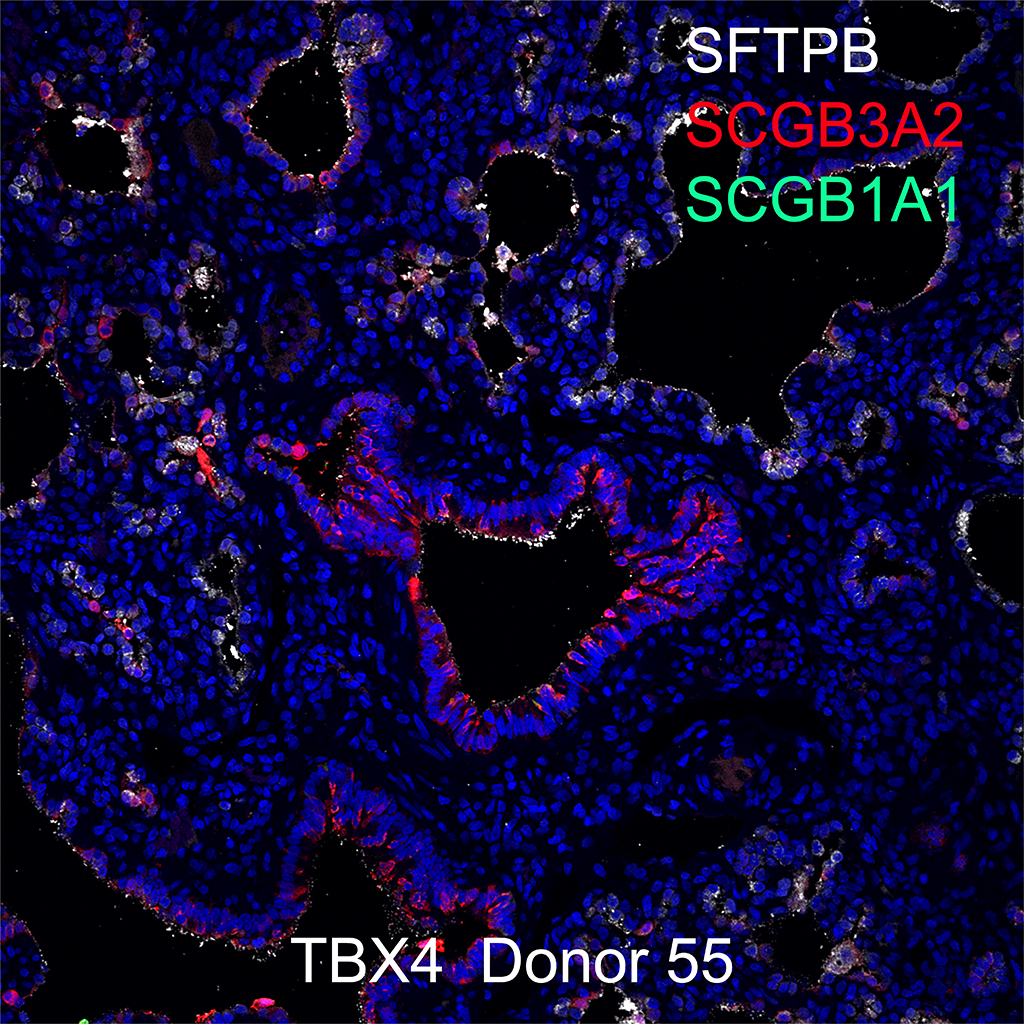
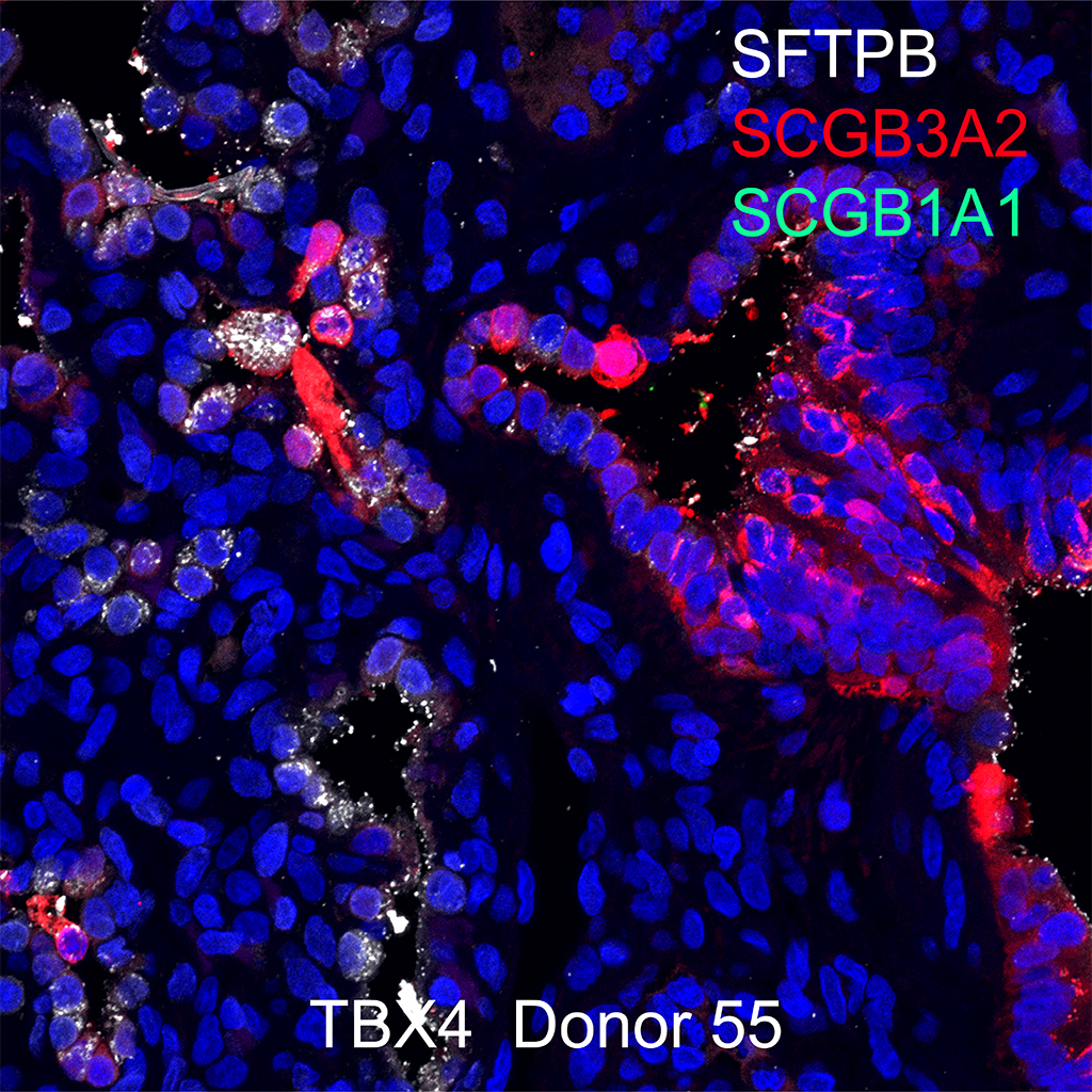
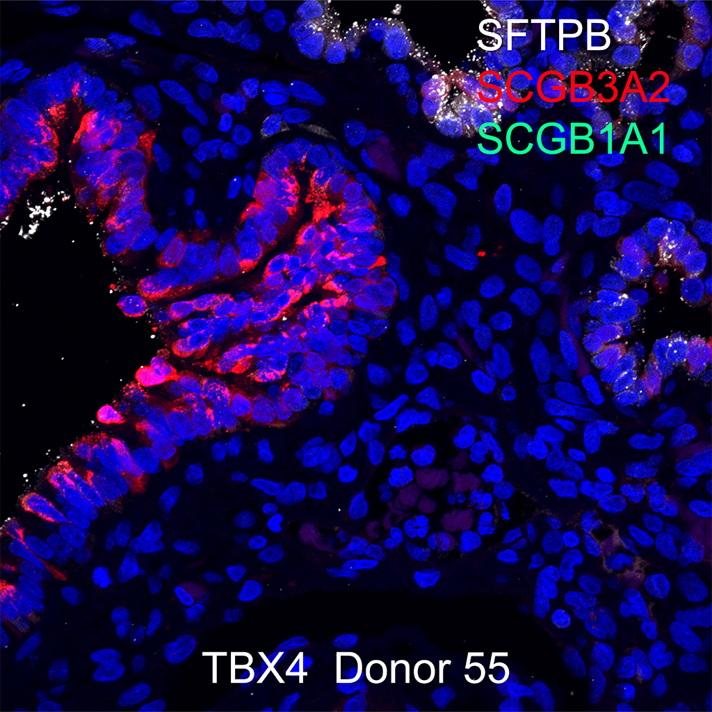
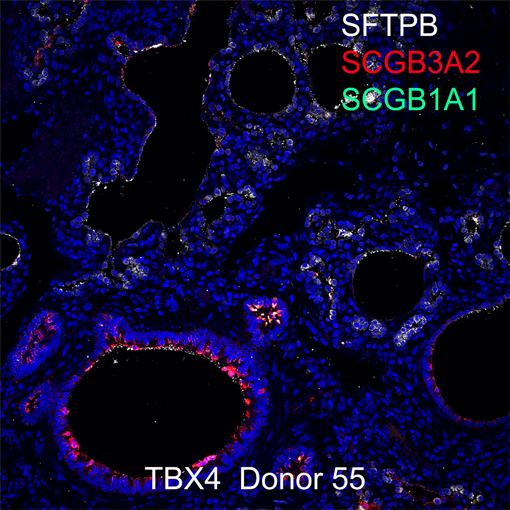
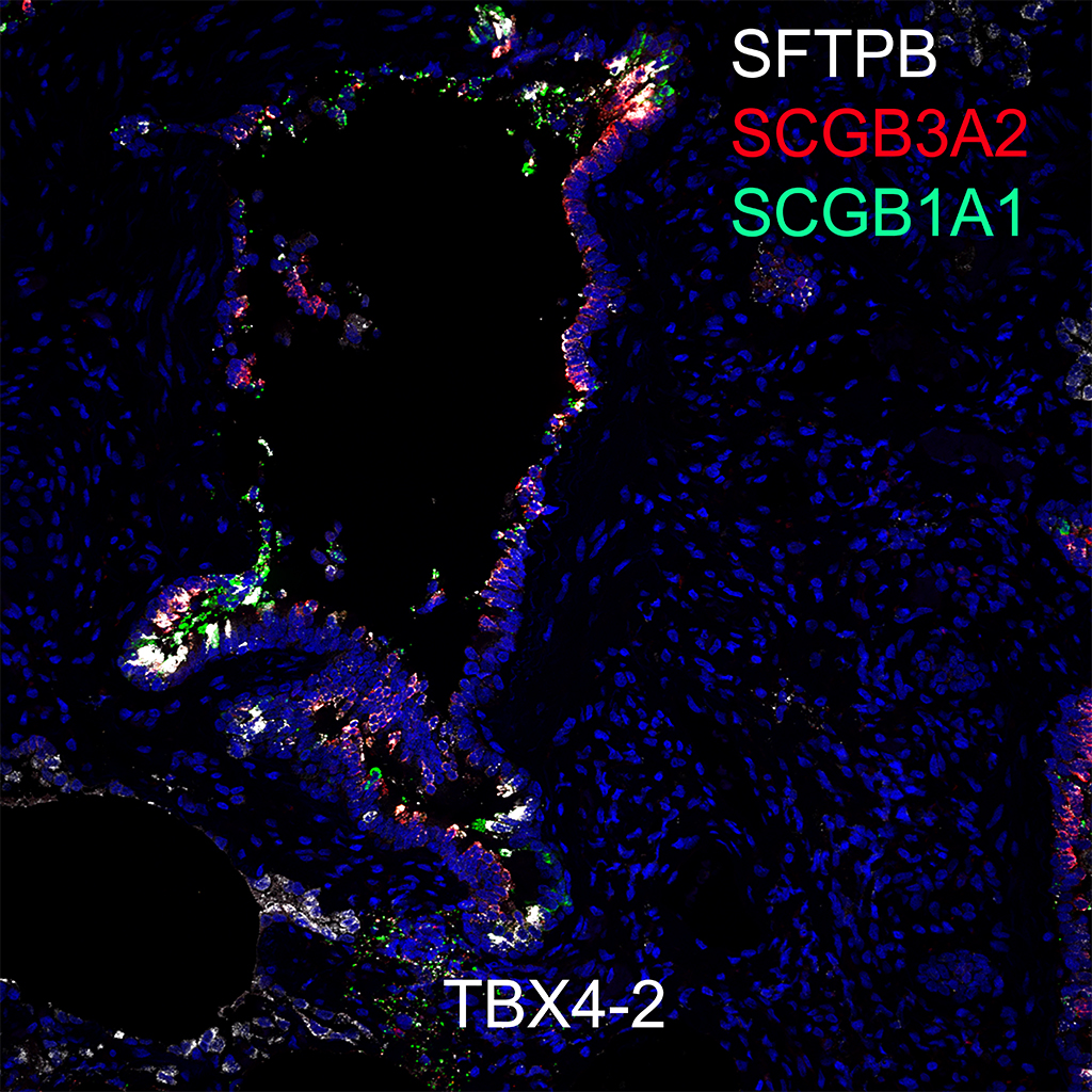
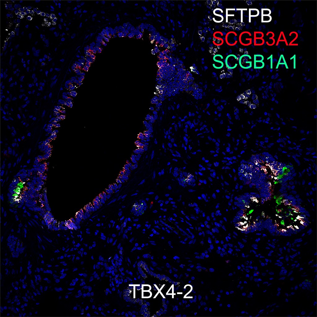
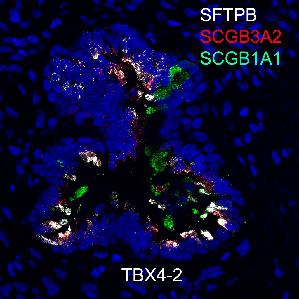
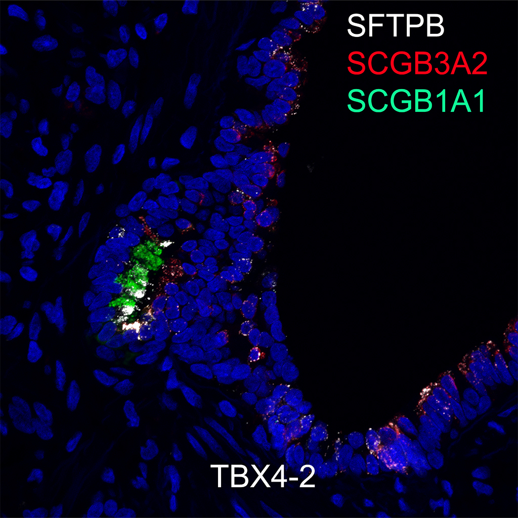
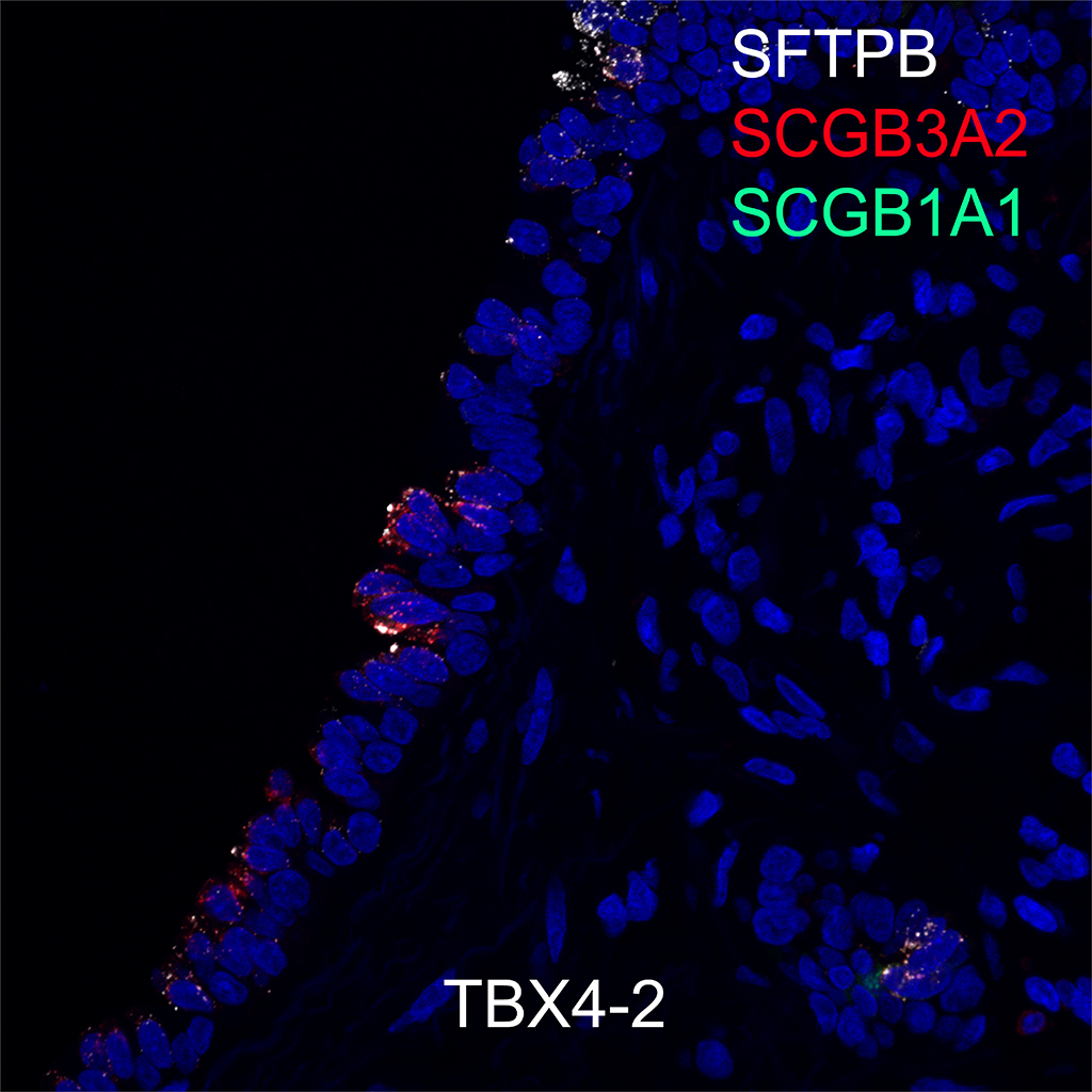
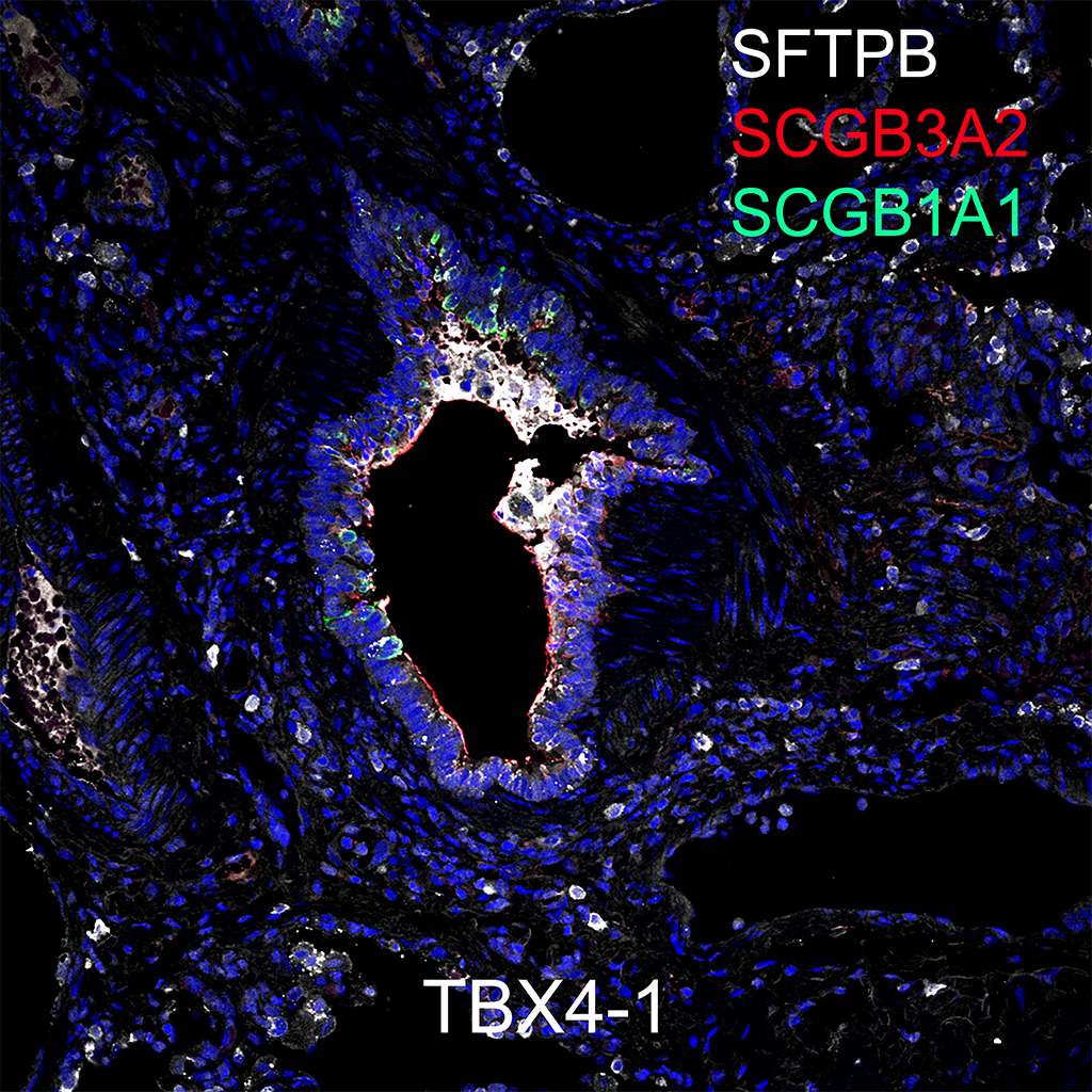
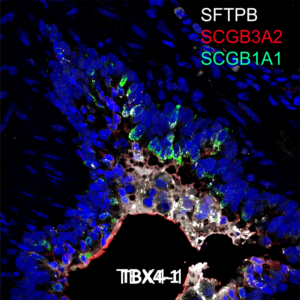
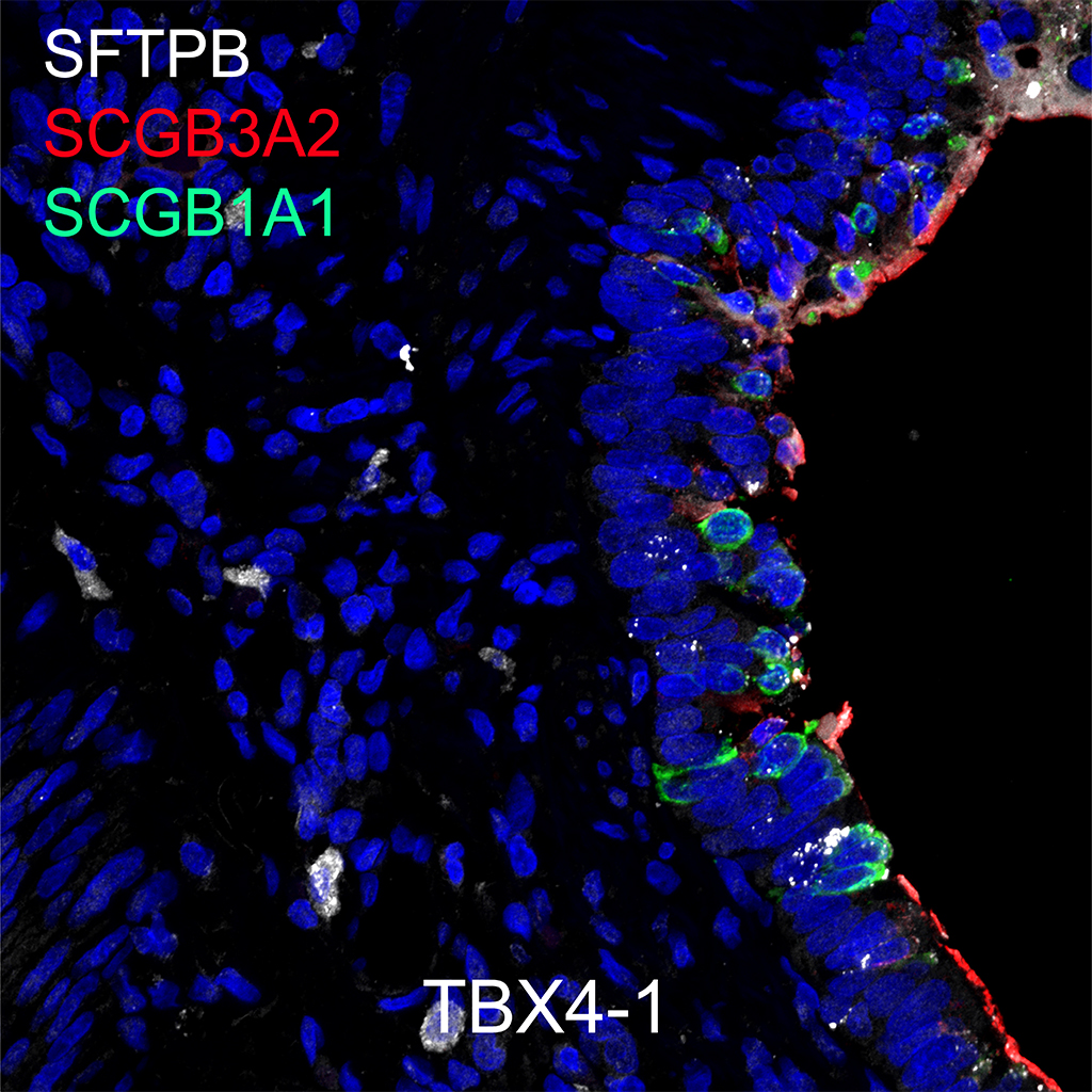
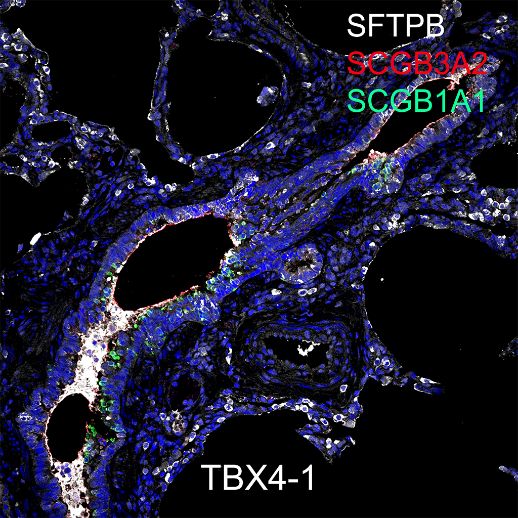
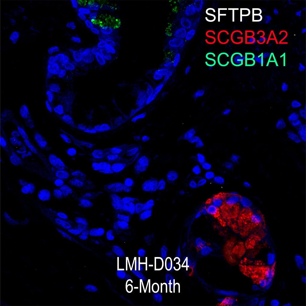
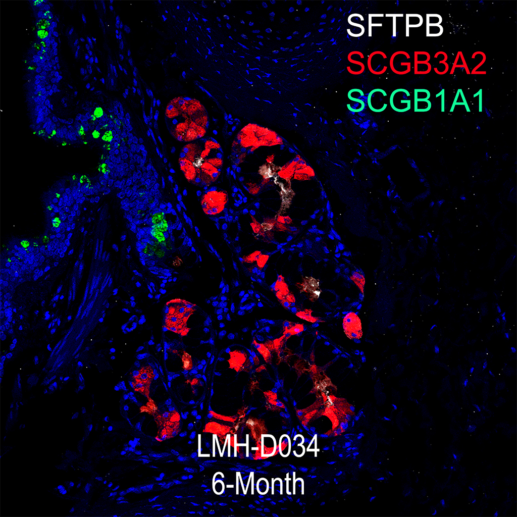
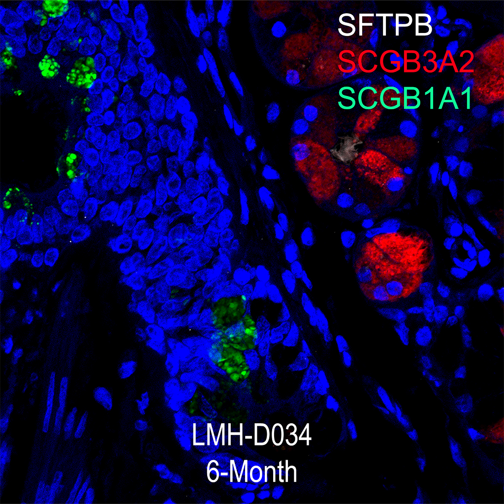
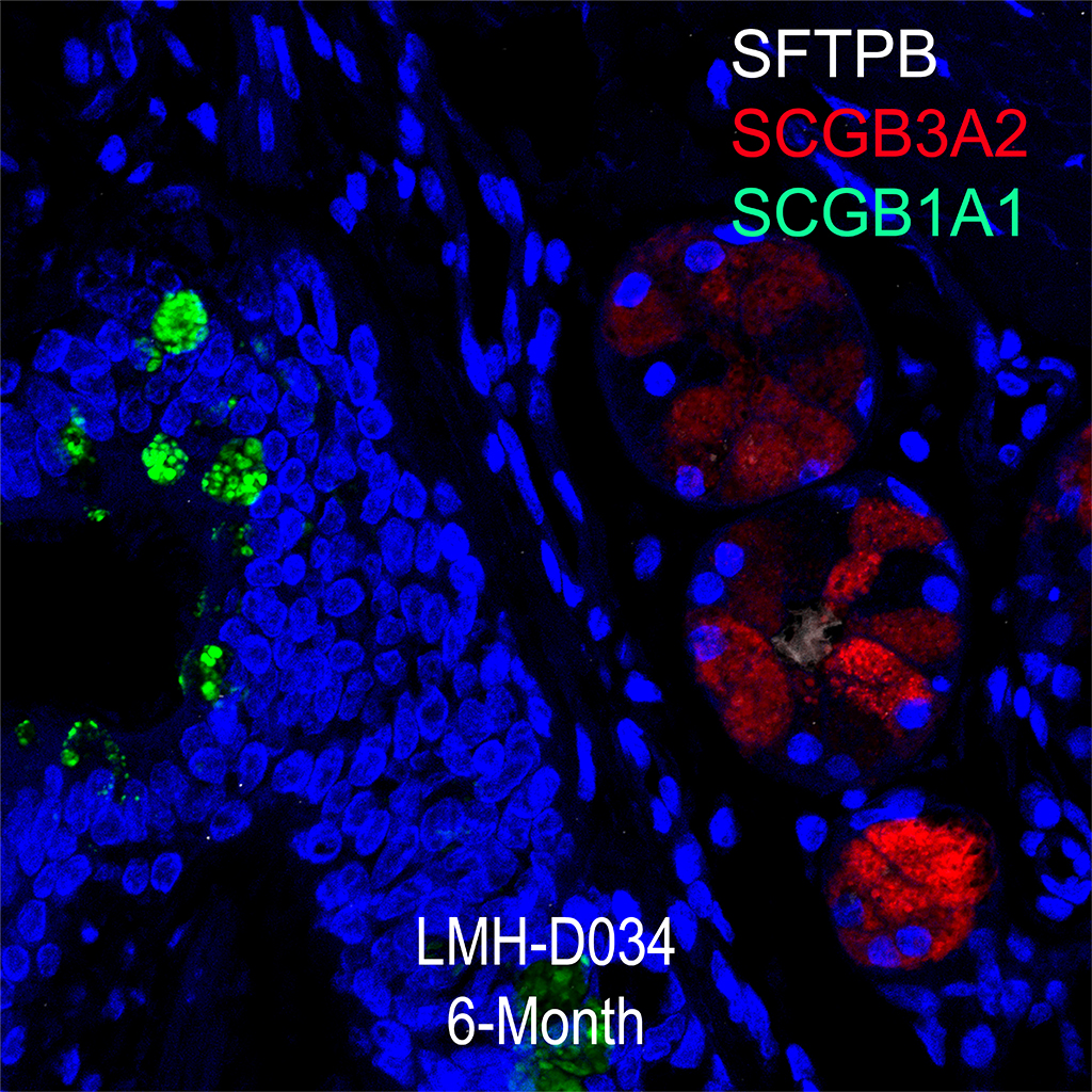
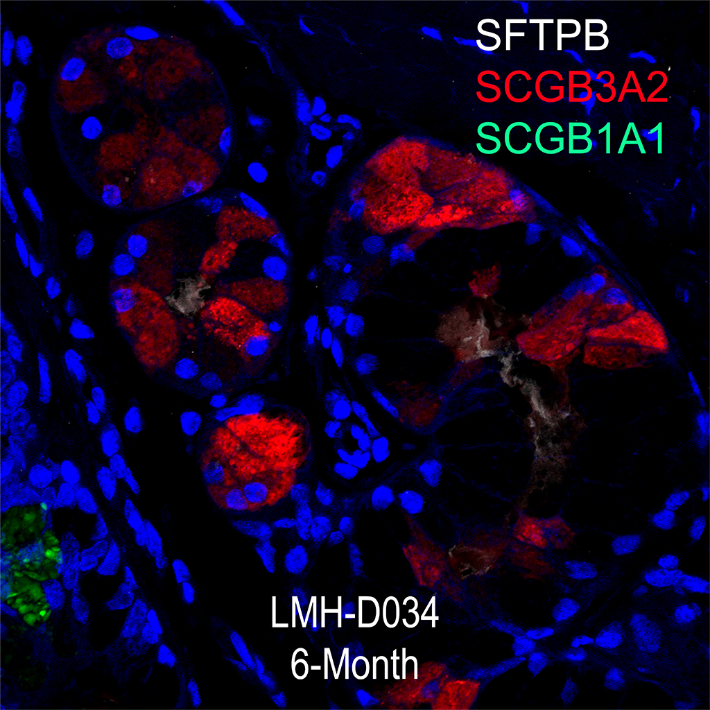
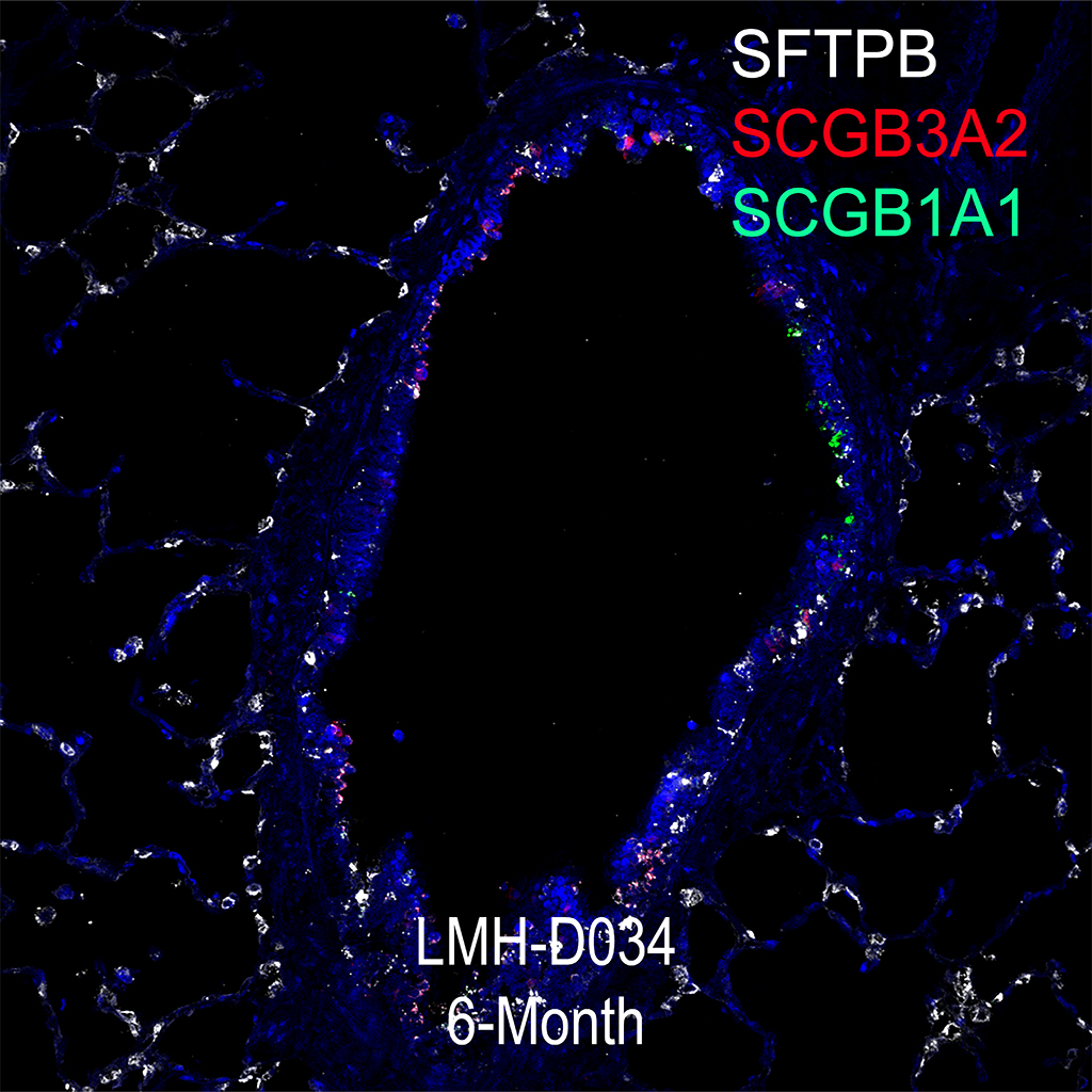
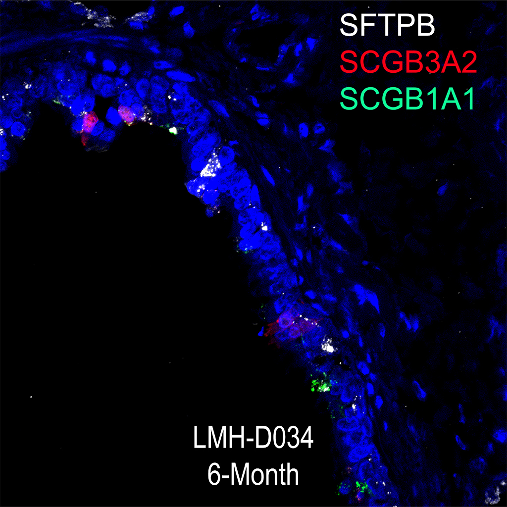
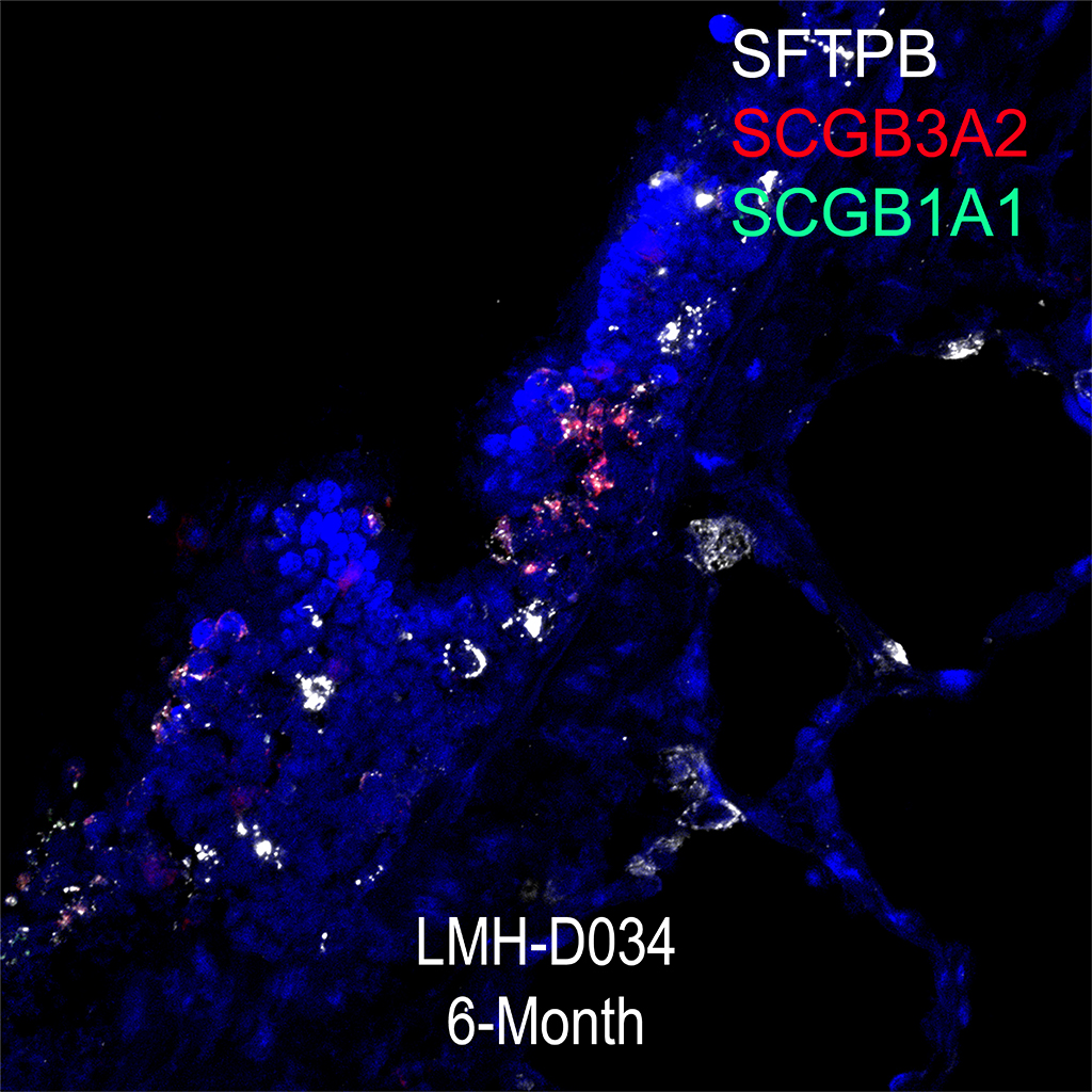
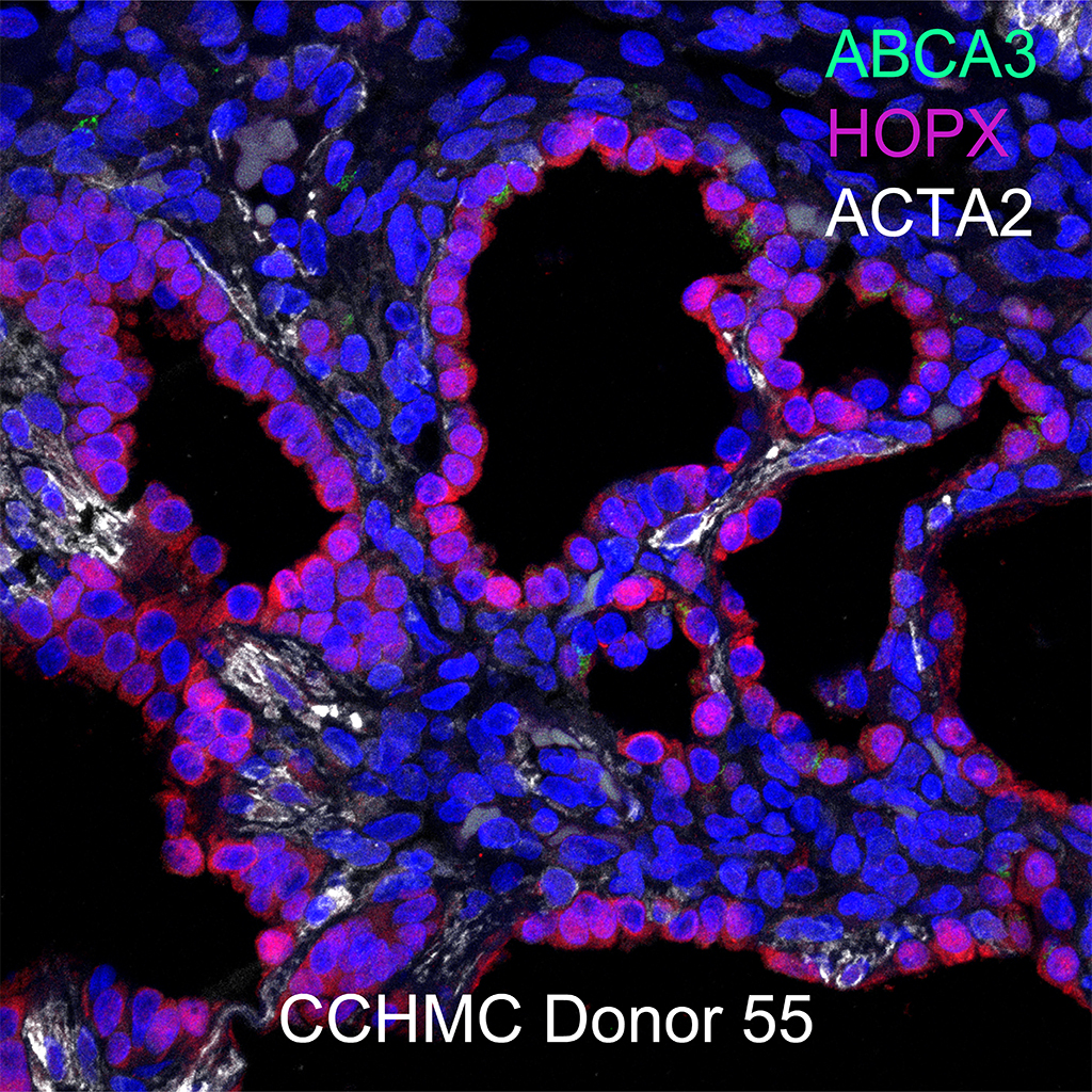
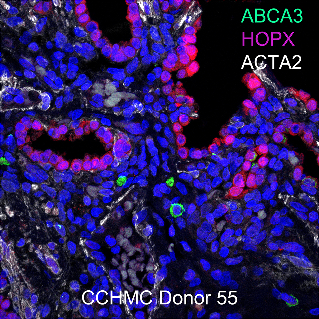
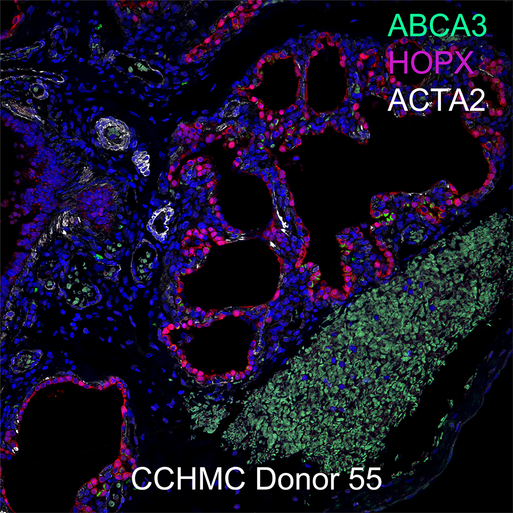
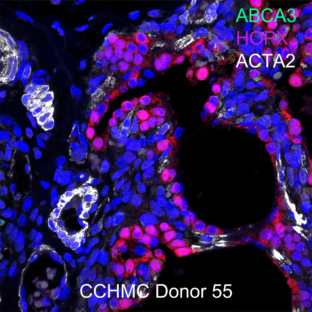
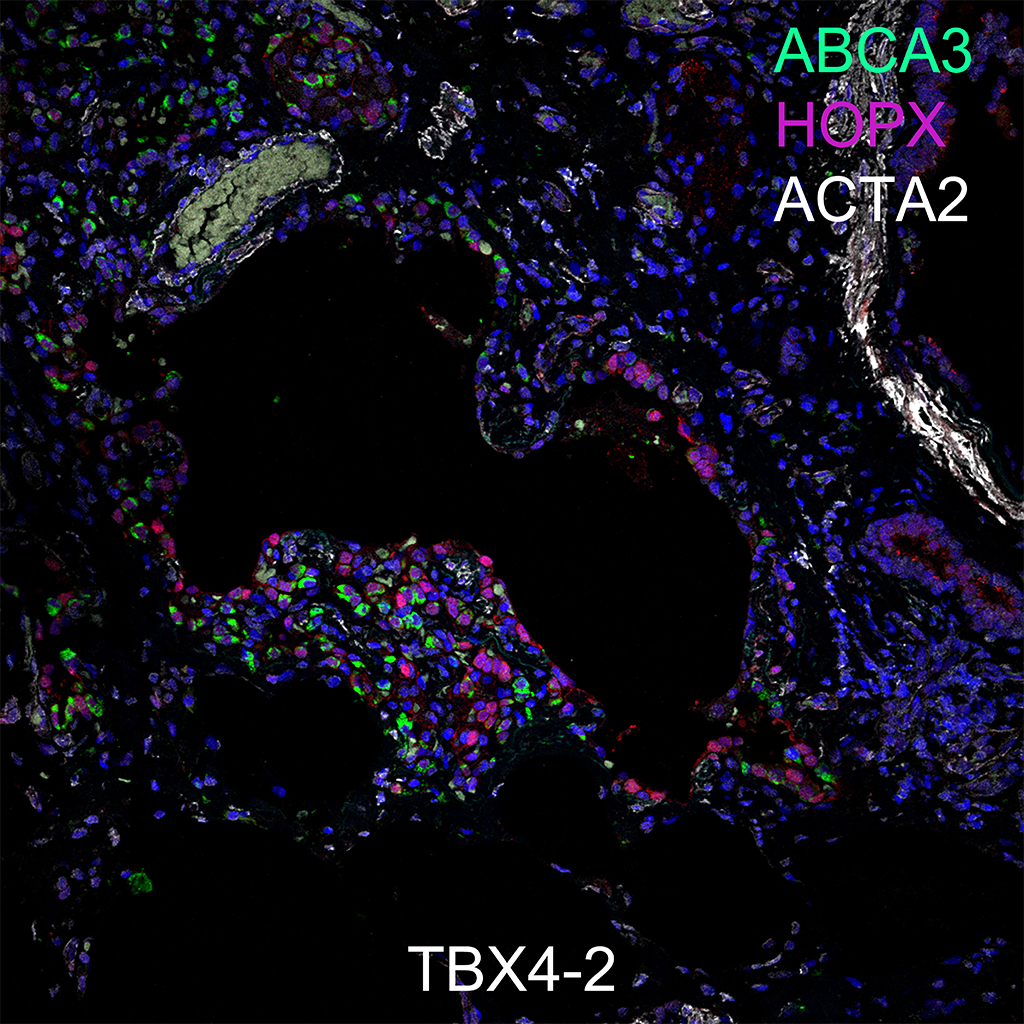
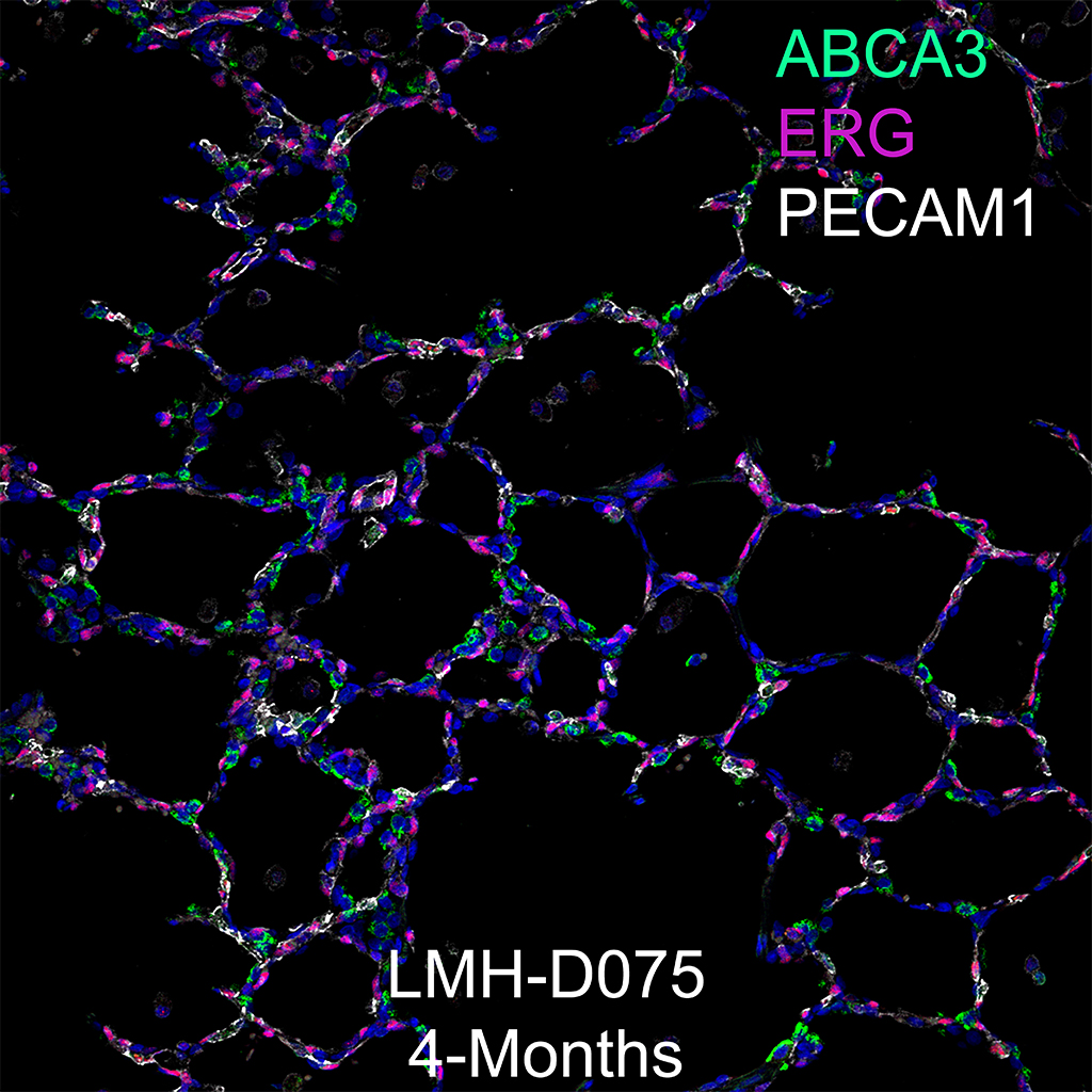
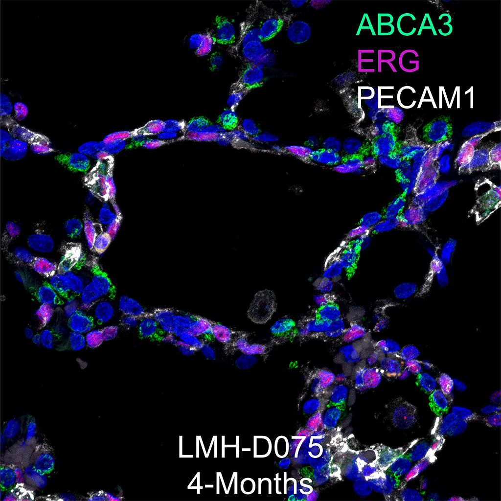
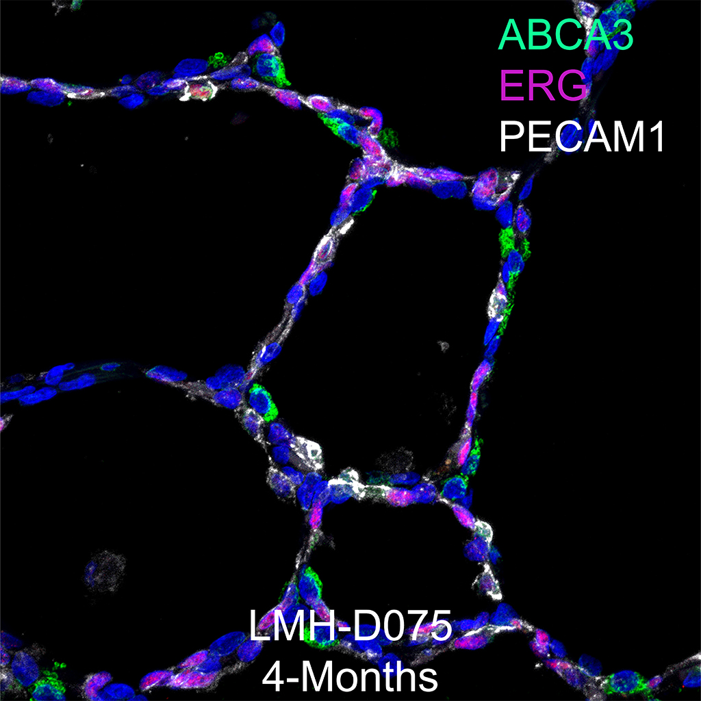
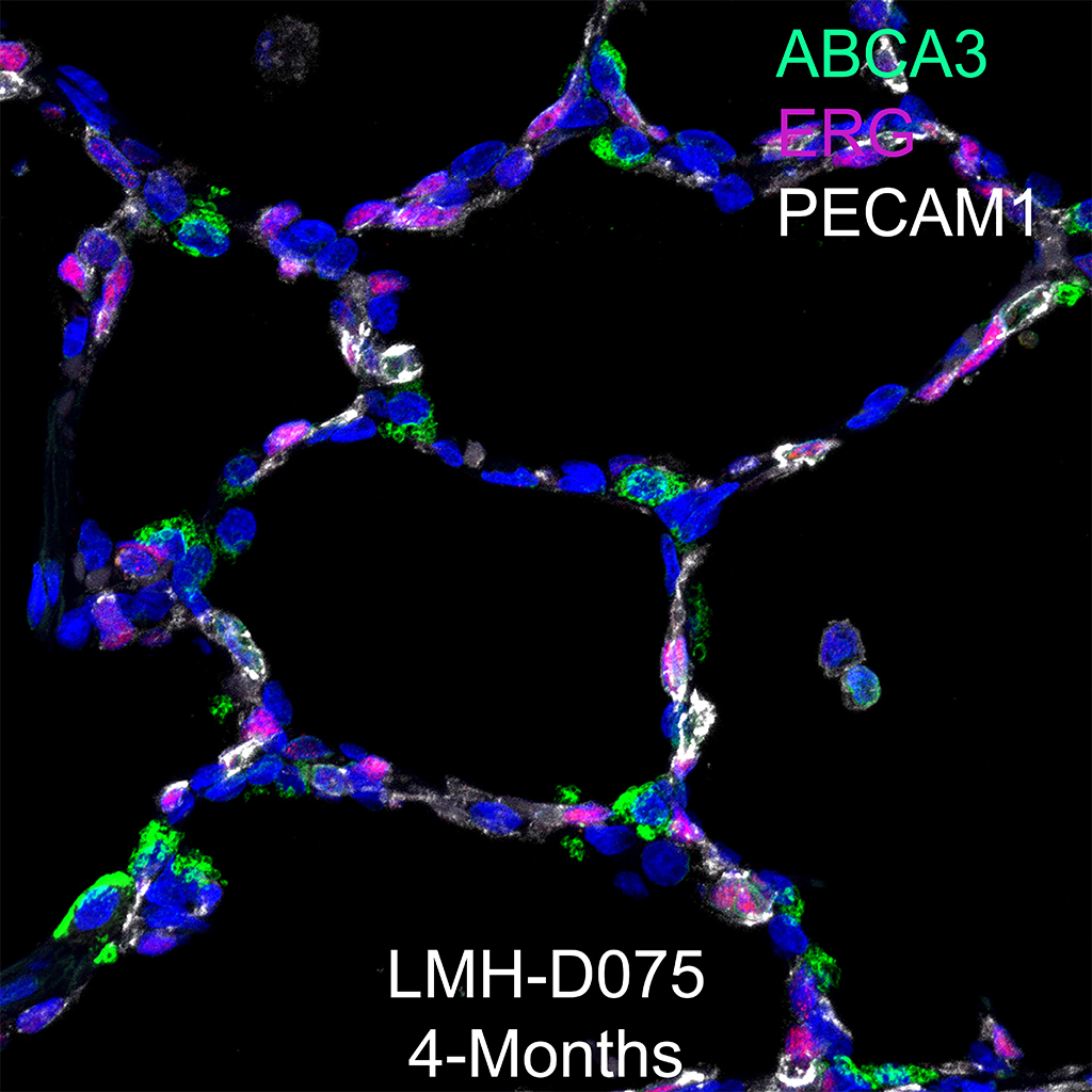
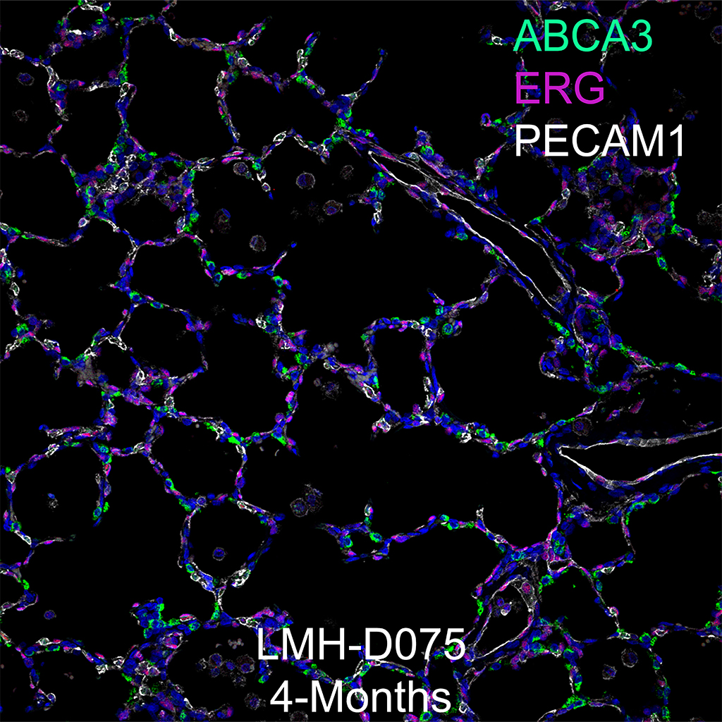
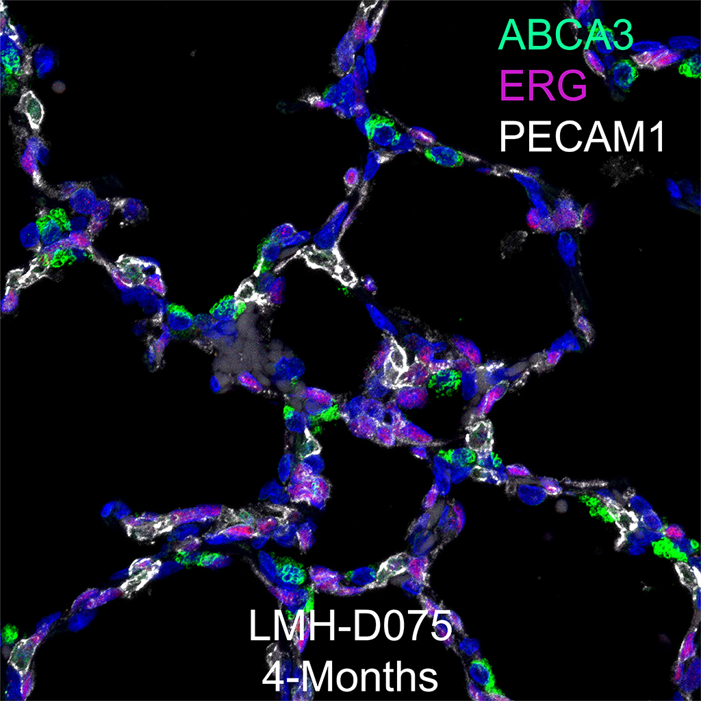
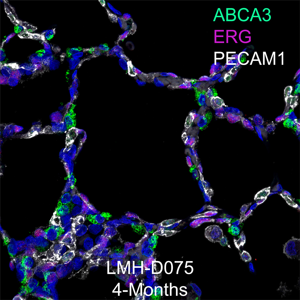
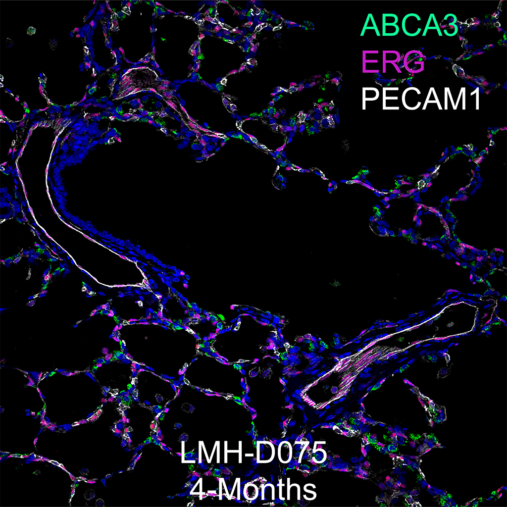
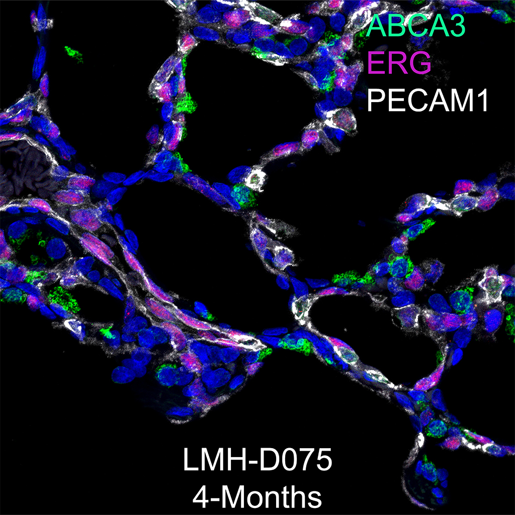
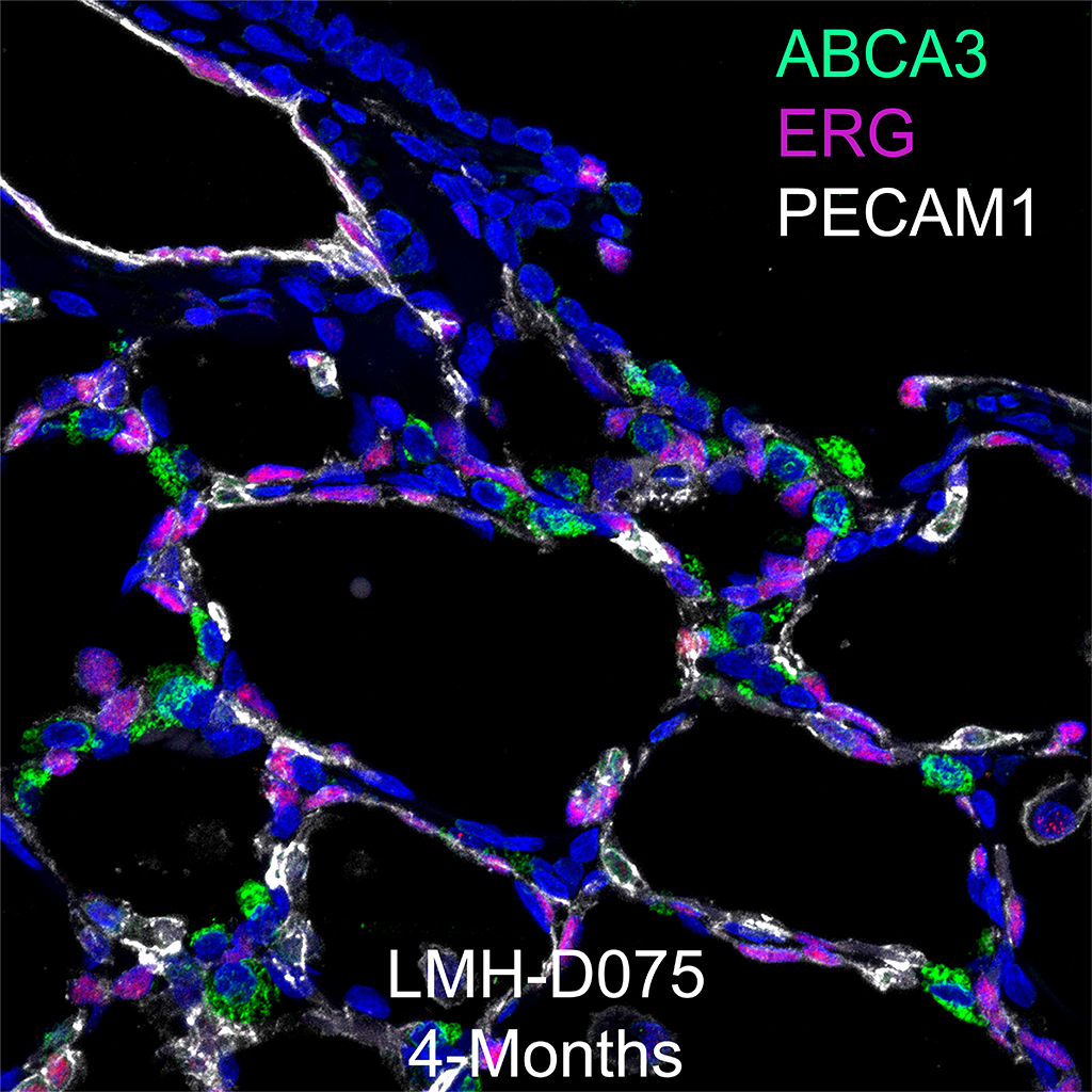
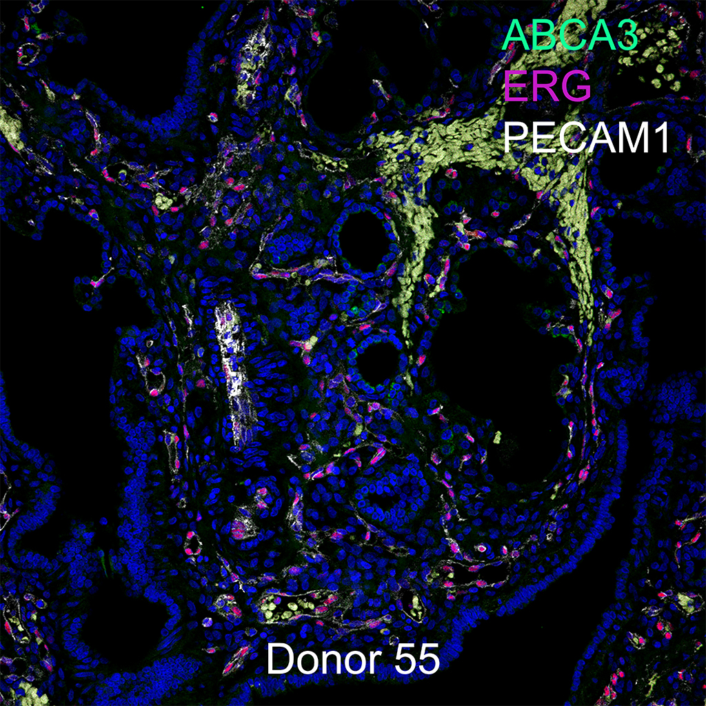
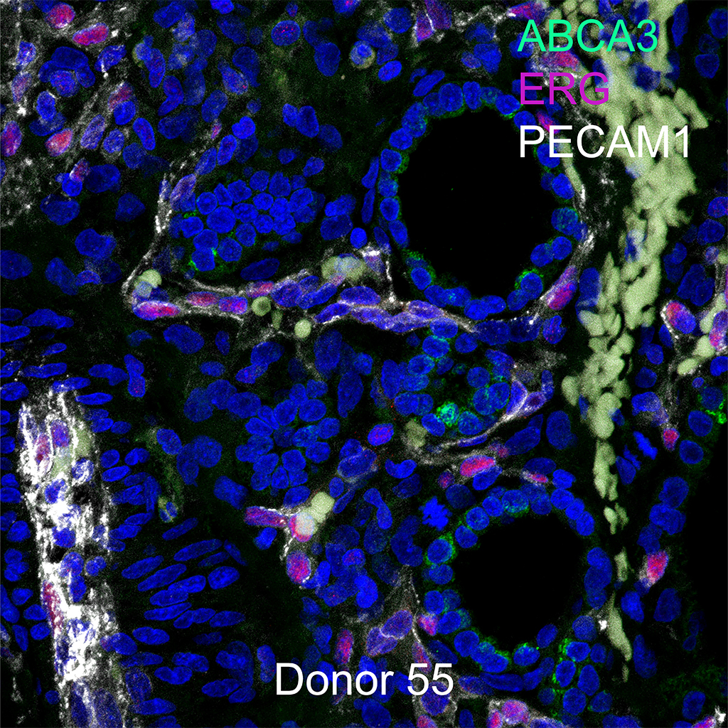
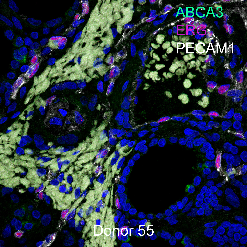
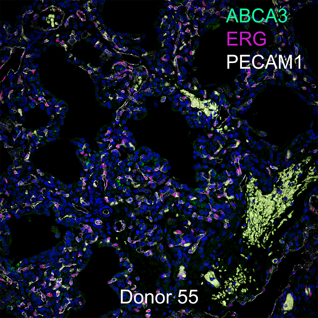
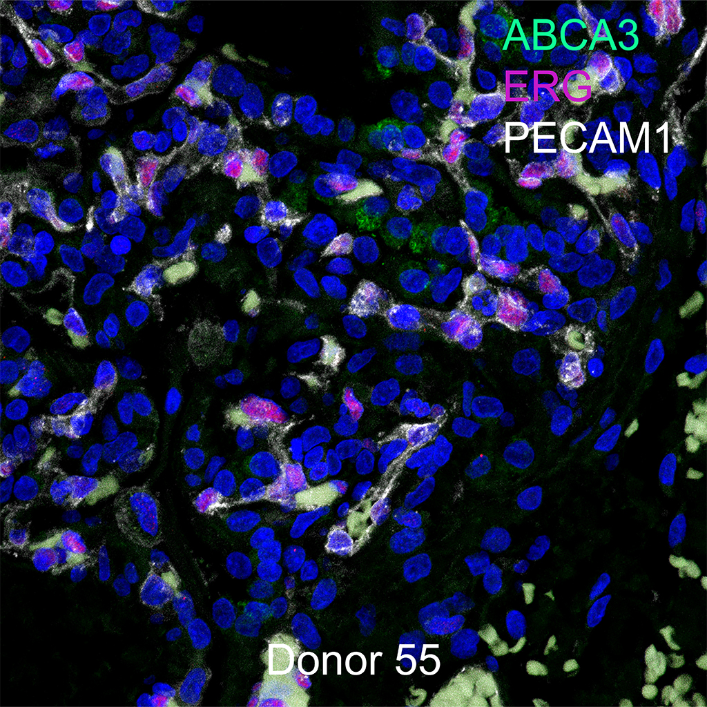
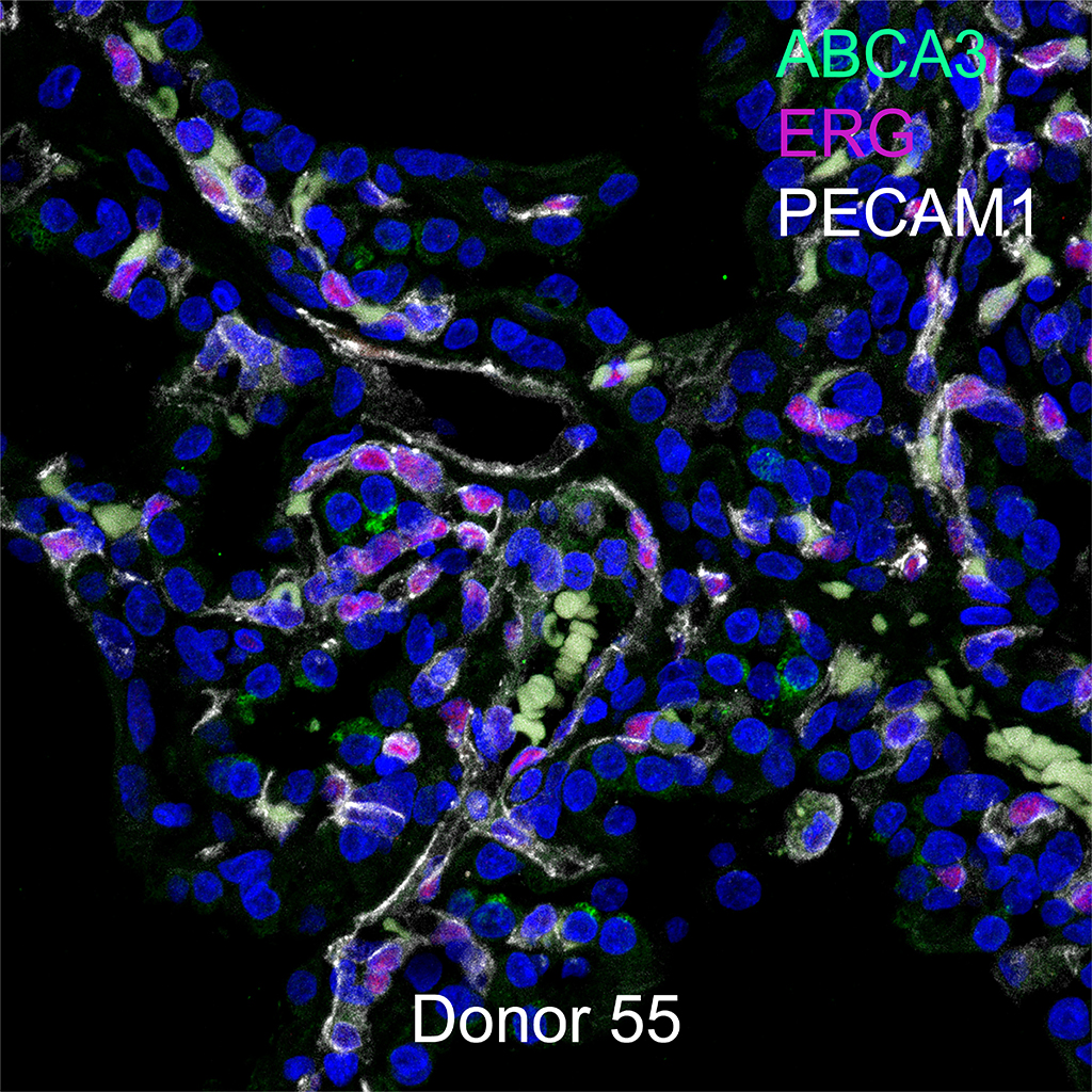
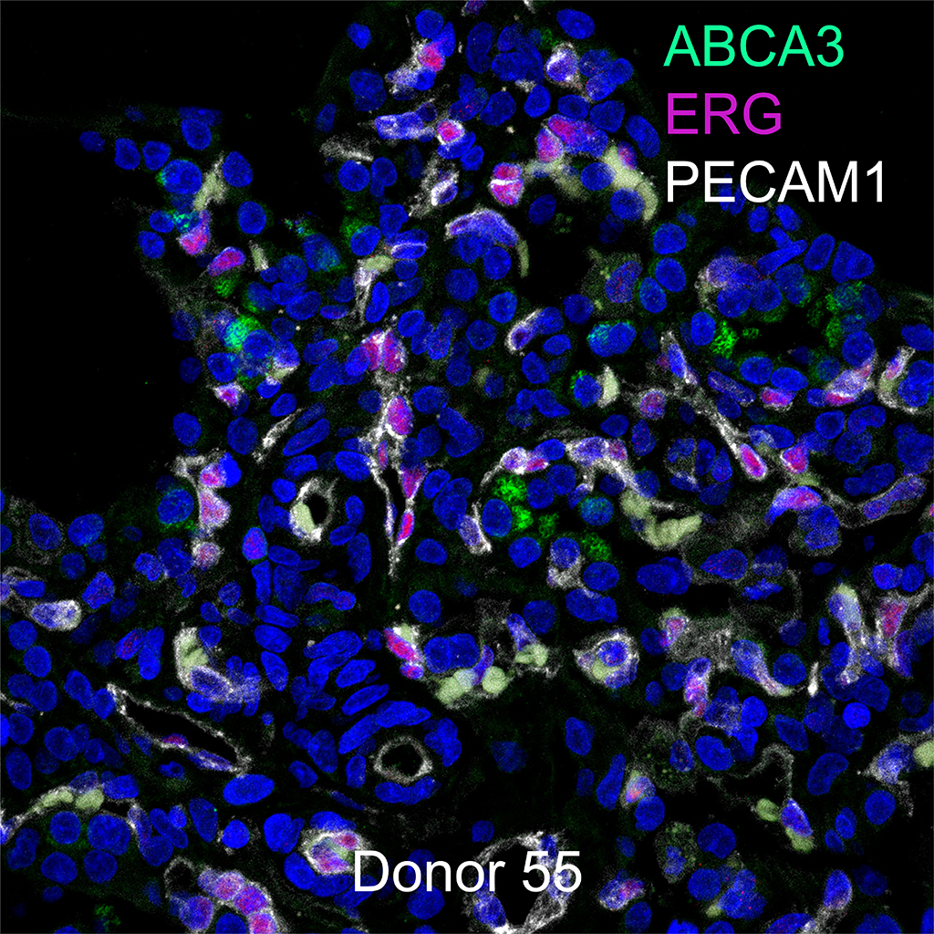
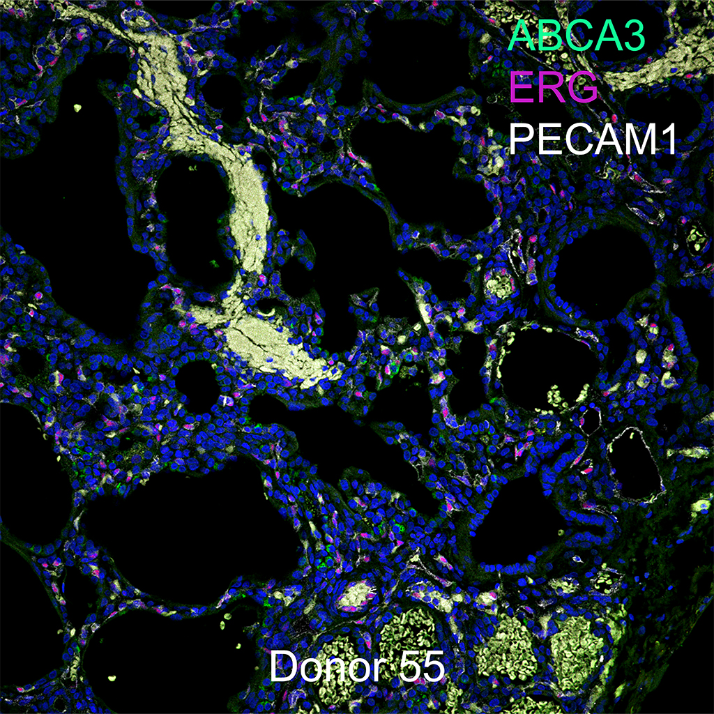
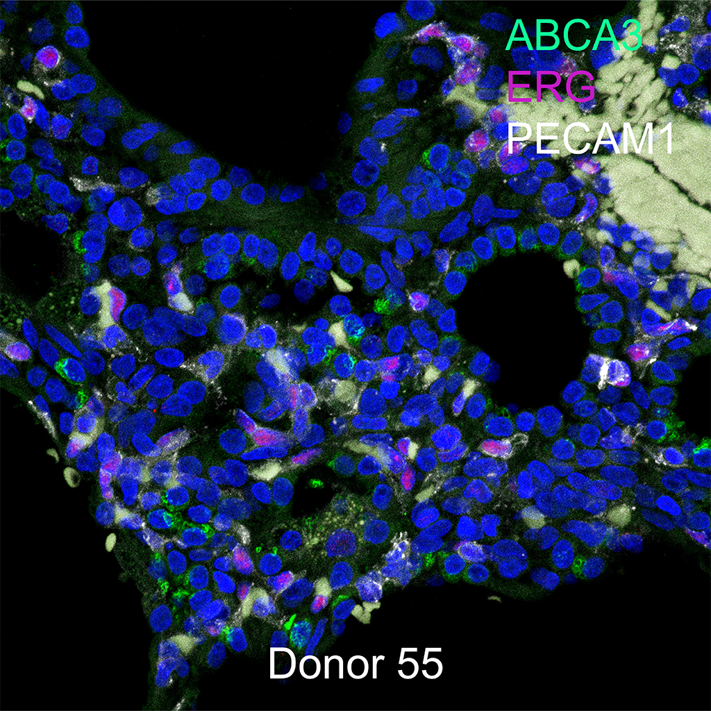
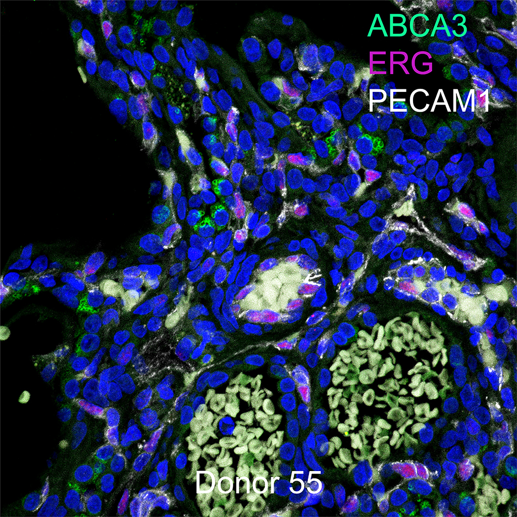
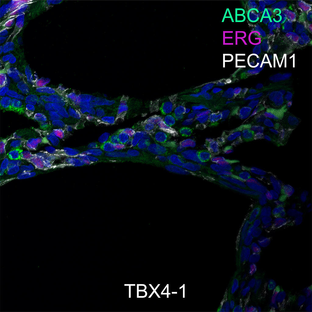
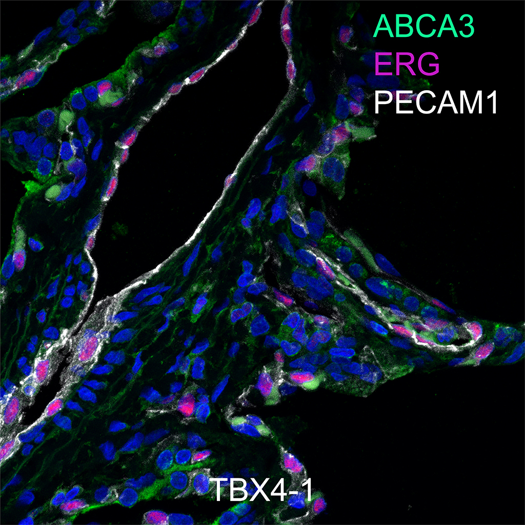
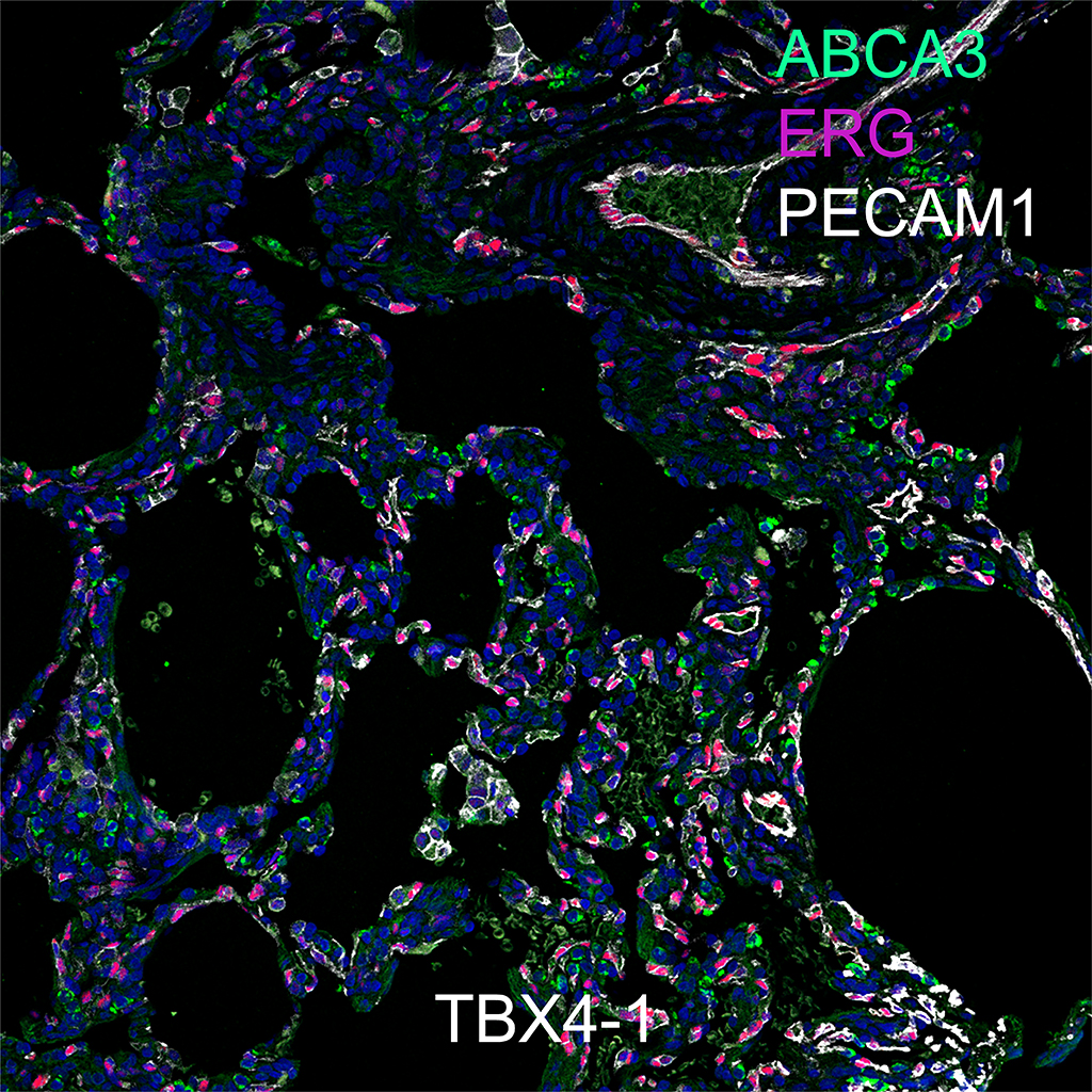
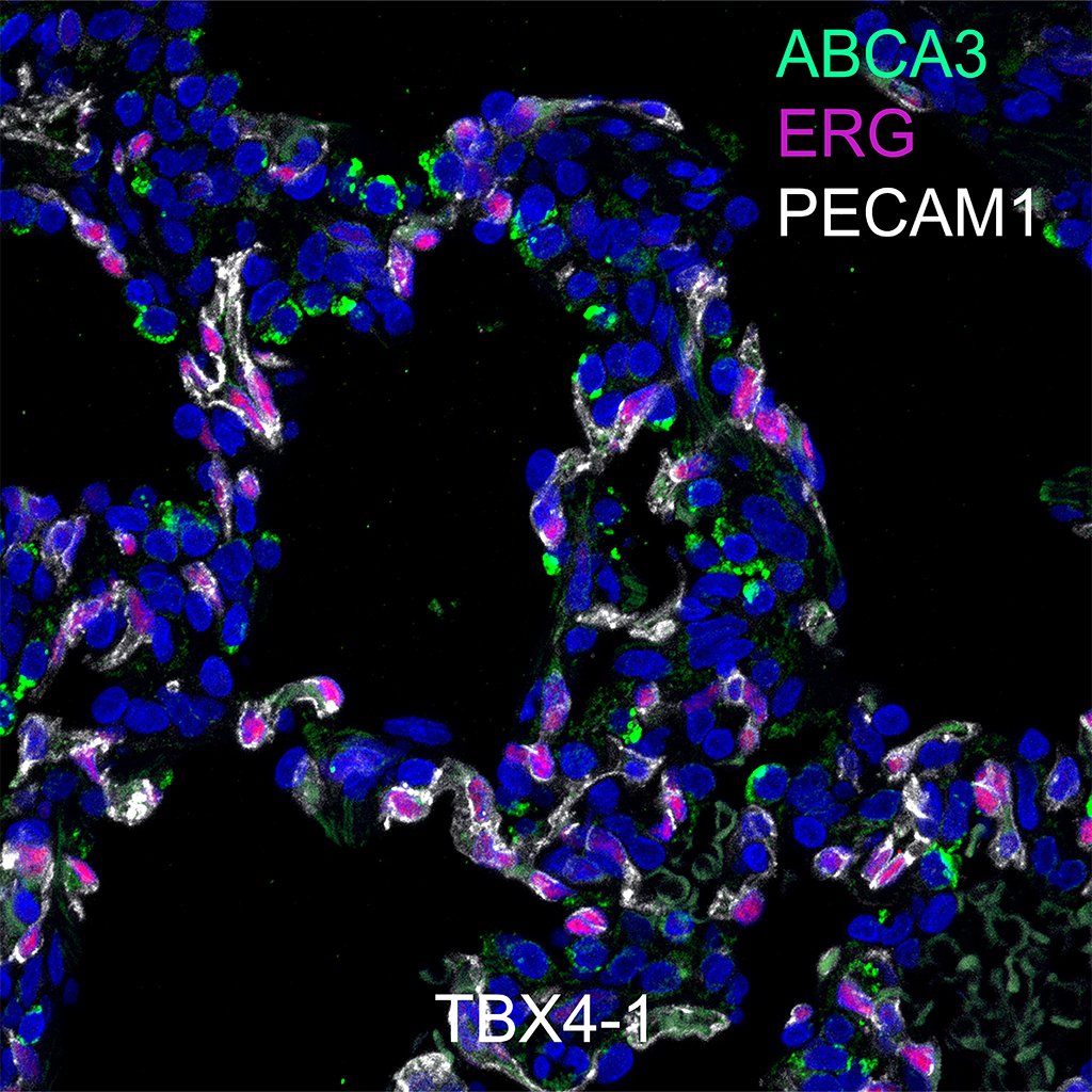
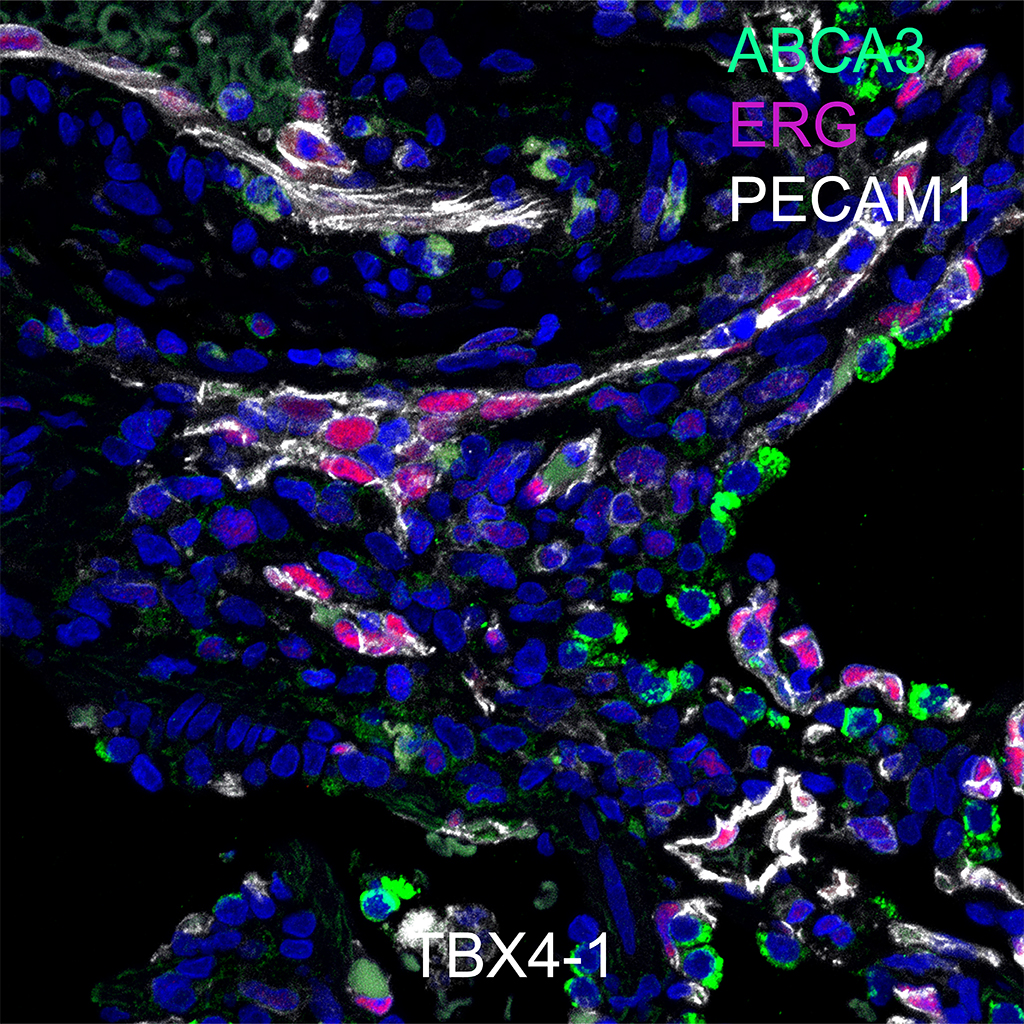
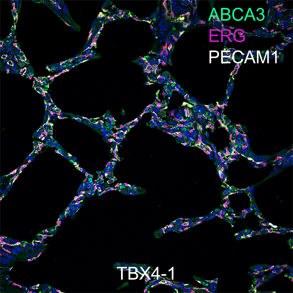
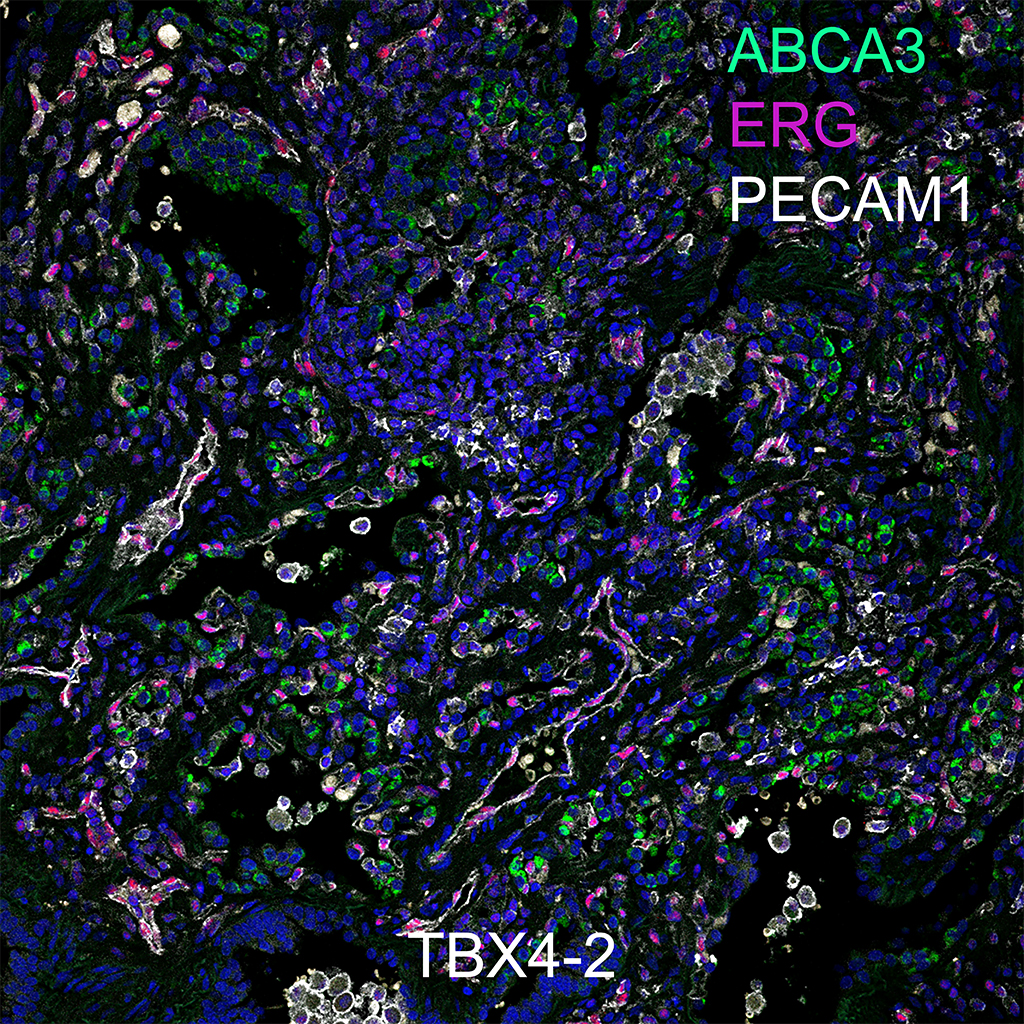
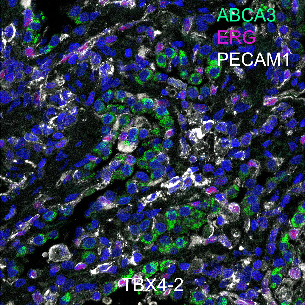
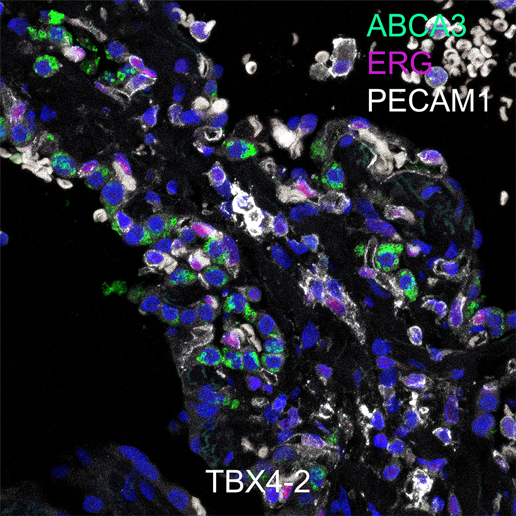
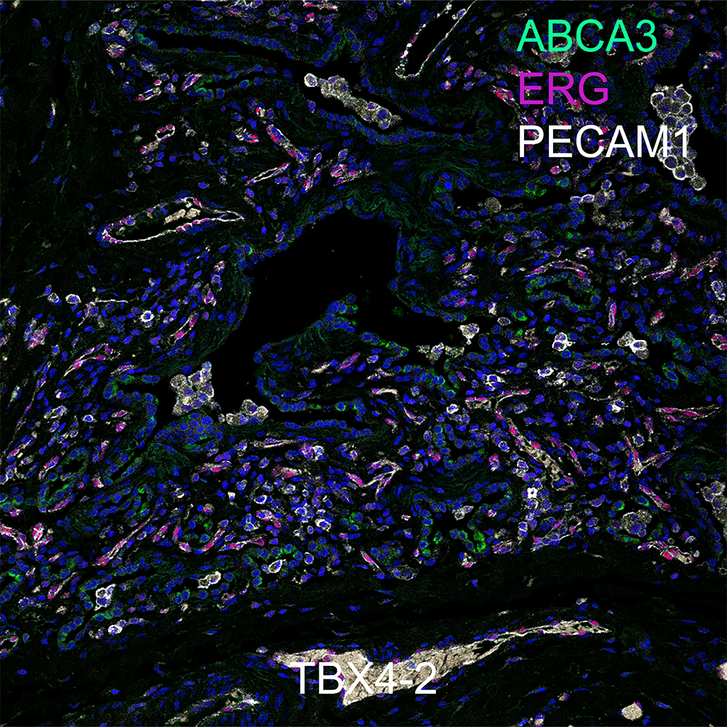
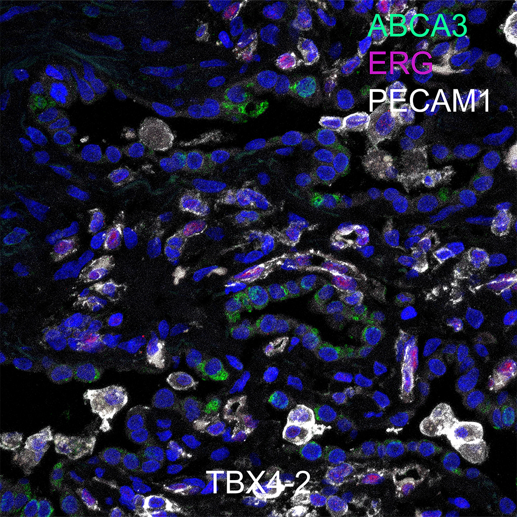
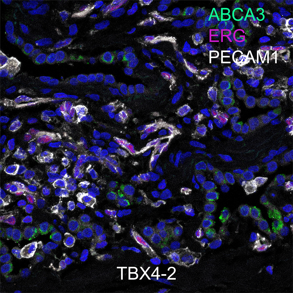
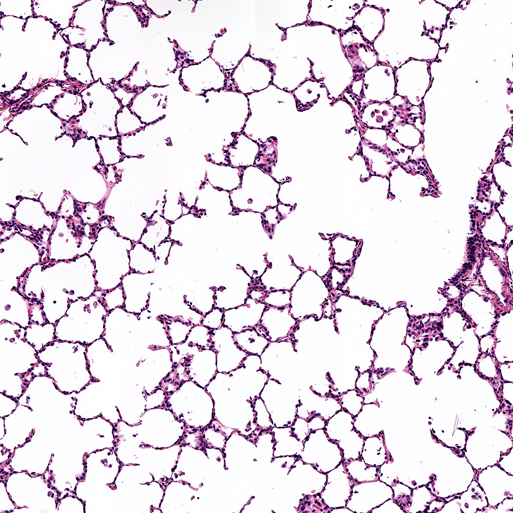
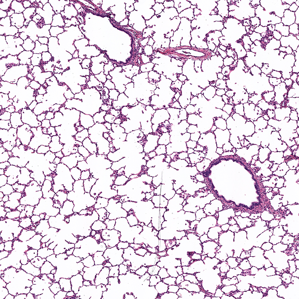
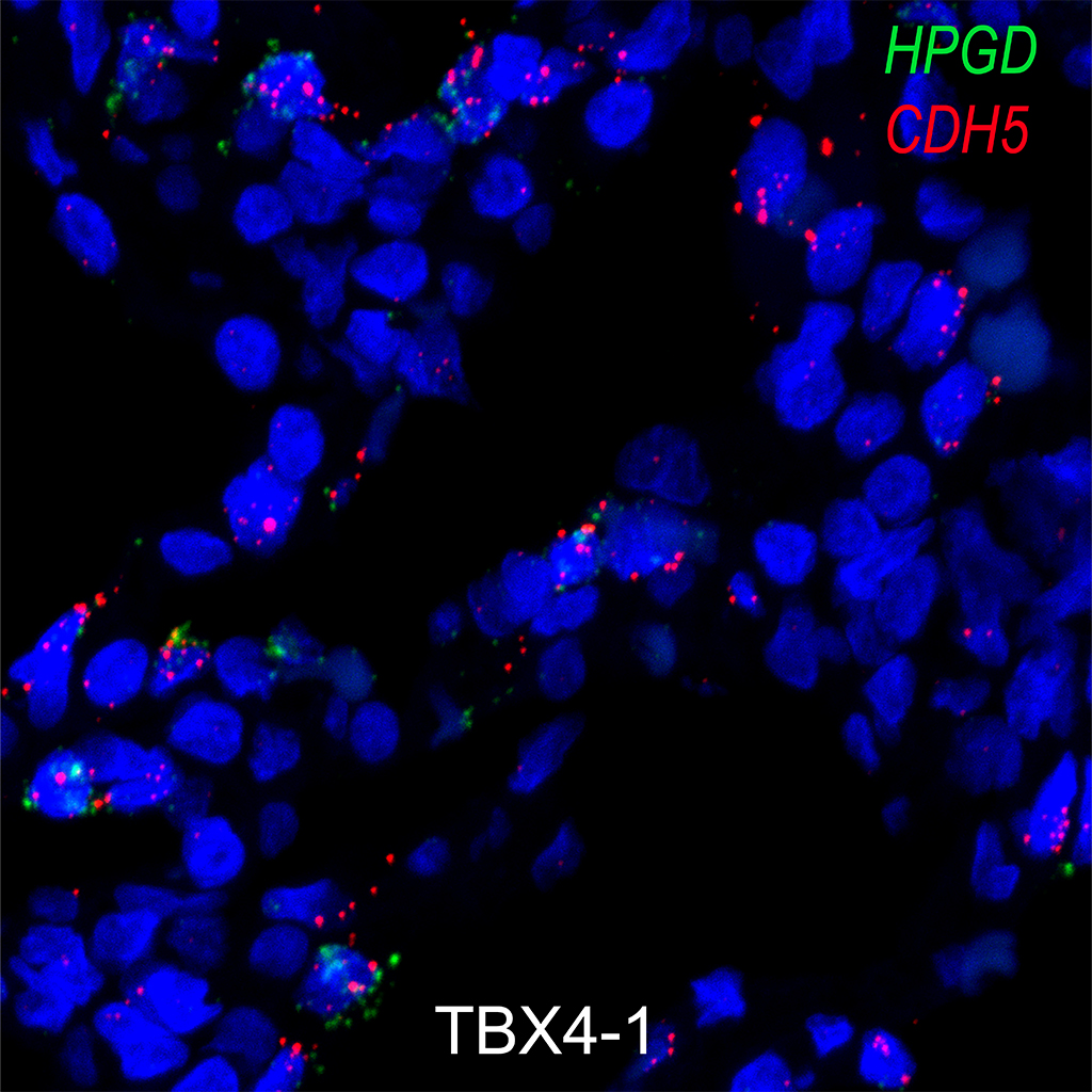
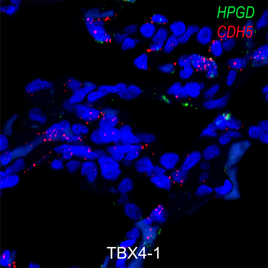
Alveolar type II cells, also known as type II pneumocytes, are specialized epithelial cells found in the alveoli of the lungs, which are the tiny air sacs responsible for gas exchange. These cells play a crucial role in maintaining the structure and function of the respiratory system.
Alveolar type II cells are located within the alveolar walls of the lungs.
Alveolar type II cells are cuboidal or squamous in shape and are smaller than alveolar type I cells, which are responsible for gas exchange. They are characterized by the presence of microvilli on their apical surface, which increases their surface area for various functions.
One of the most important functions of alveolar type II cells is the production and secretion of pulmonary surfactant. Surfactant is a complex mixture of lipids and proteins that reduces the surface tension of the alveolar fluid, preventing the collapse of alveoli during exhalation. This property is crucial for maintaining the stability of the alveoli and preventing lung collapse, particularly at the end of expiration.
Alveolar type II cells serve as progenitor cells for alveolar type I cells. They have the ability to self-renew and differentiate into type I cells, helping in the repair and regeneration of the alveolar epithelium after injury or damage. This regenerative capacity is essential for maintaining the integrity of the respiratory barrier.
Alveolar type II cells also play a role in the innate immune response of the lungs. They can produce and secrete various immune molecules, including cytokines, chemokines, and antimicrobial peptides, in response to pathogens or inflammatory stimuli. These molecules help to recruit immune cells to the site of infection and promote the clearance of pathogens from the lungs.
Alveolar type II cells are involved in the regulation of ion and fluid balance within the alveoli. They express various ion channels and transporters that regulate the movement of ions, such as sodium and chloride, across the epithelial barrier. This process is important for maintaining the osmotic balance of the alveolar fluid and preventing the accumulation of fluid in the lungs (pulmonary edema).
The surfactant produced by alveolar type II cells consists mainly of phospholipids (such as dipalmitoylphosphatidylcholine, or DPPC) and surfactant proteins (SP-A, SP-B, SP-C, and SP-D). These components work together to reduce surface tension and maintain the stability of the alveoli.
The production and secretion of surfactant by alveolar type II cells are regulated by various factors, including mechanical stretch, glucocorticoid hormones, and certain signaling molecules (such as epinephrine and thyroid hormones). These regulatory mechanisms ensure that surfactant production is matched to the demands of respiration and the maintenance of lung function.