
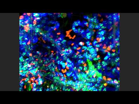

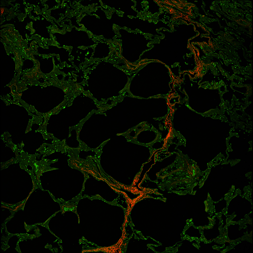
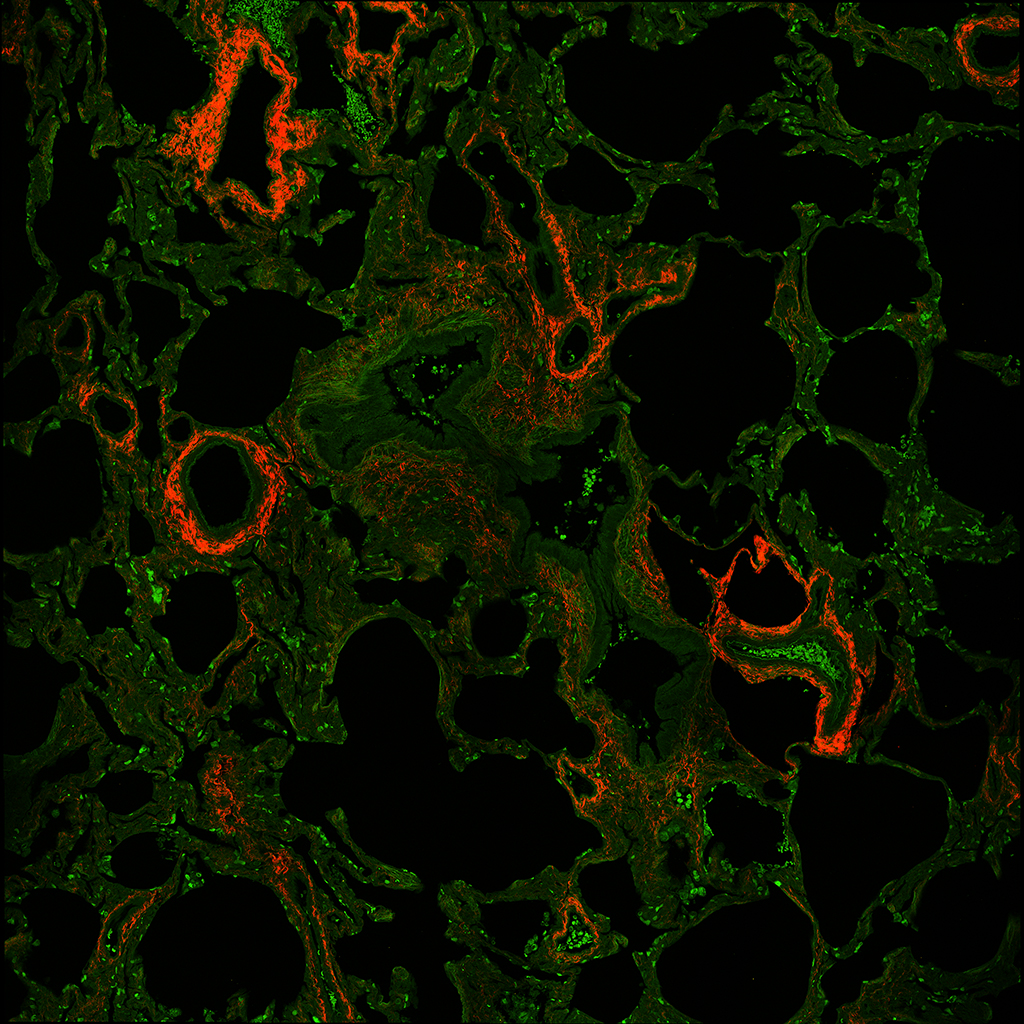
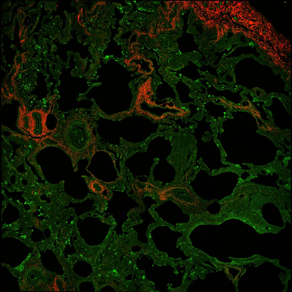
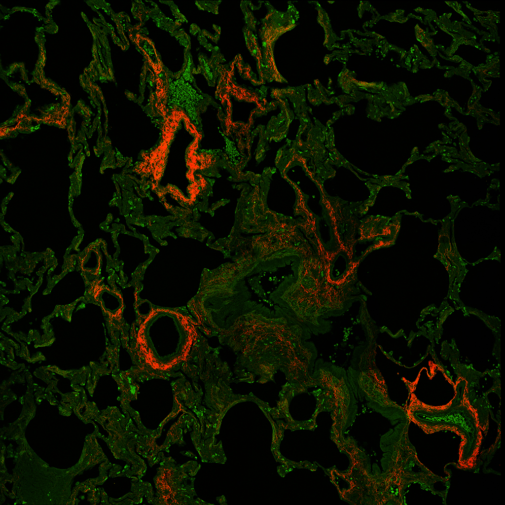
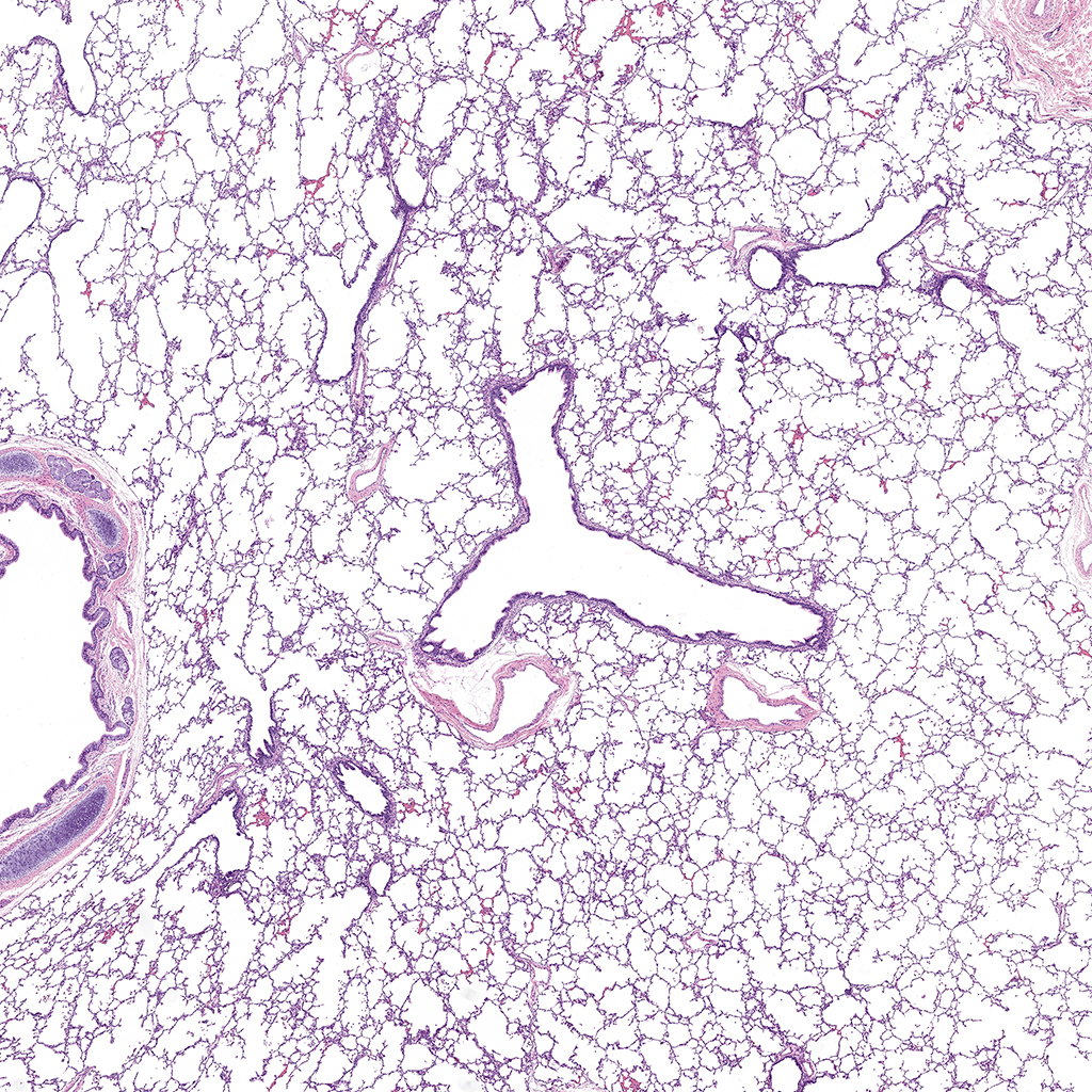
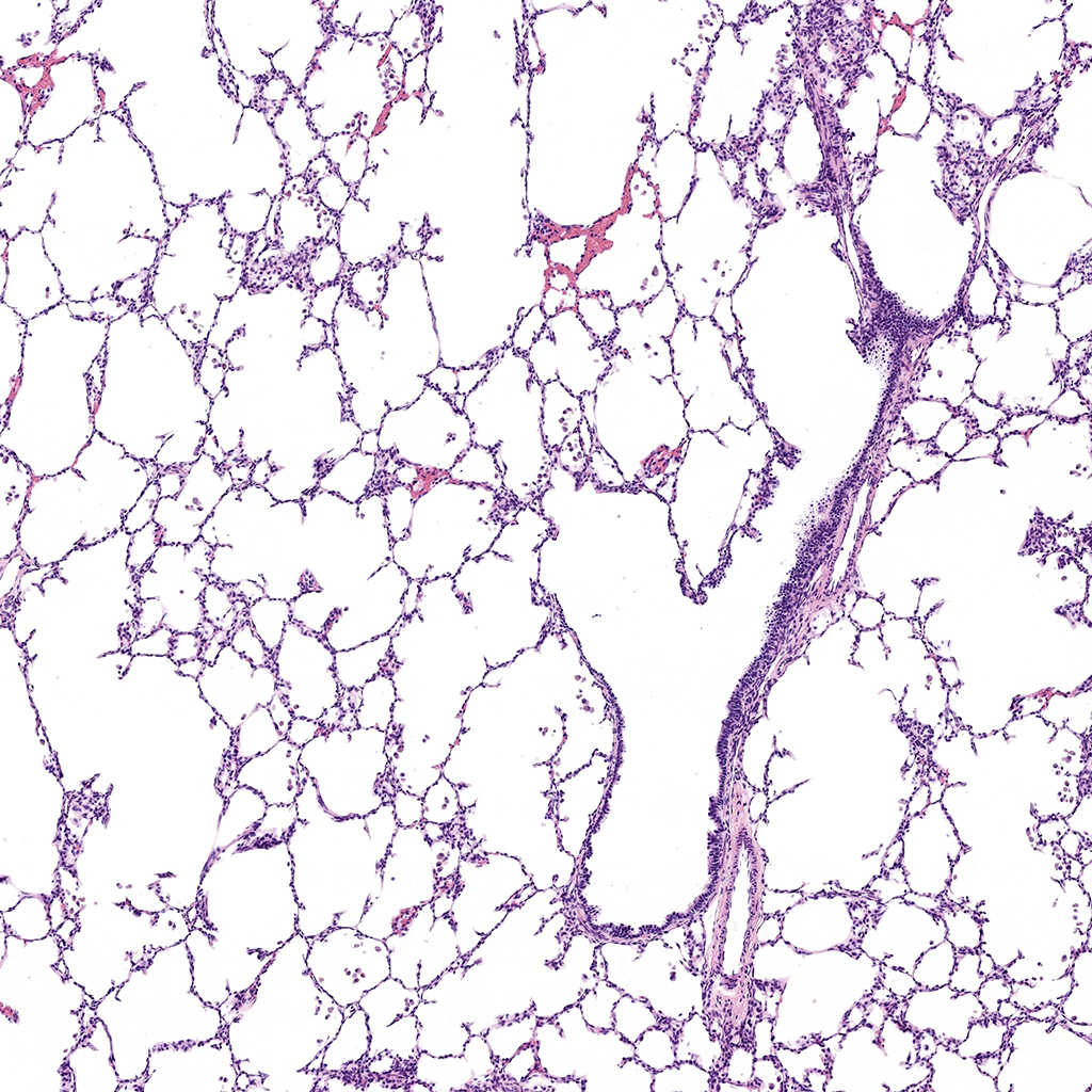
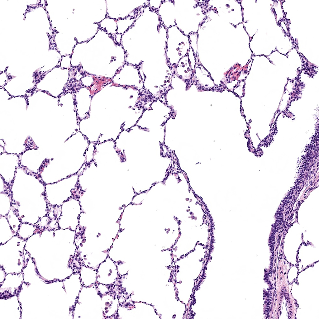
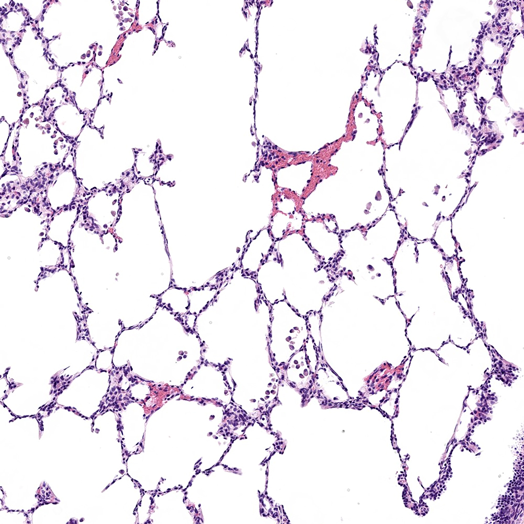
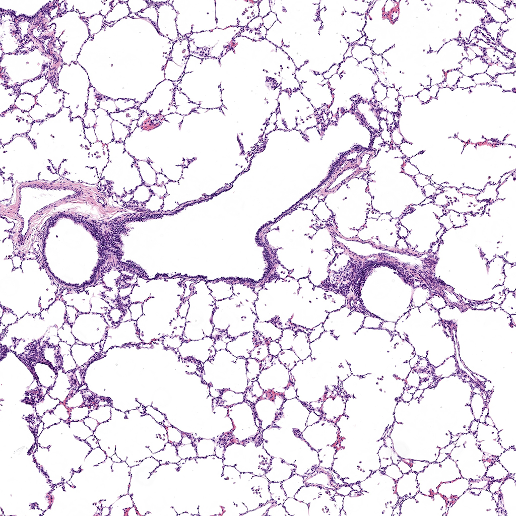
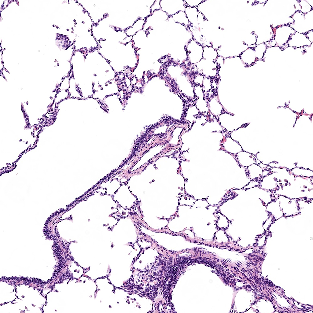
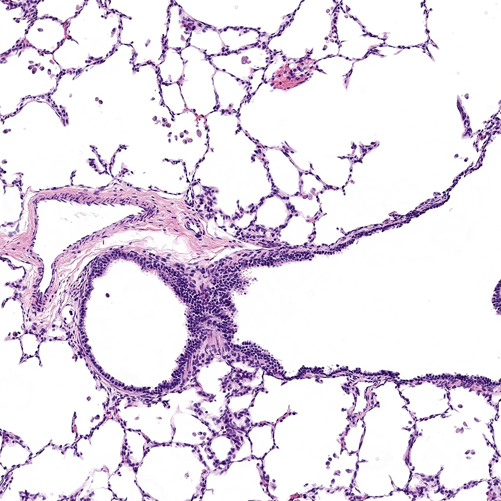
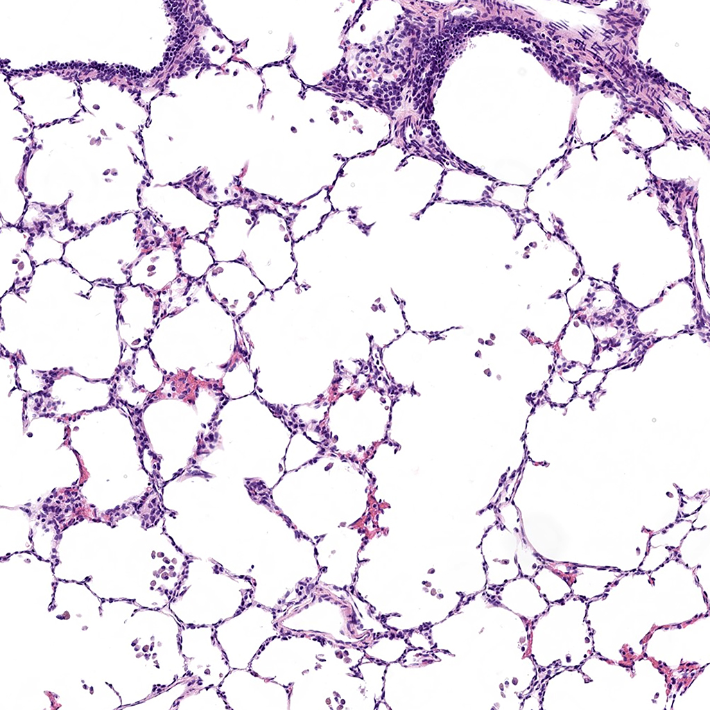
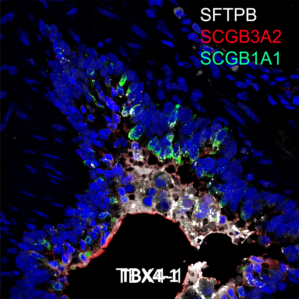
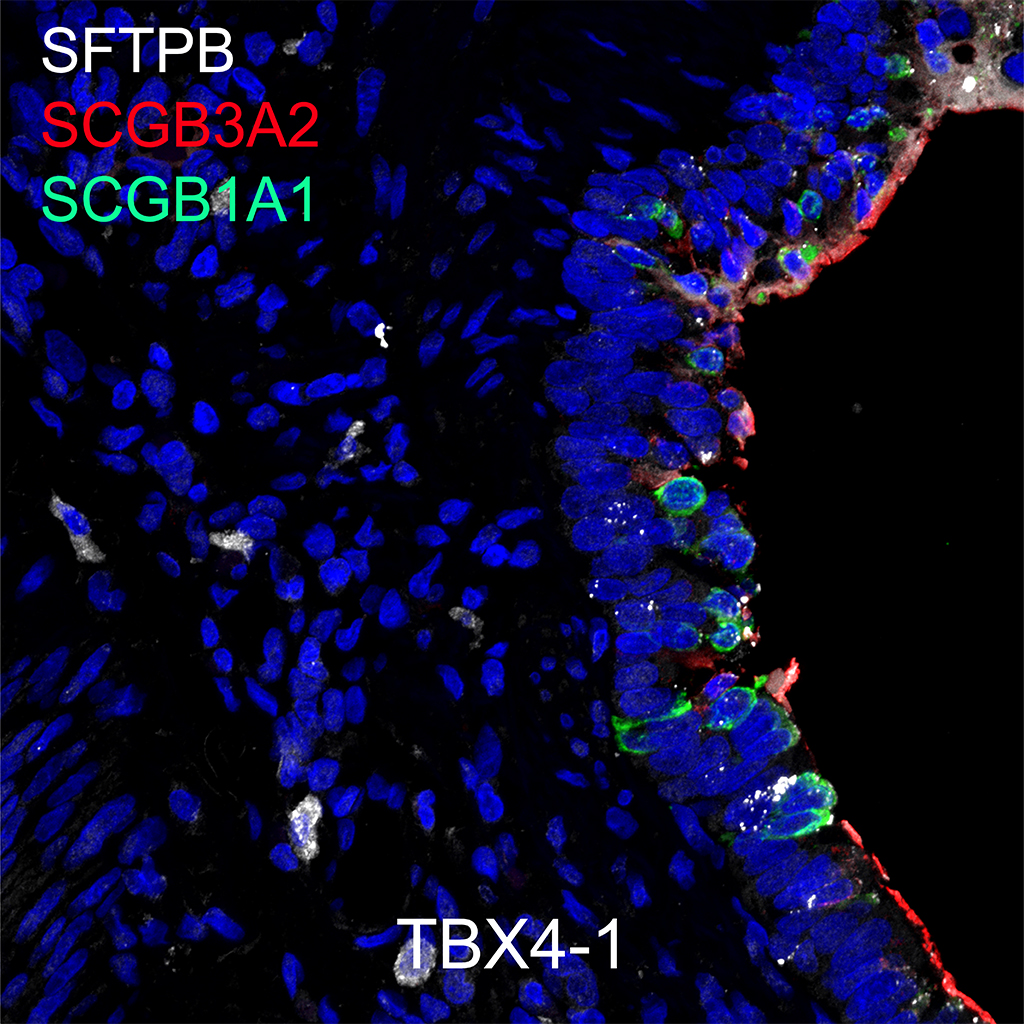
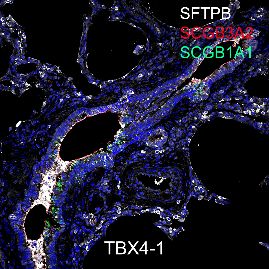
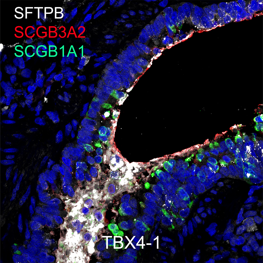
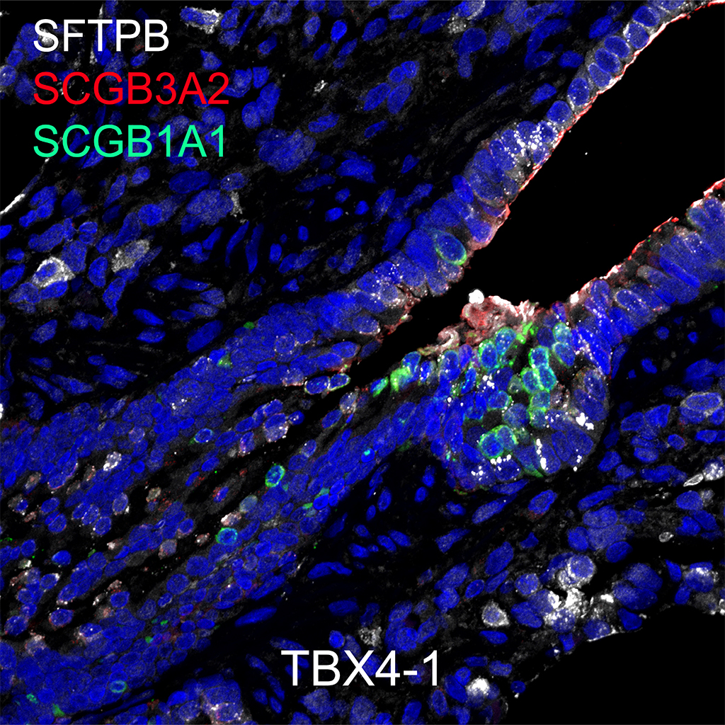
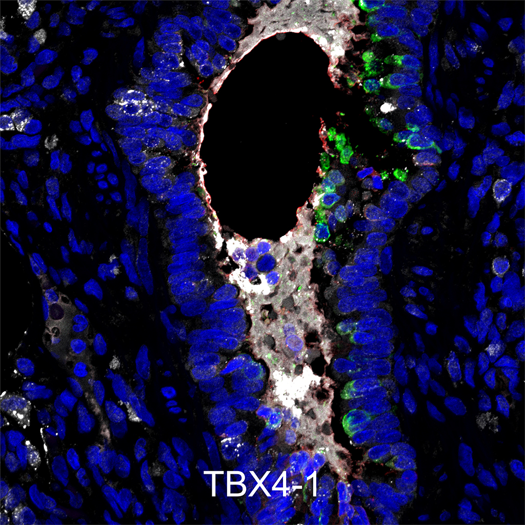
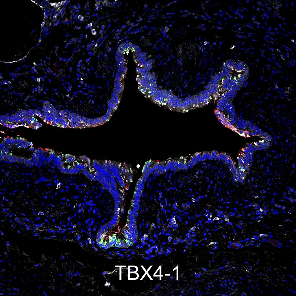
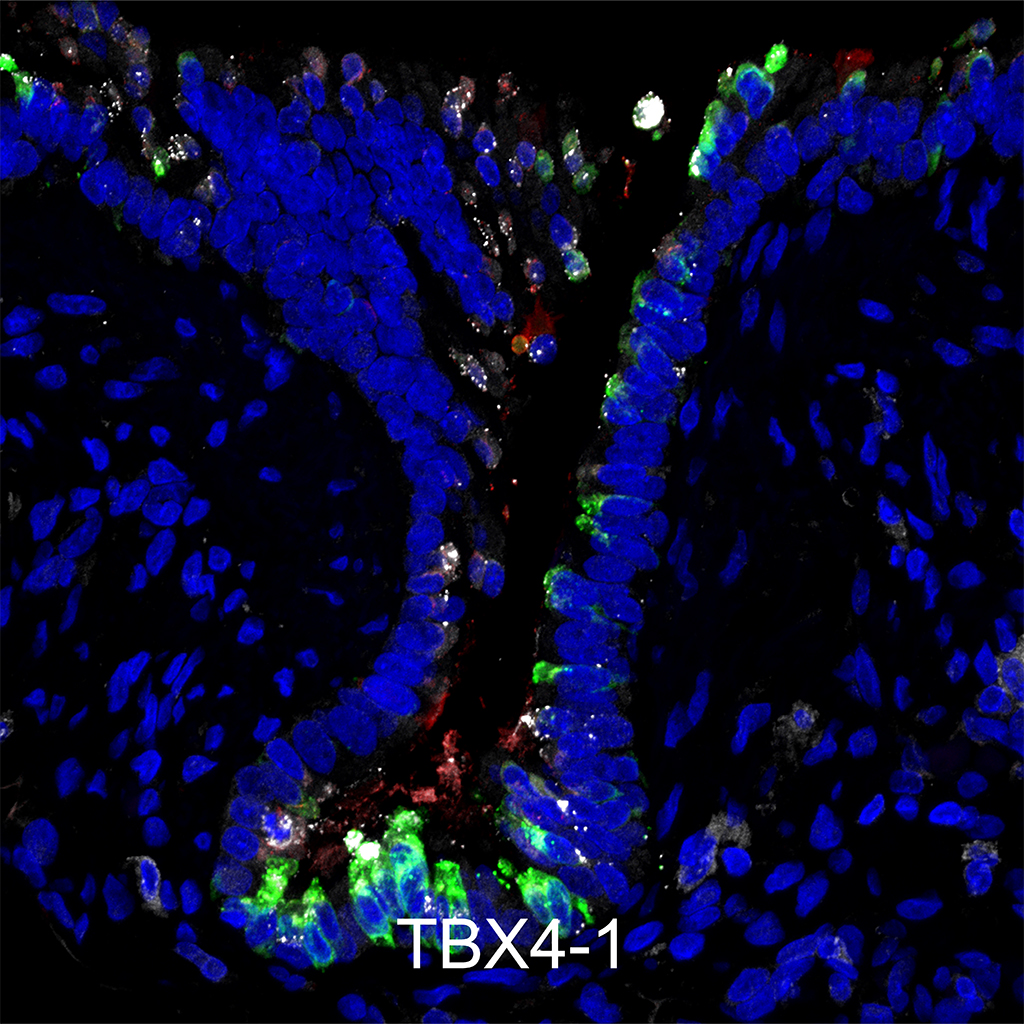
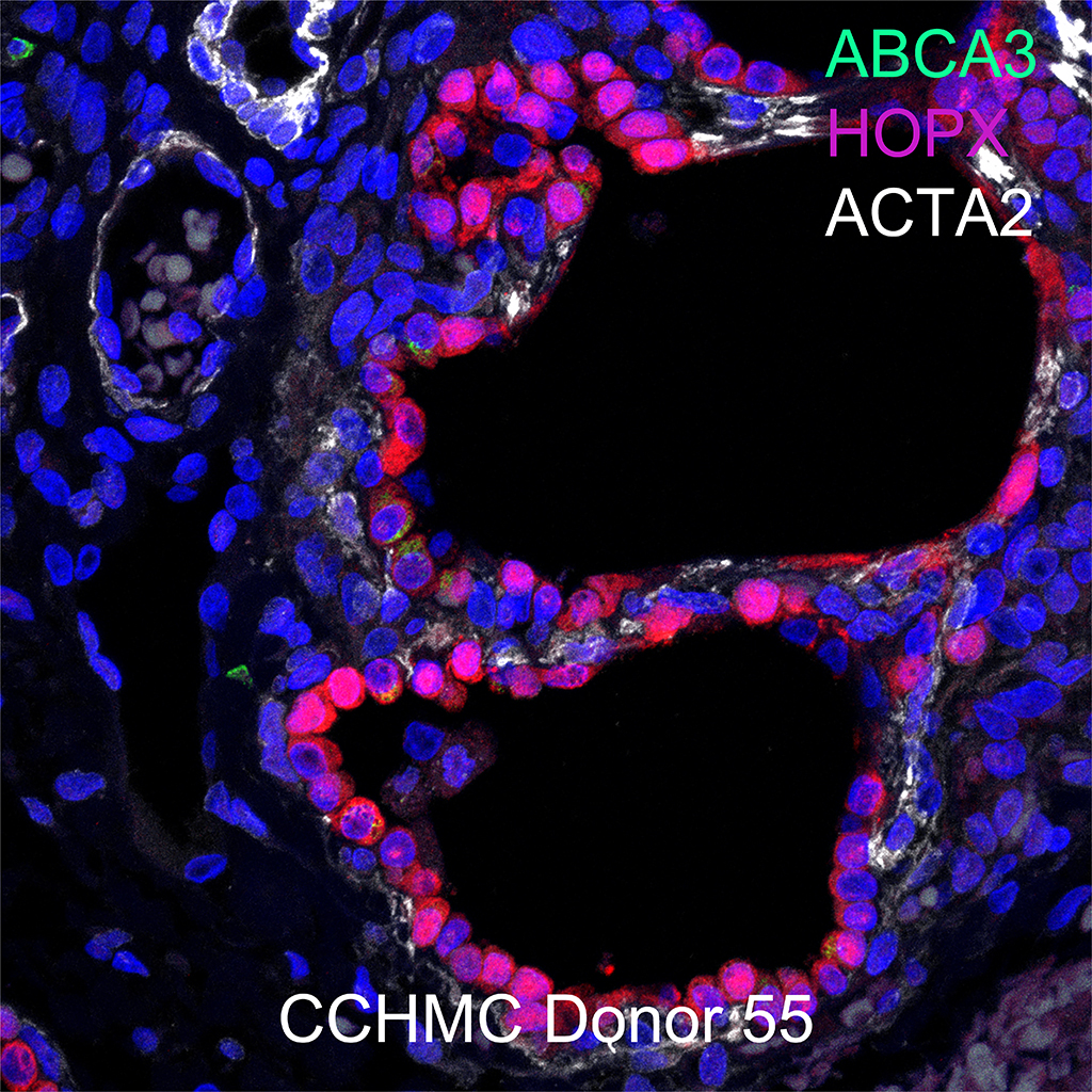
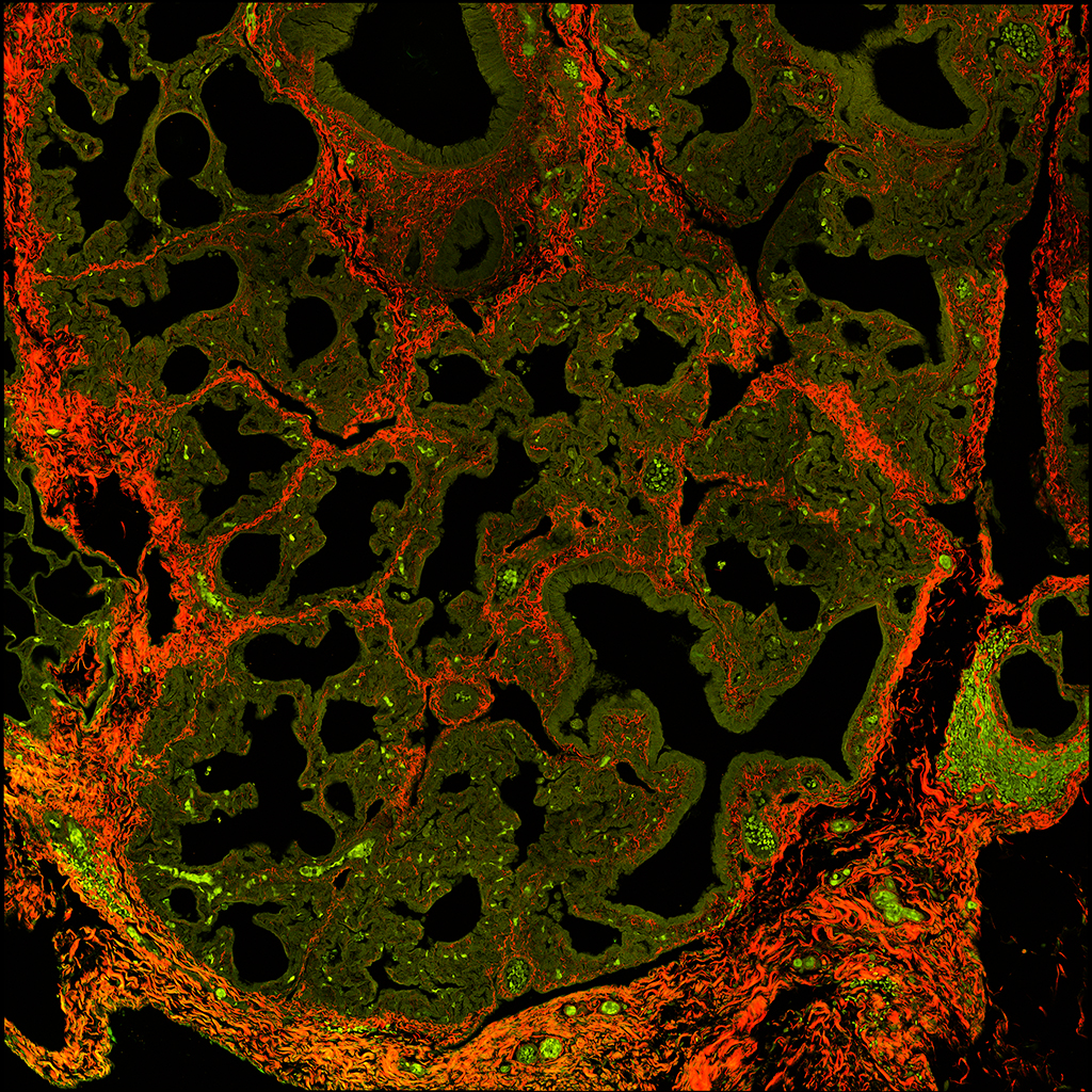
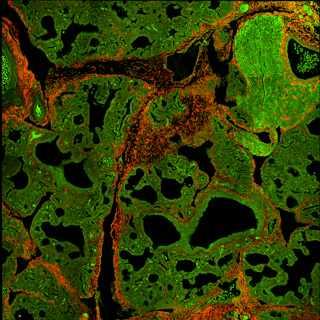
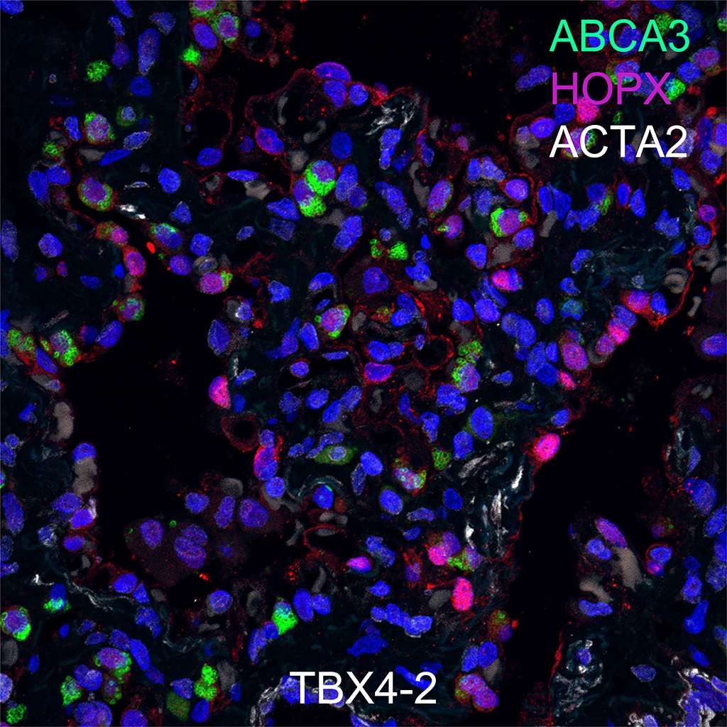
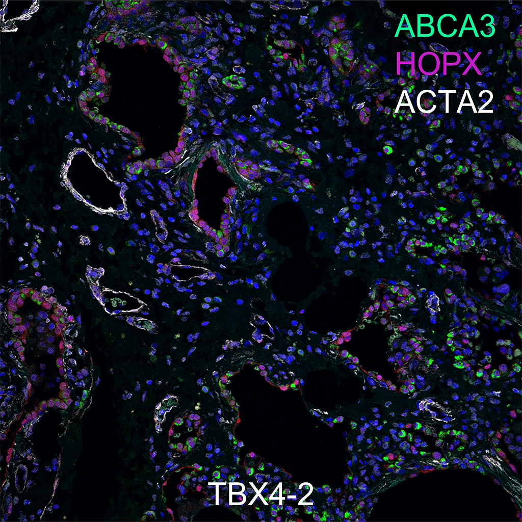
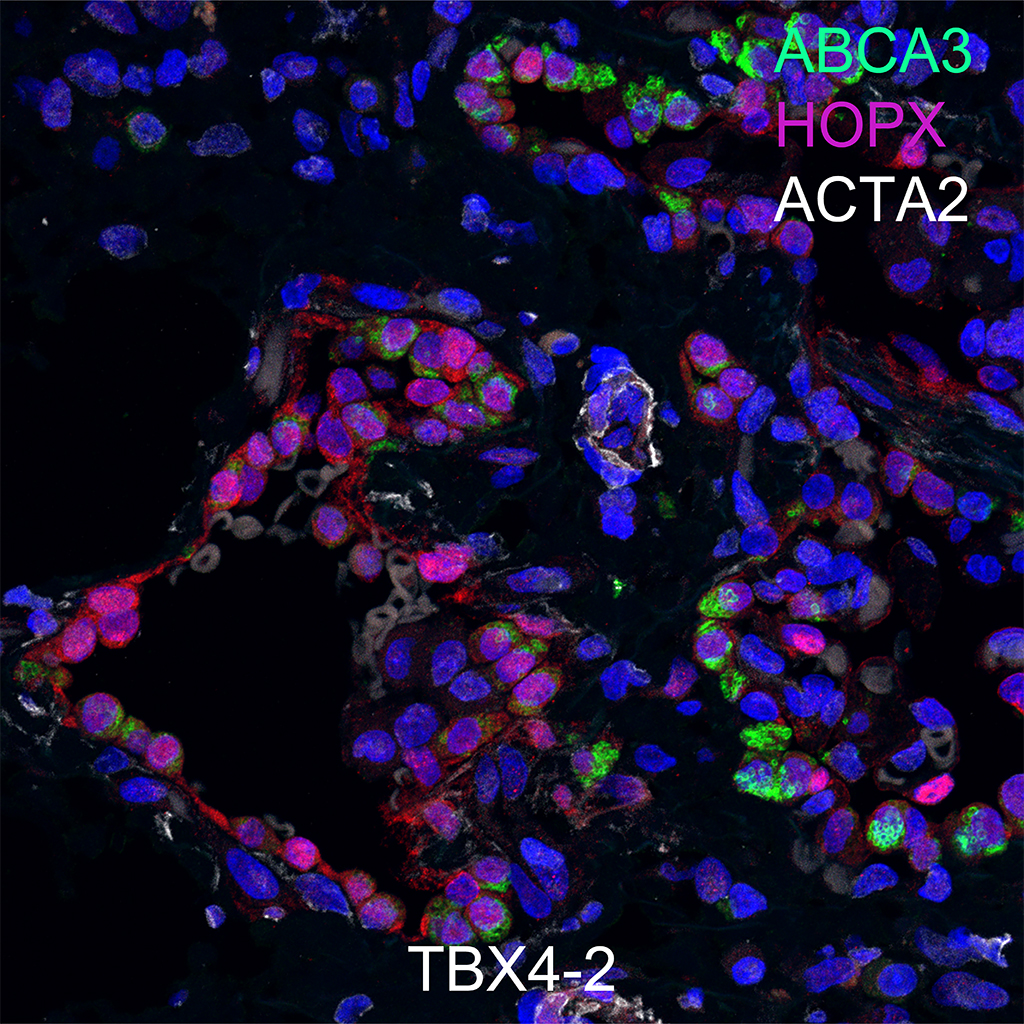
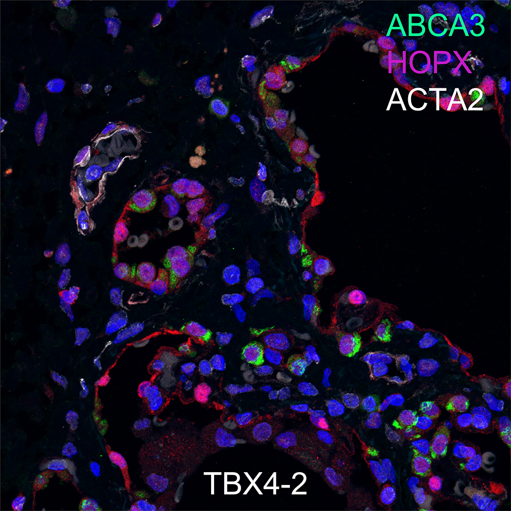
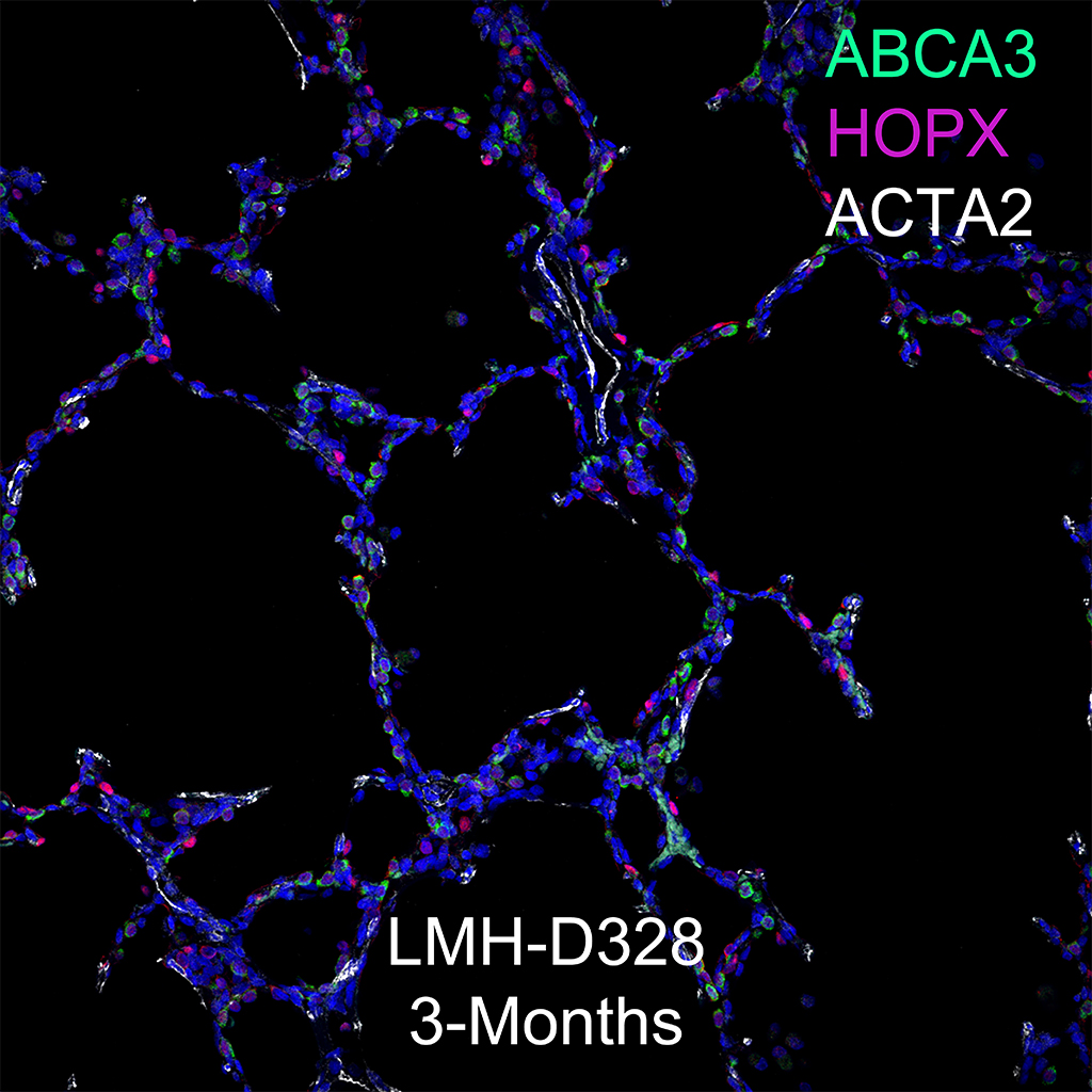
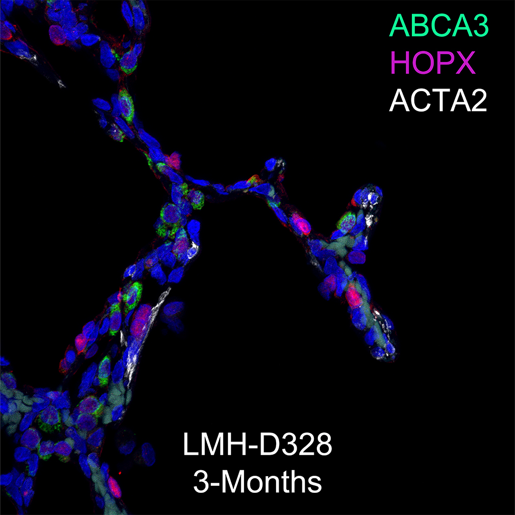
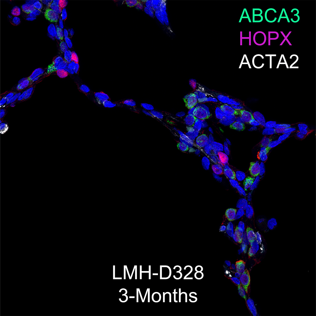
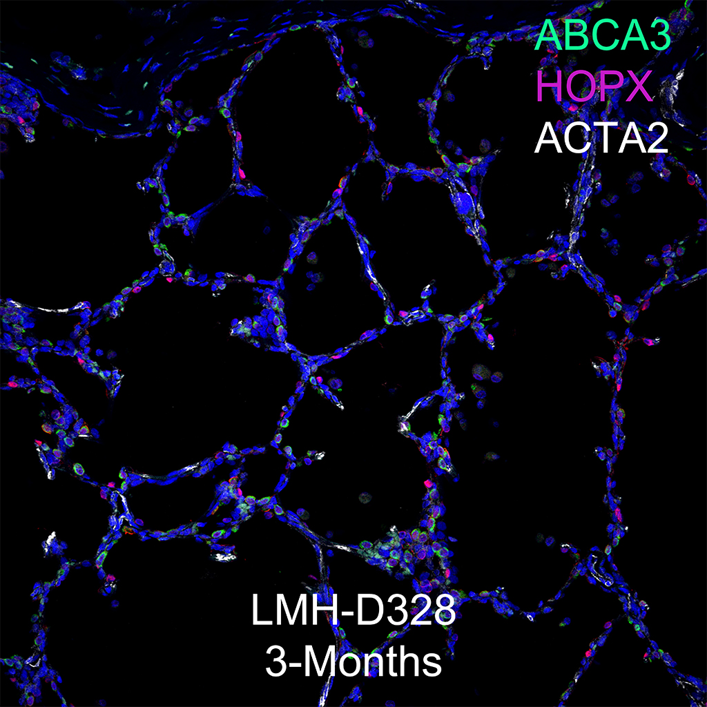
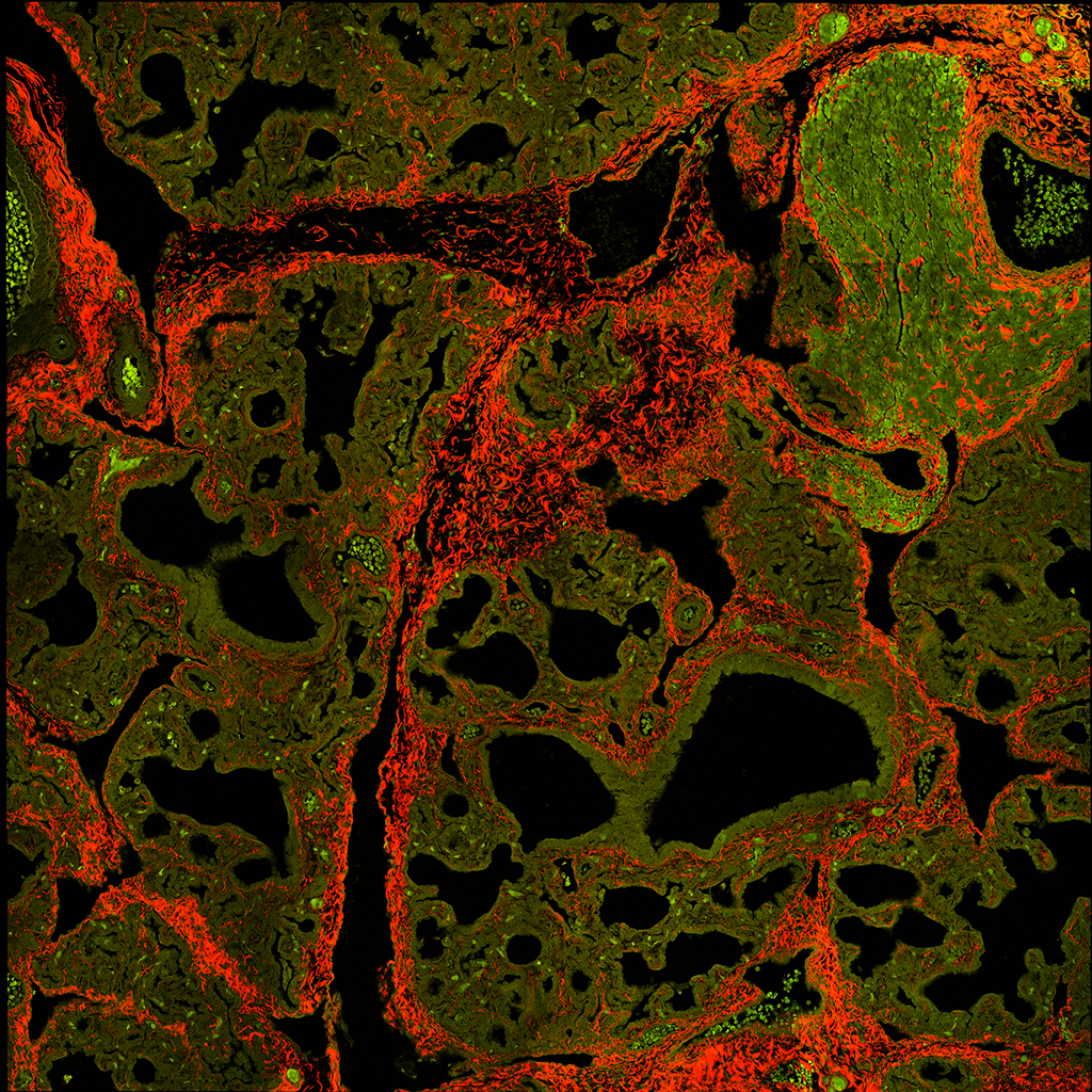
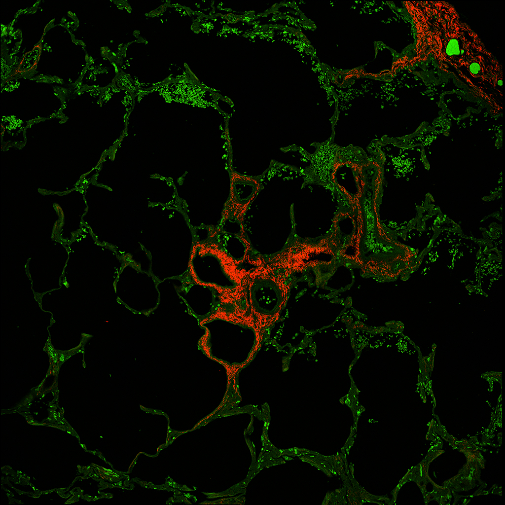
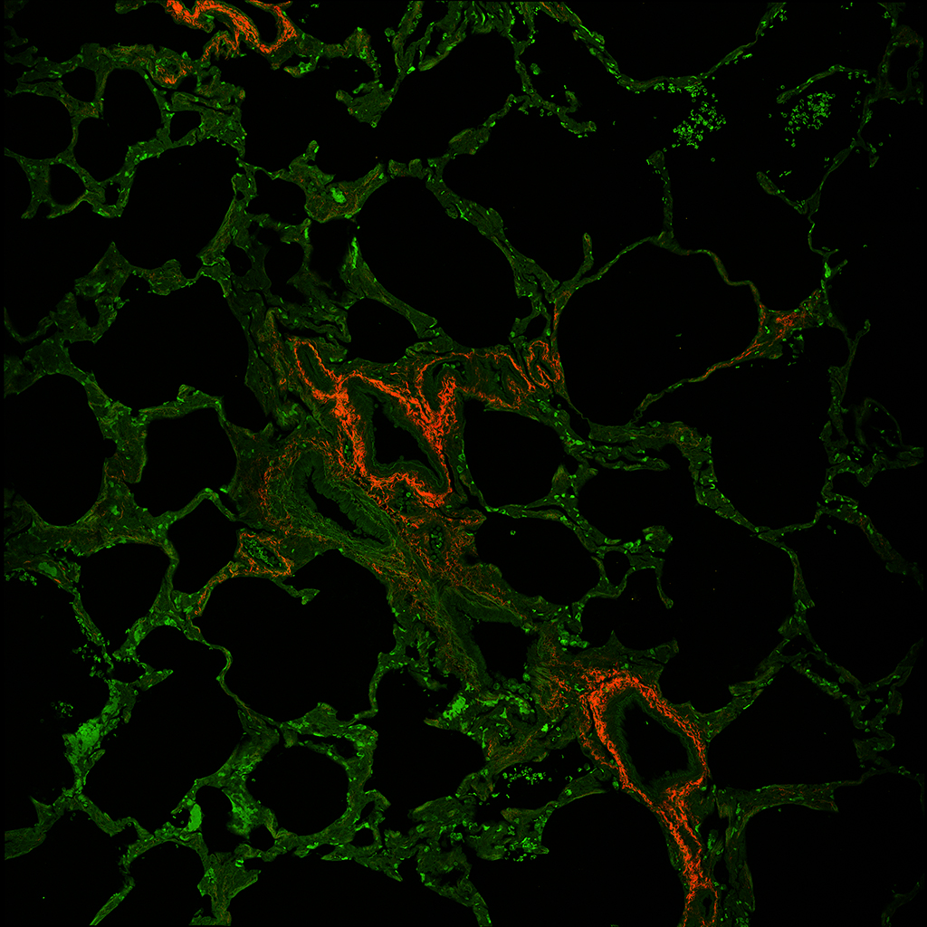
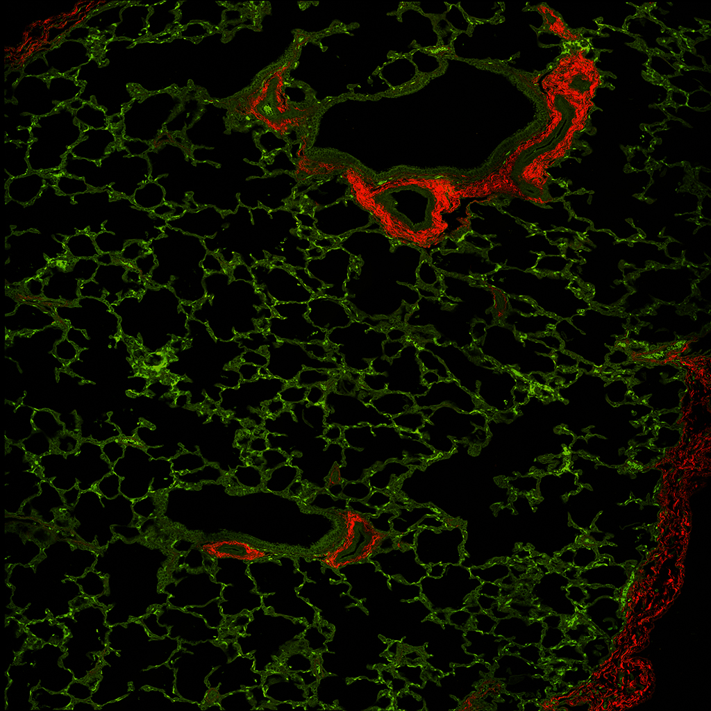
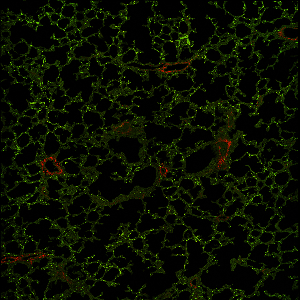
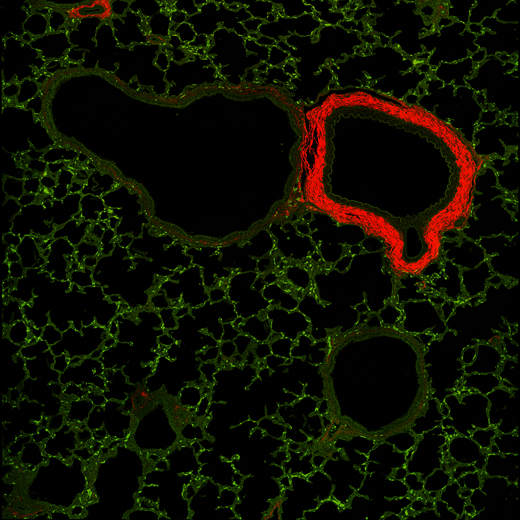
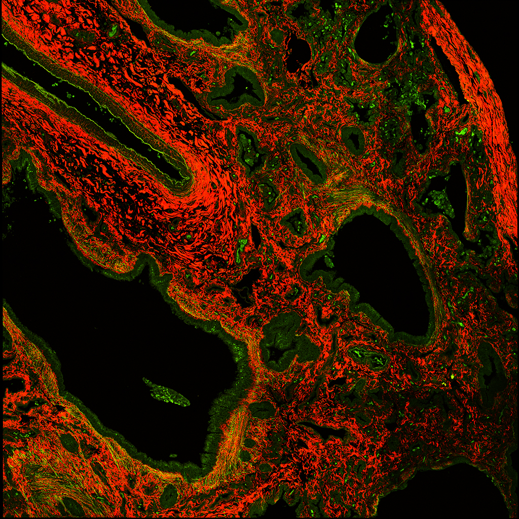
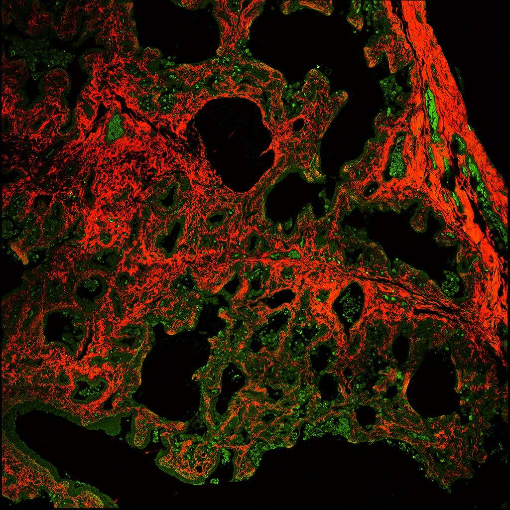
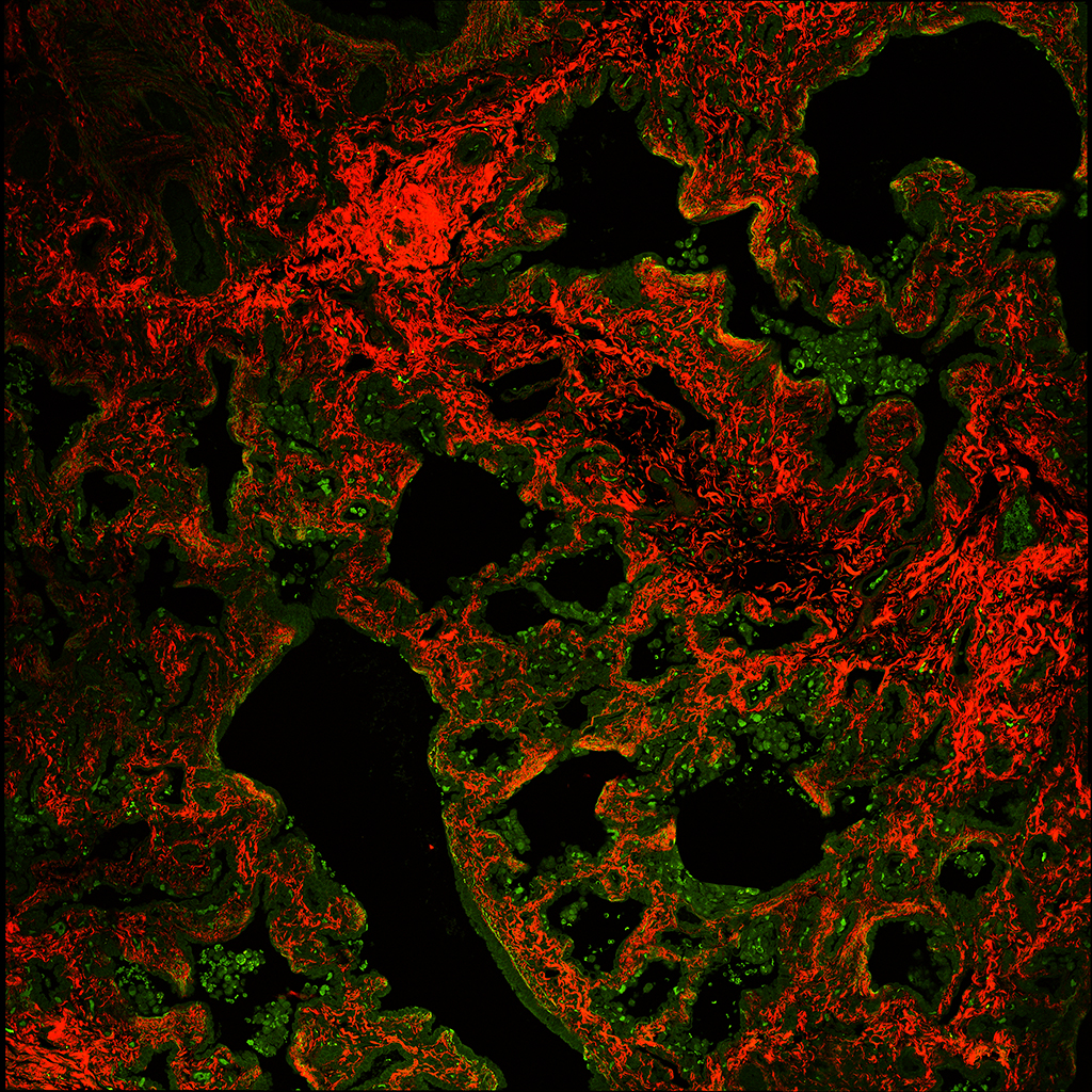
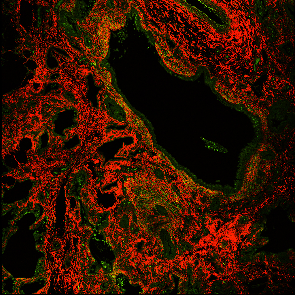
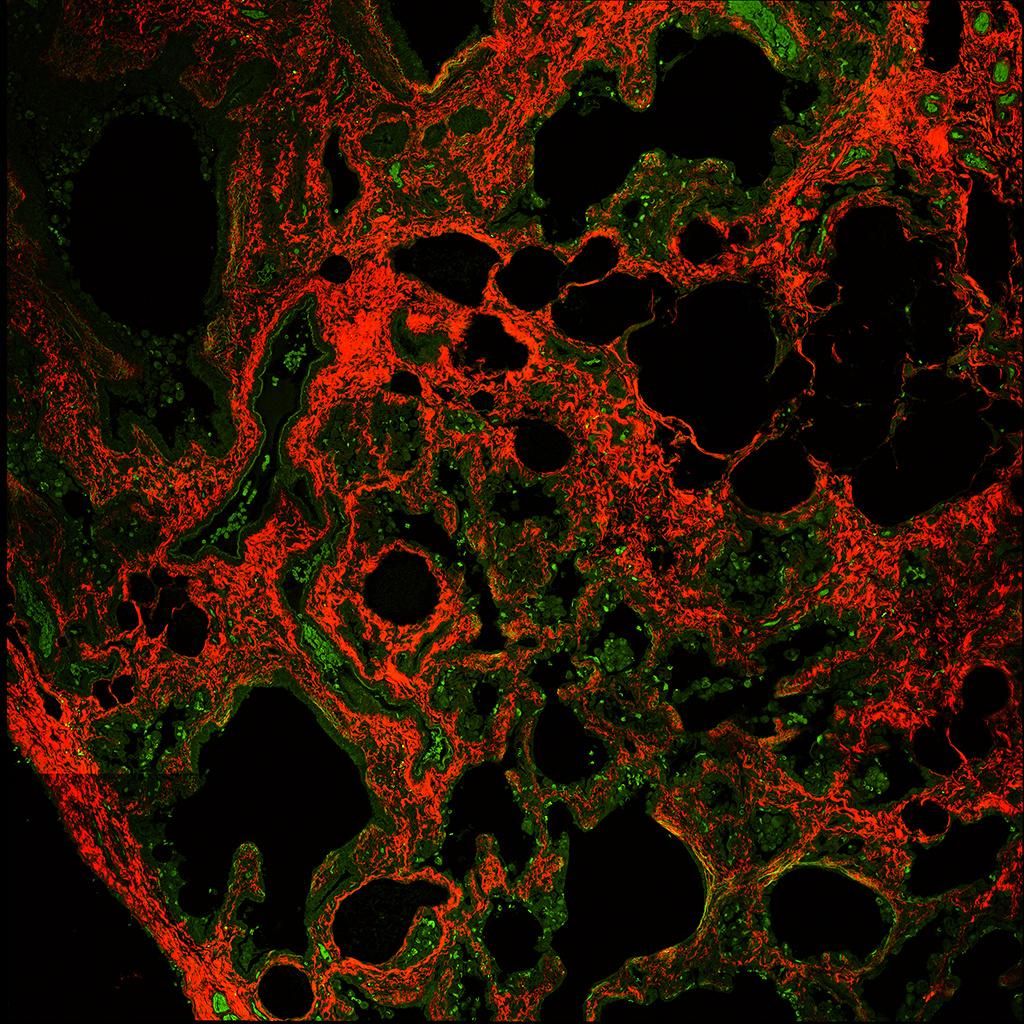
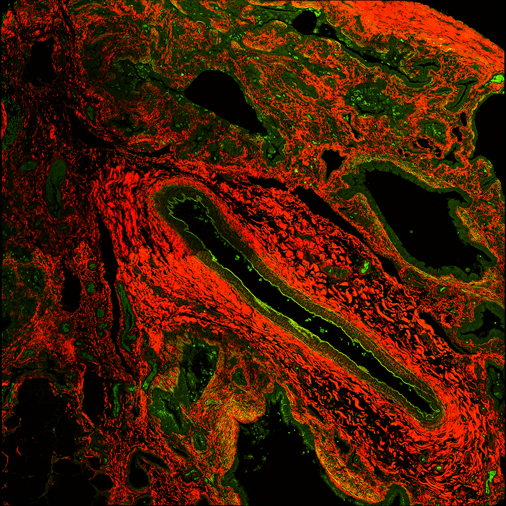
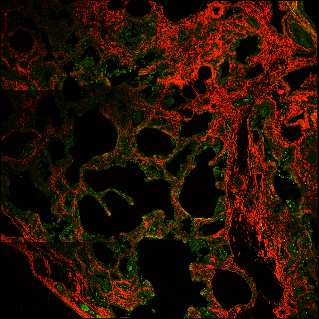
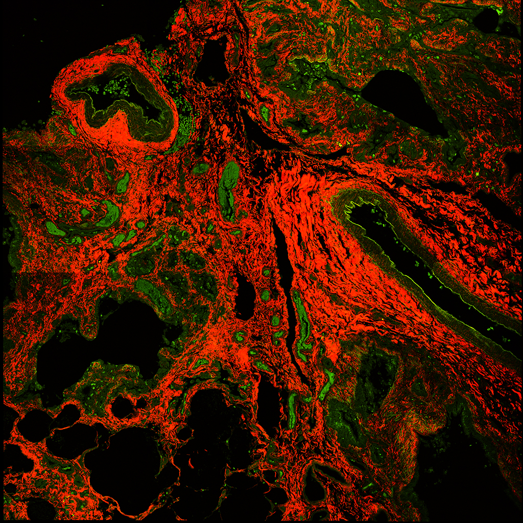
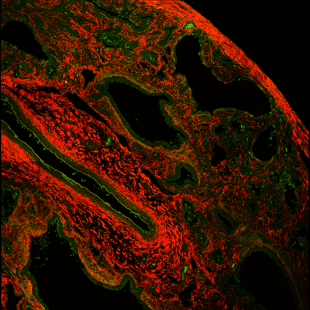
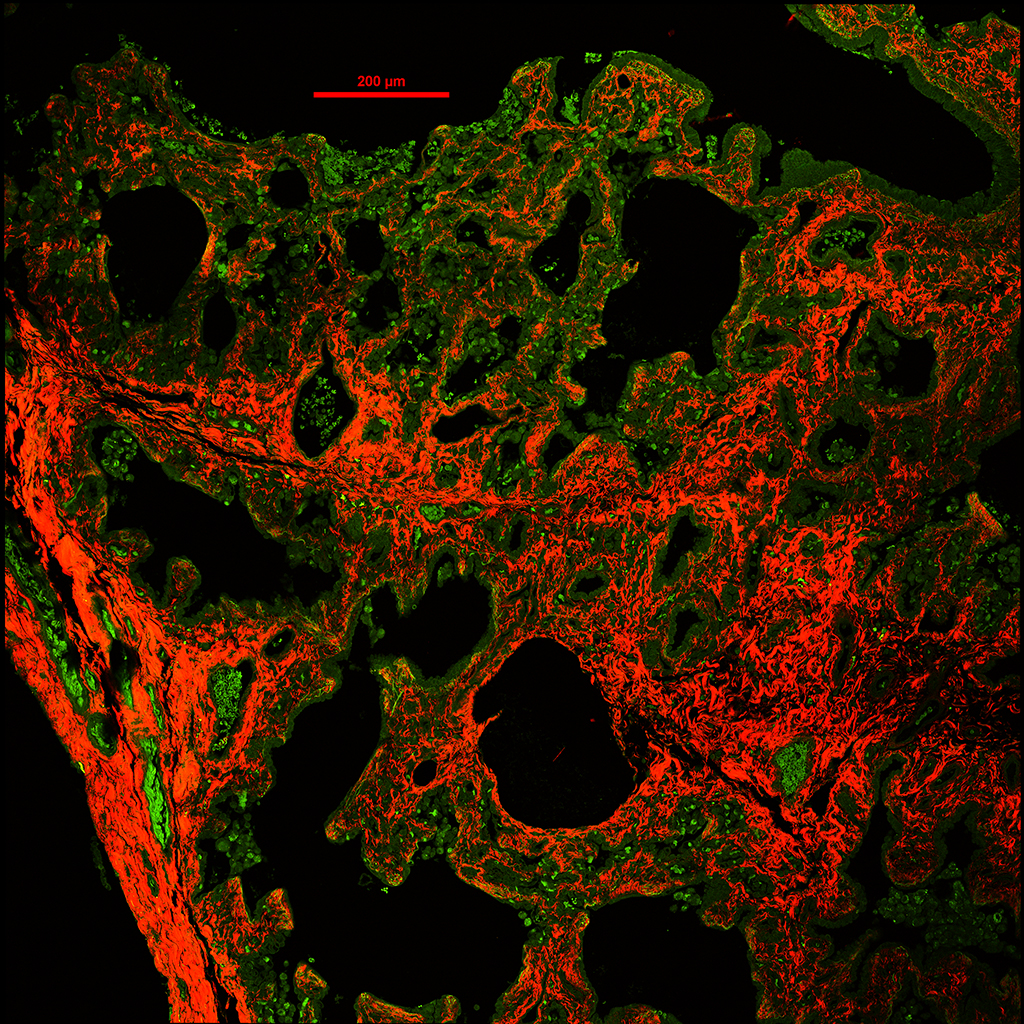
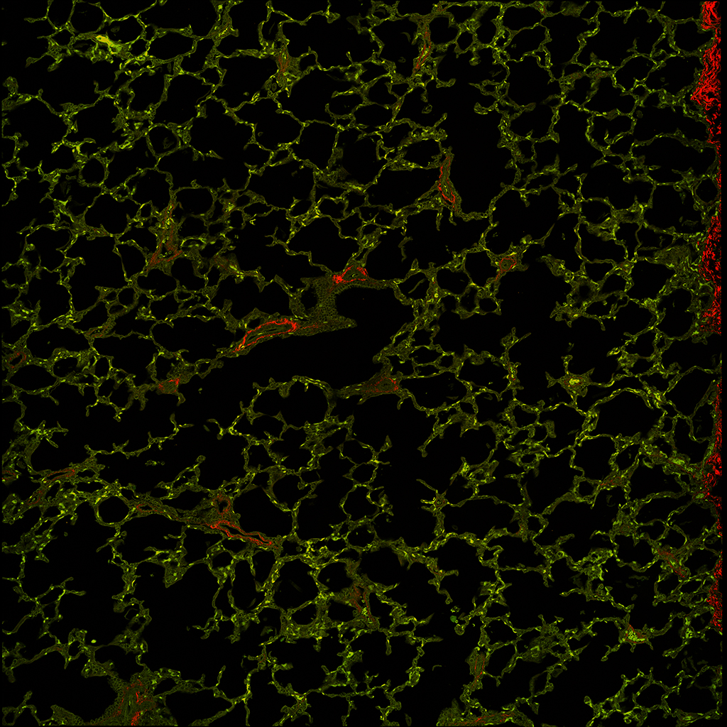
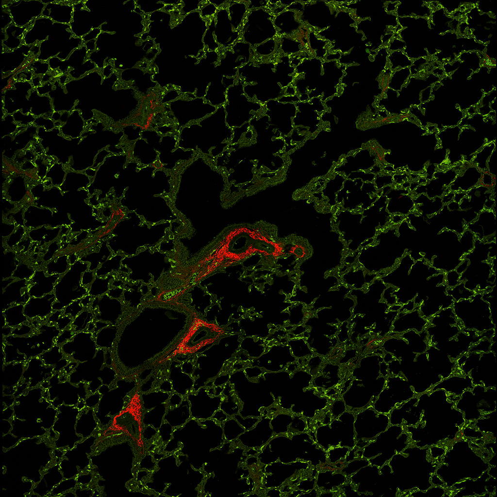
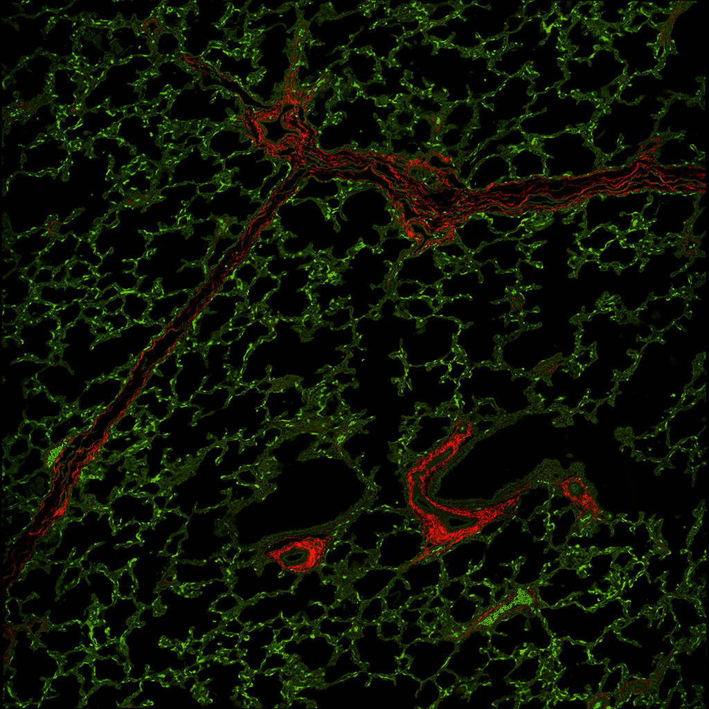
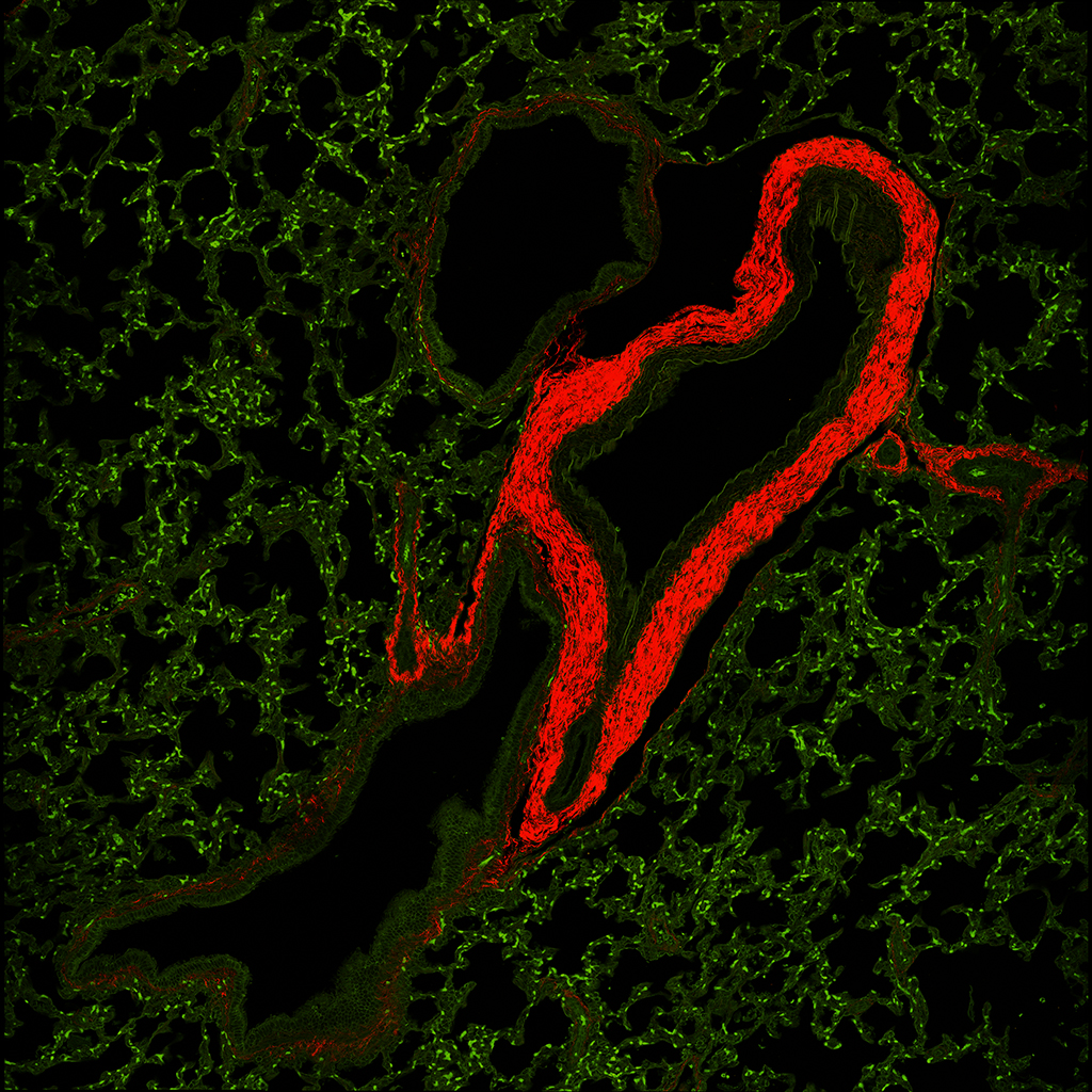
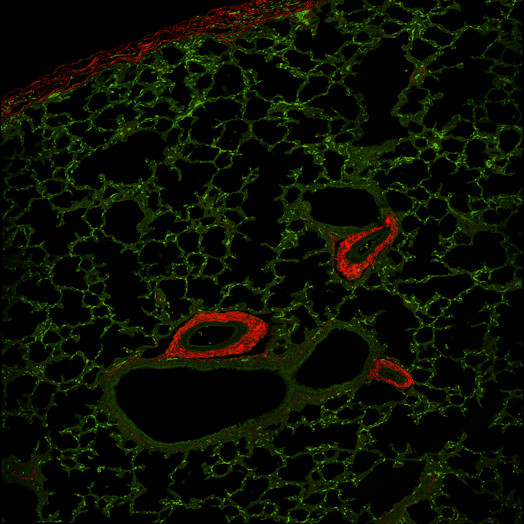
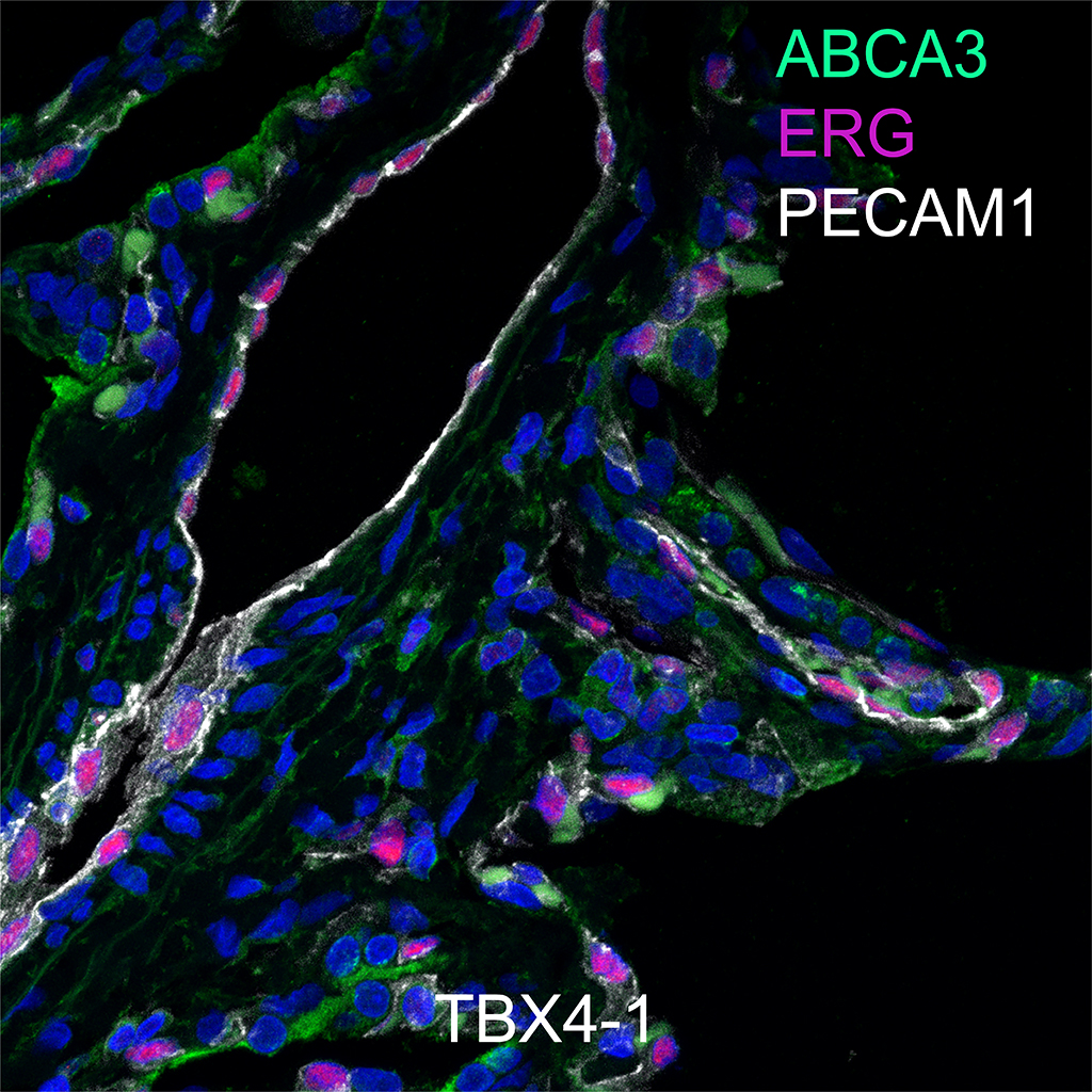
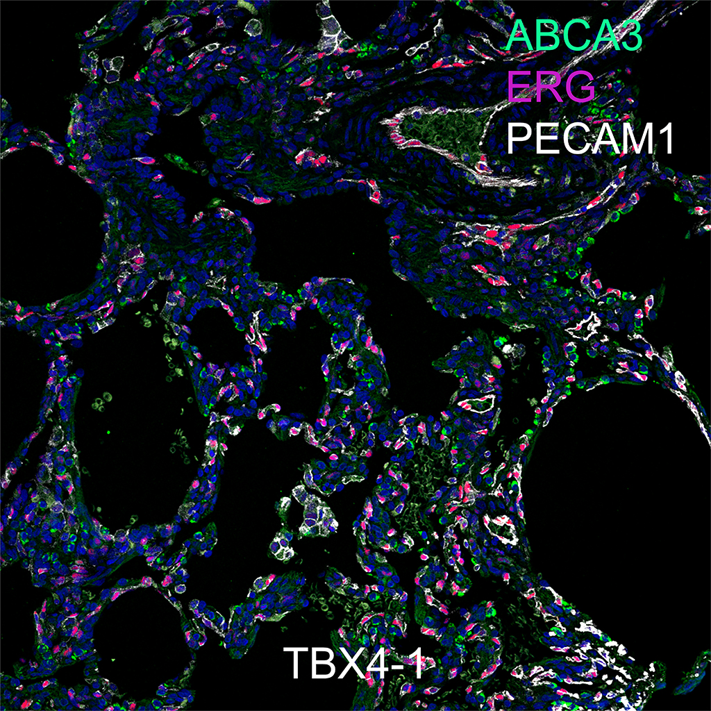
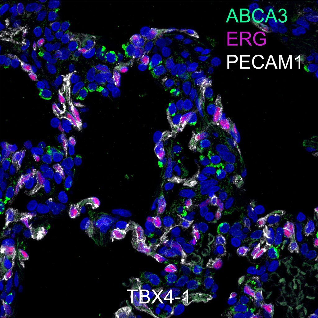
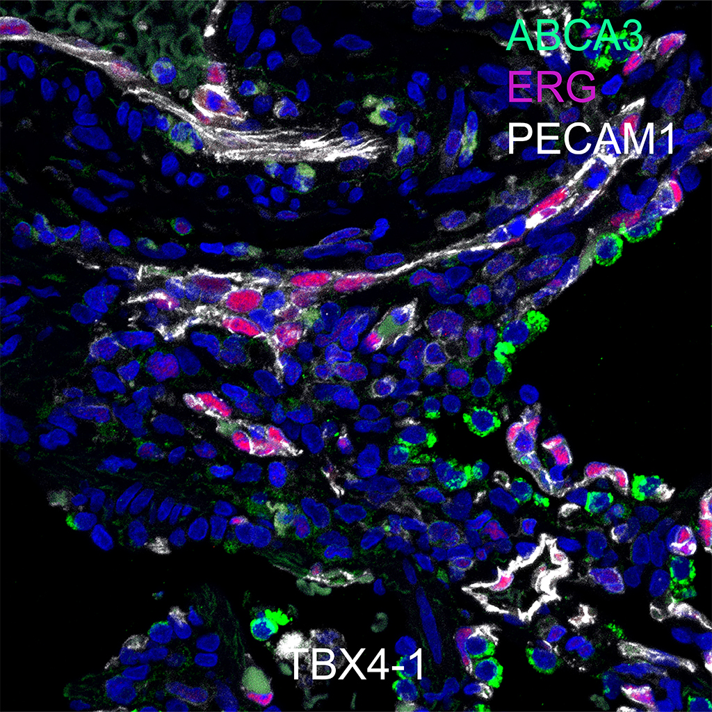
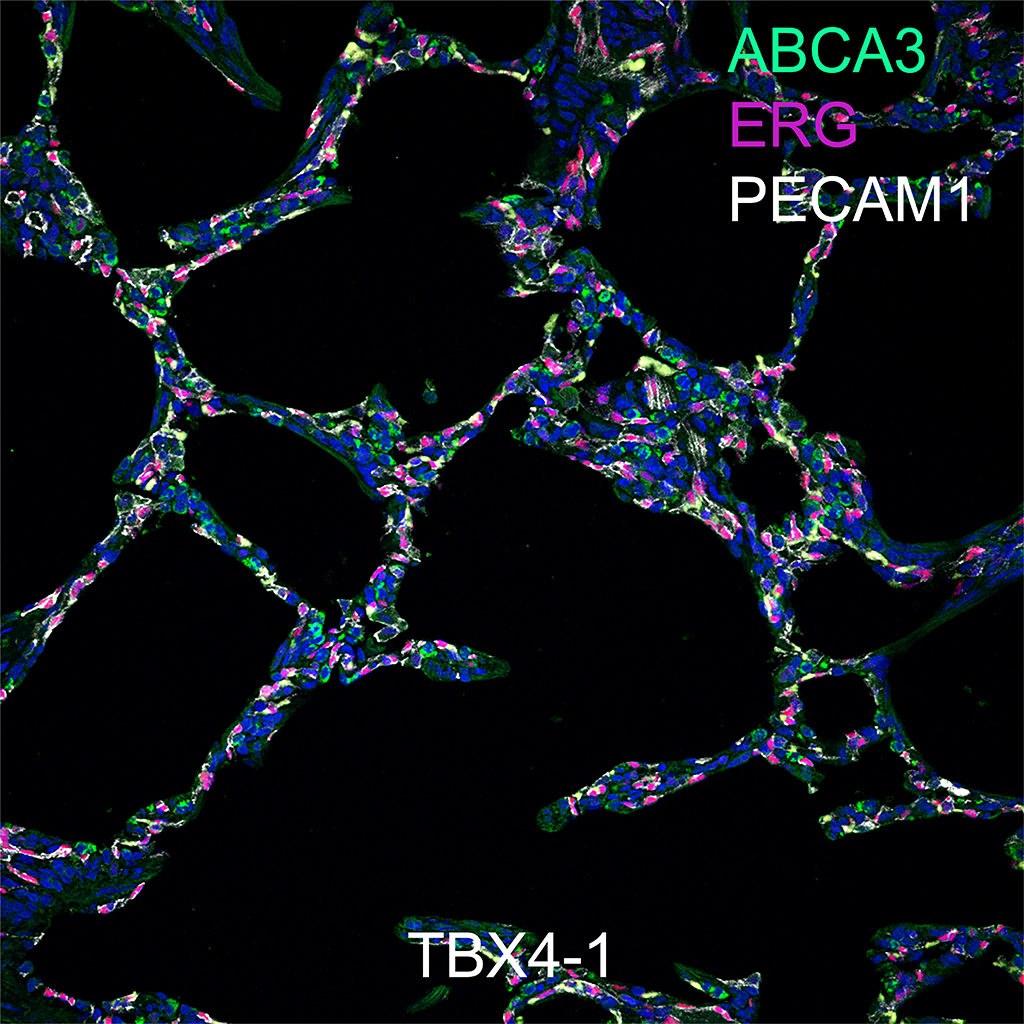
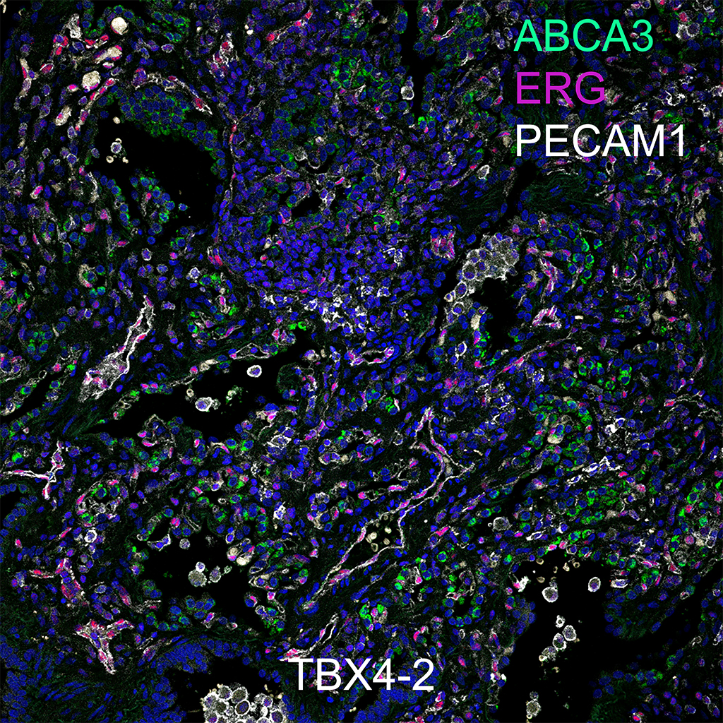
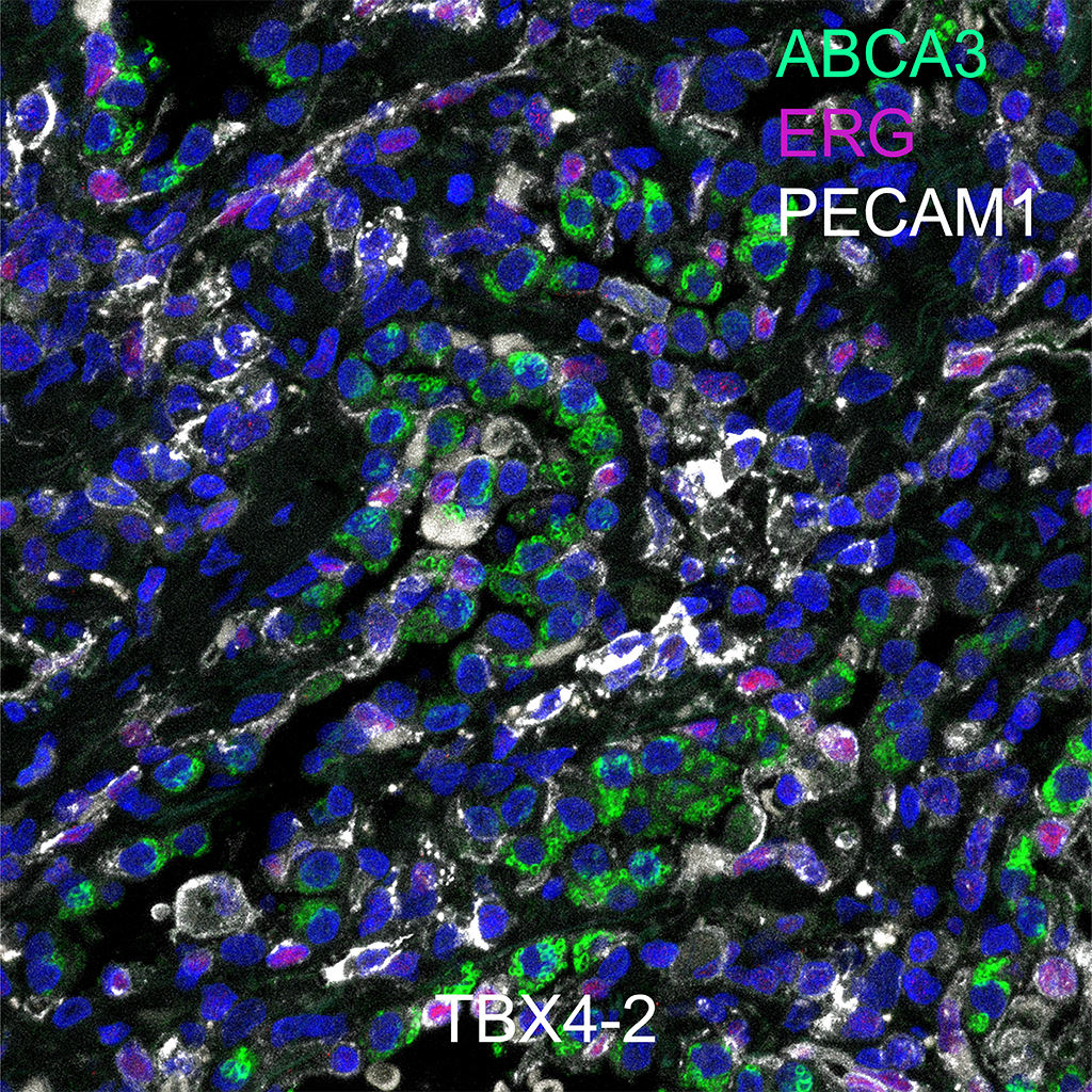
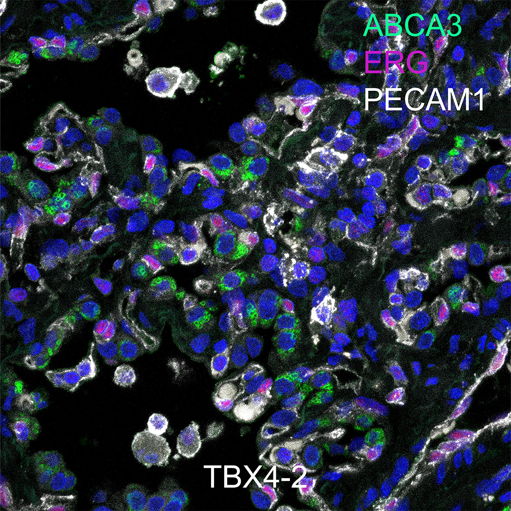
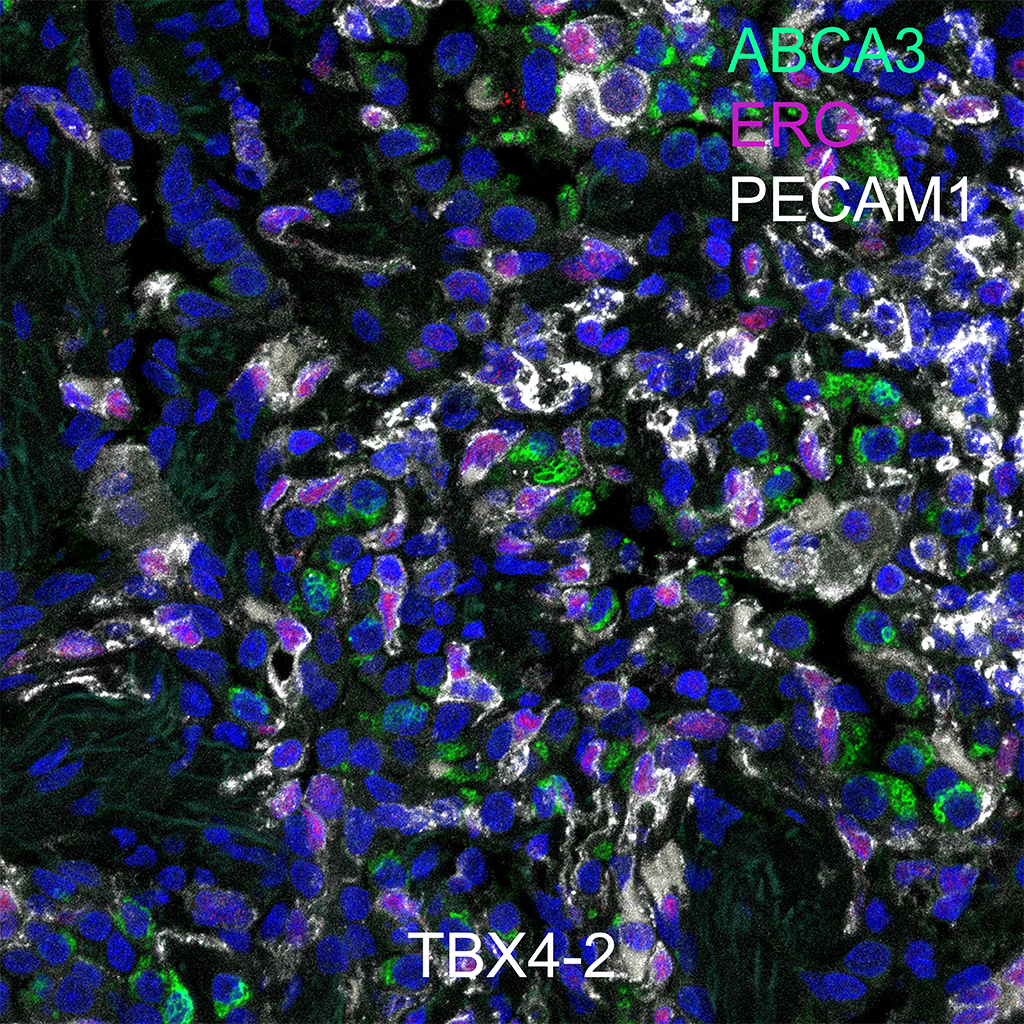
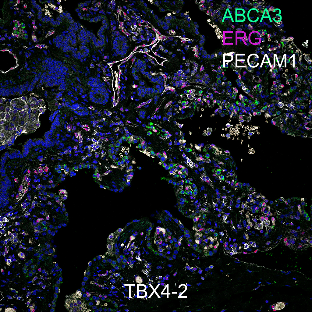
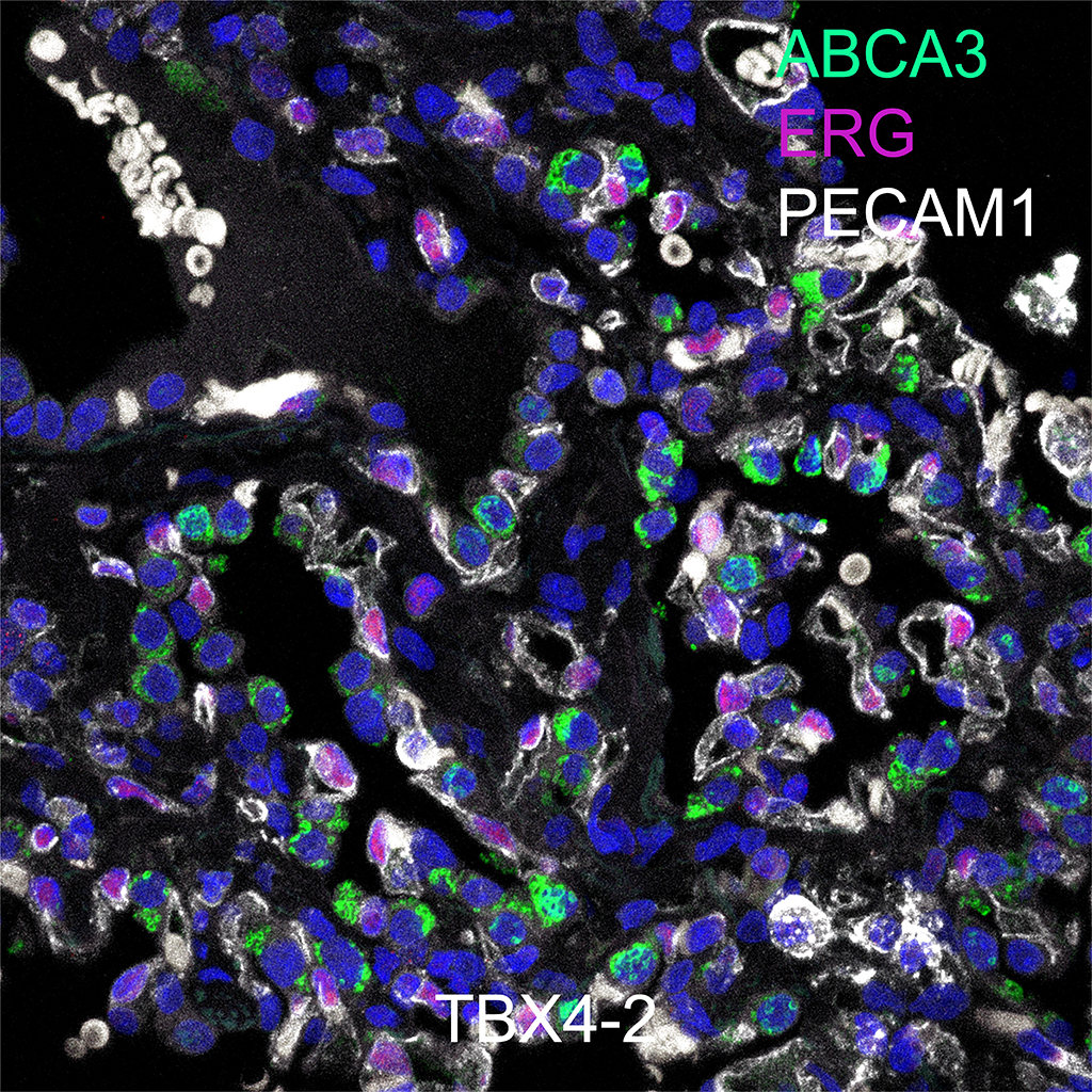
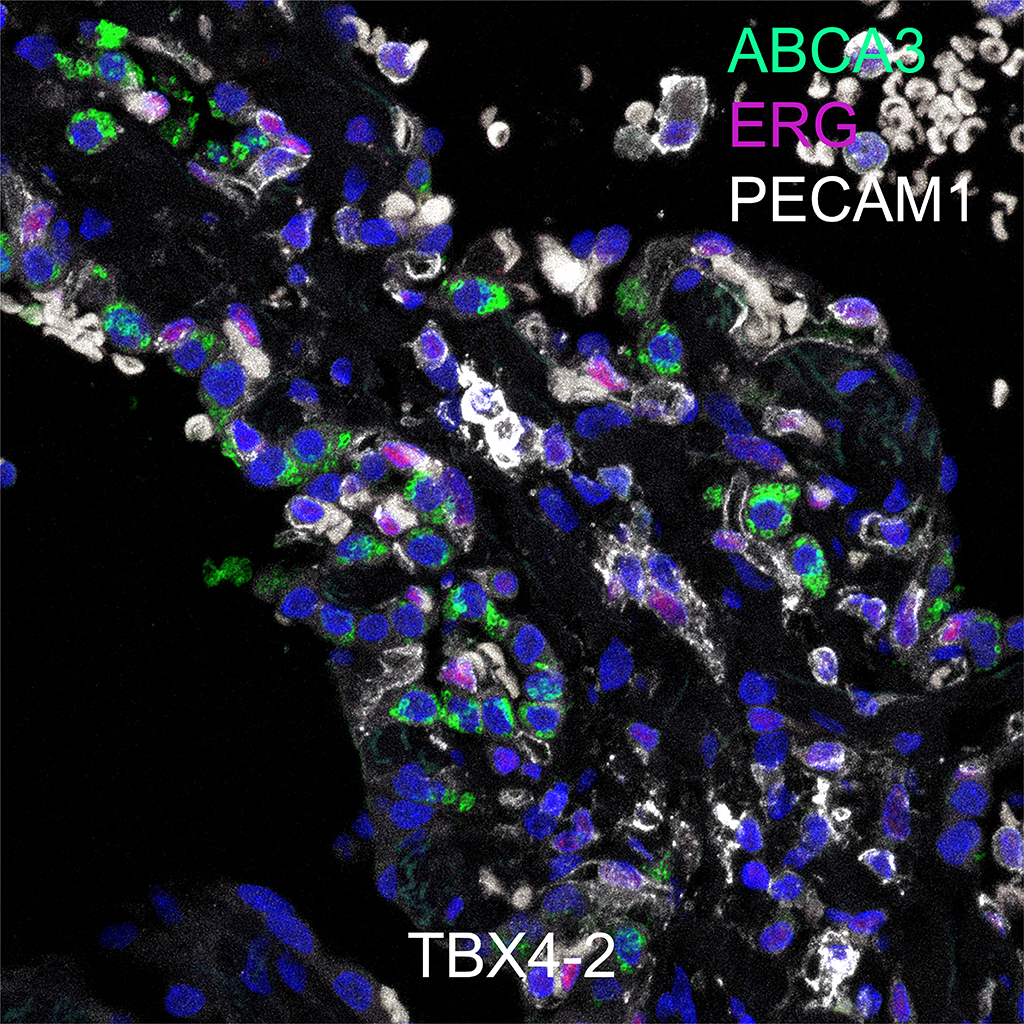
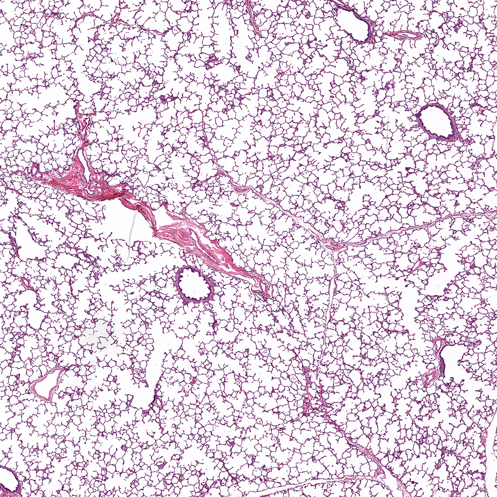
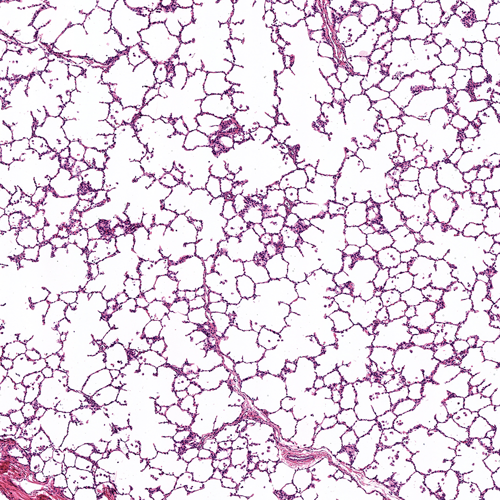
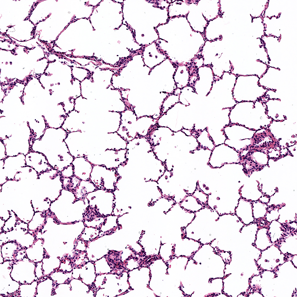
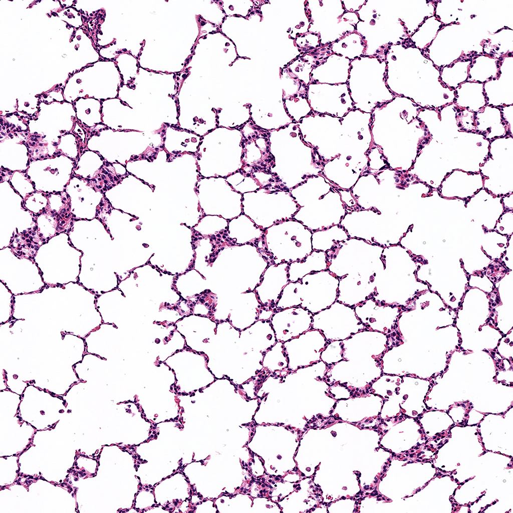
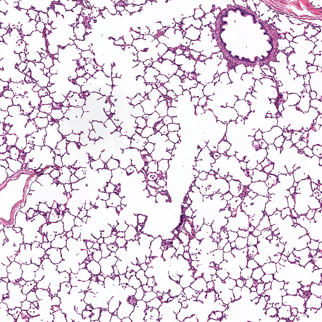
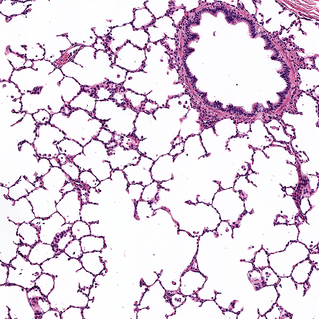
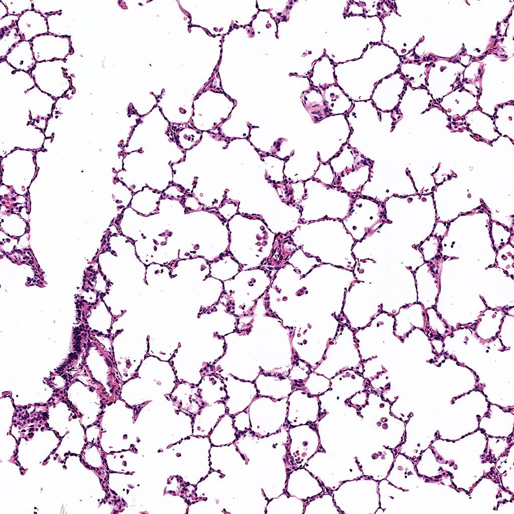
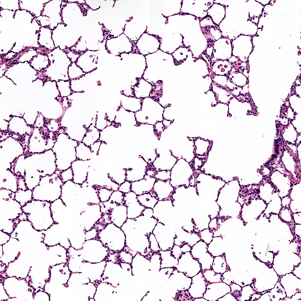
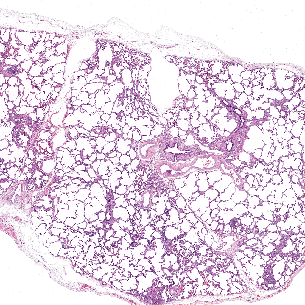
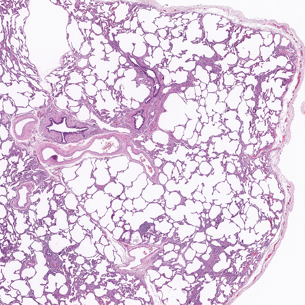
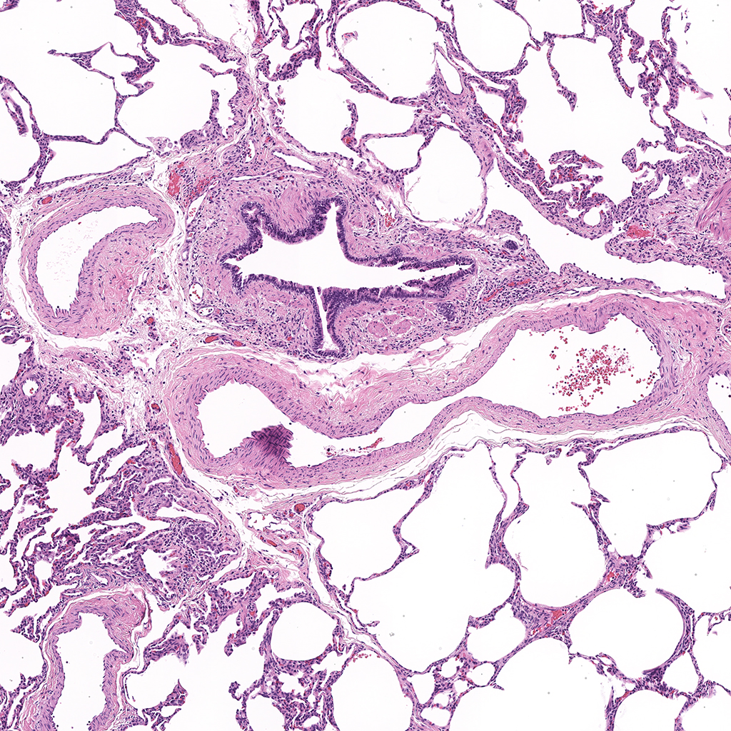
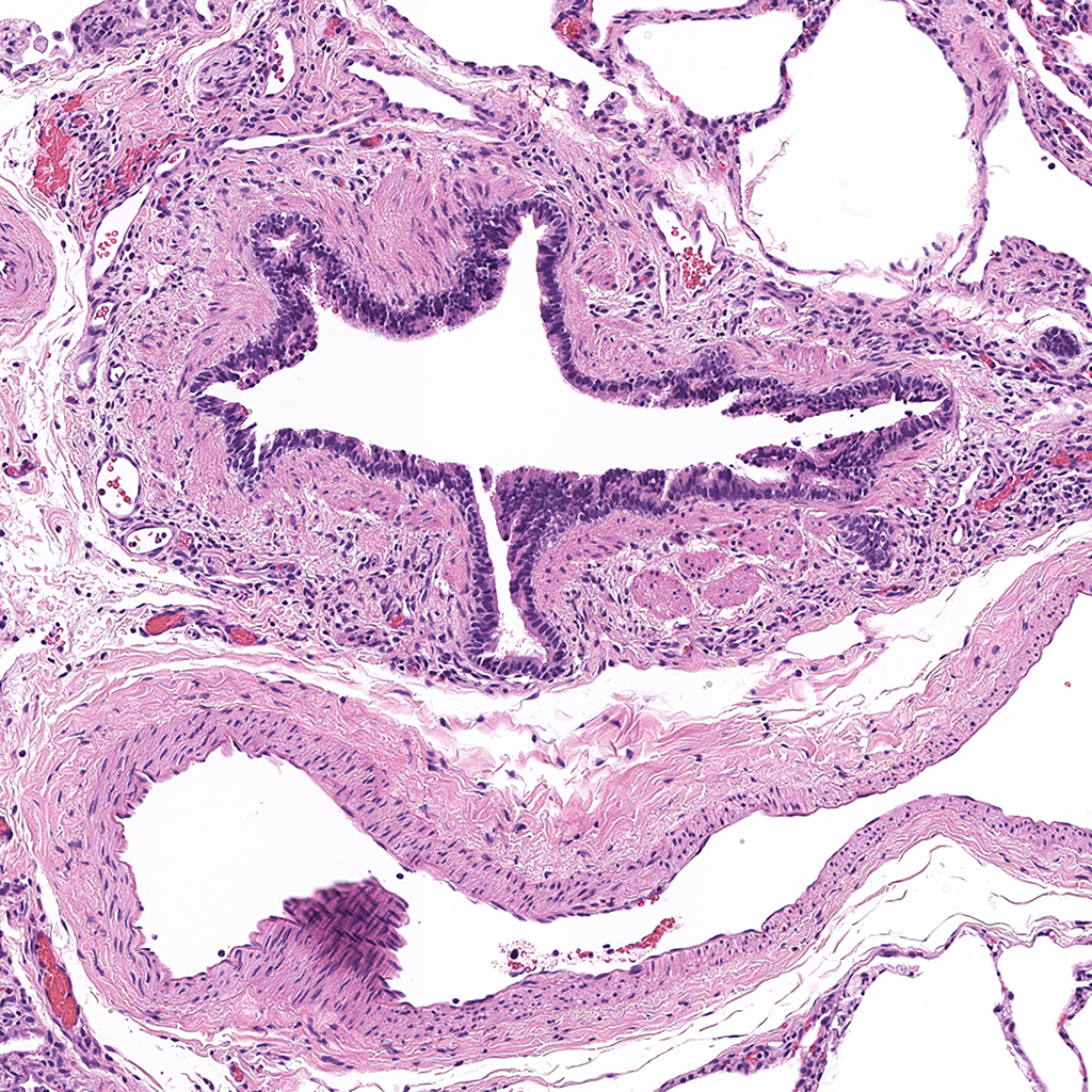
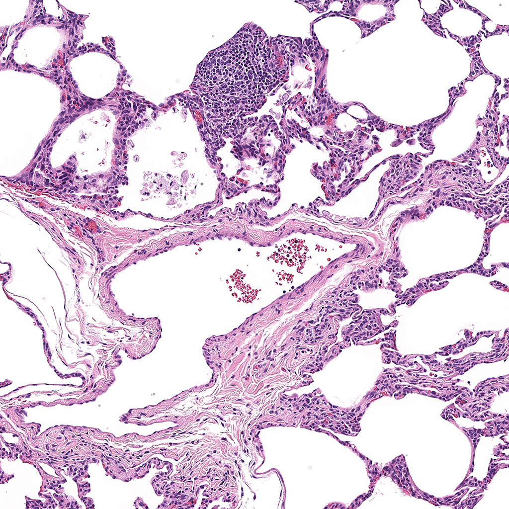
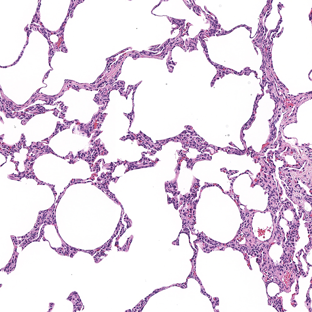
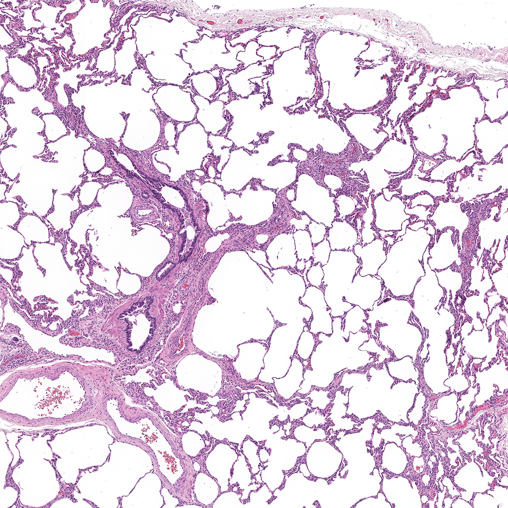
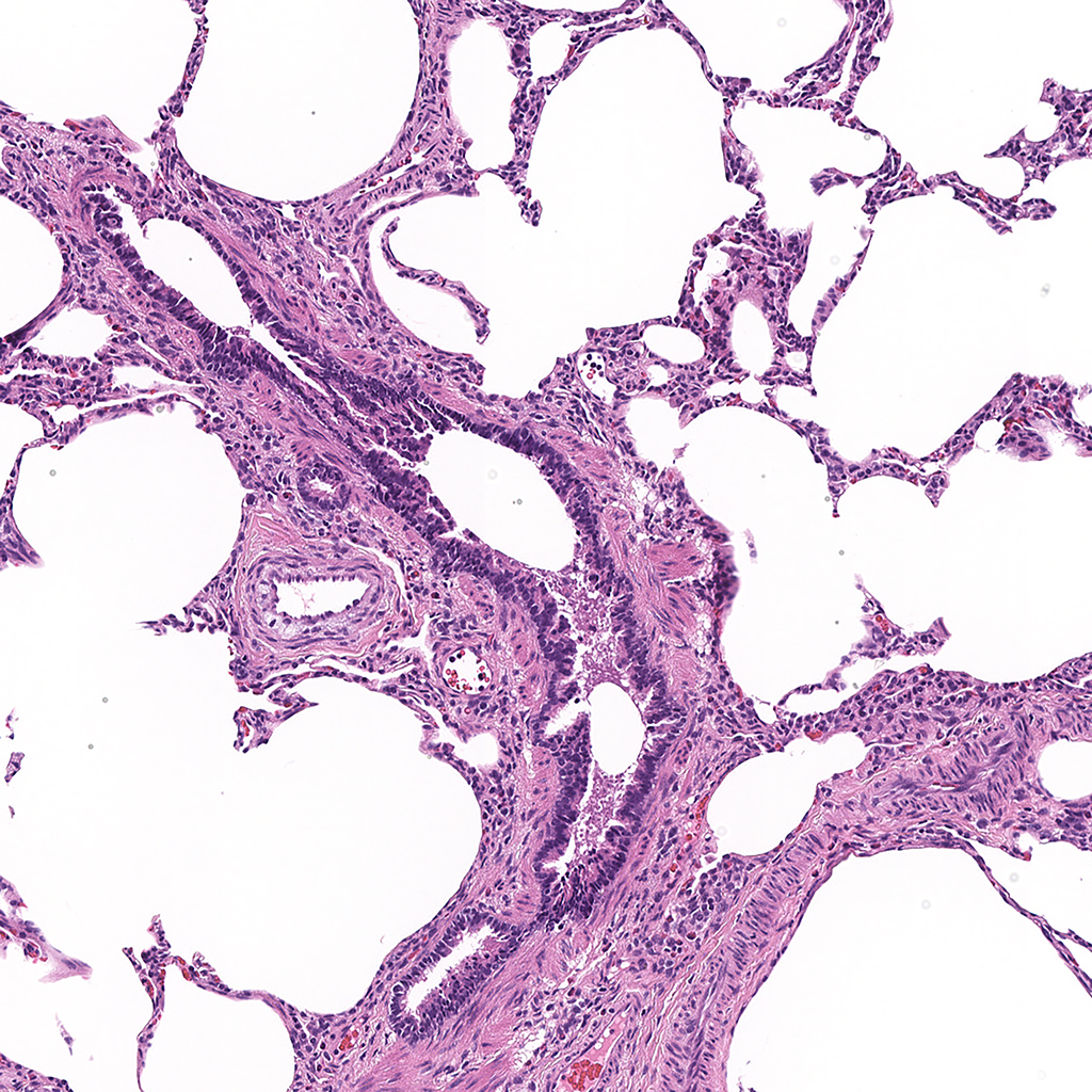
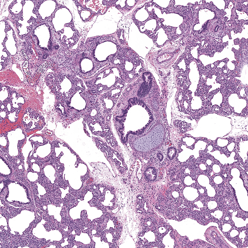
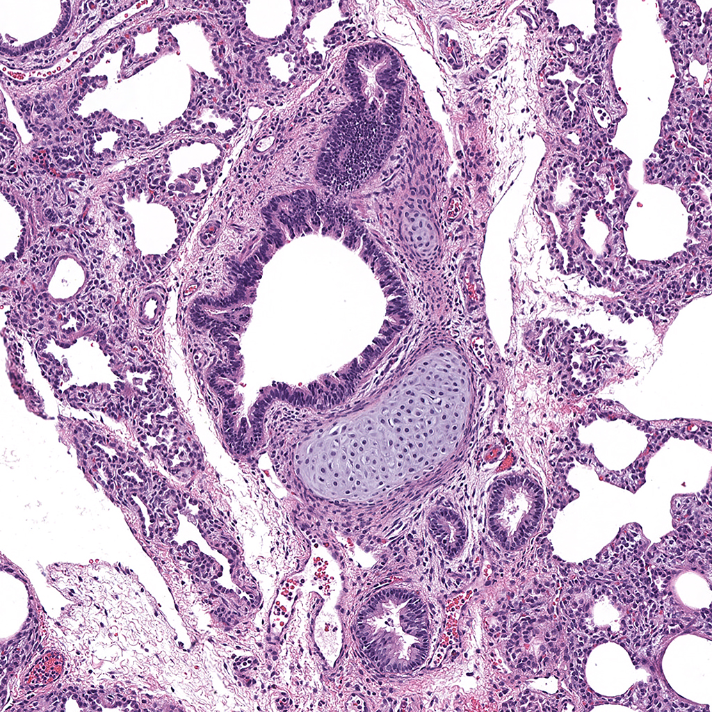
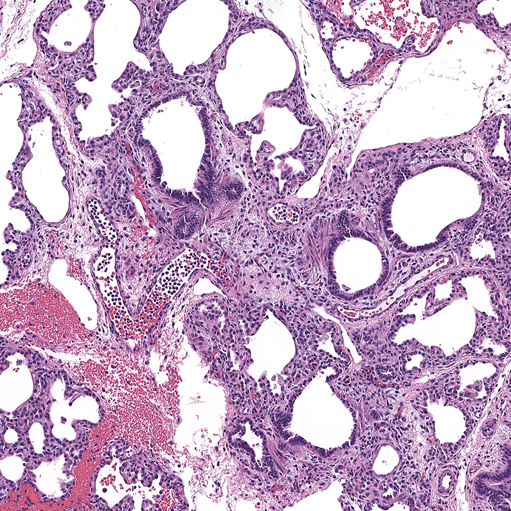
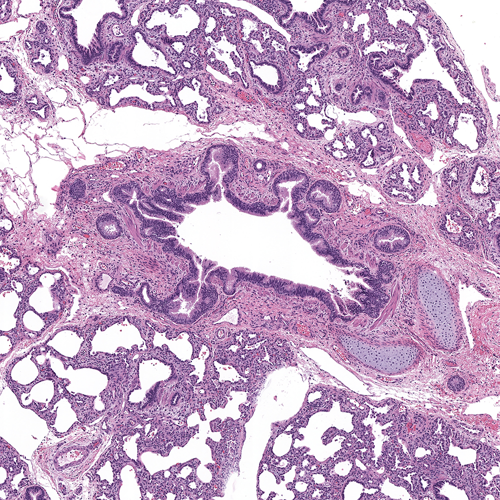
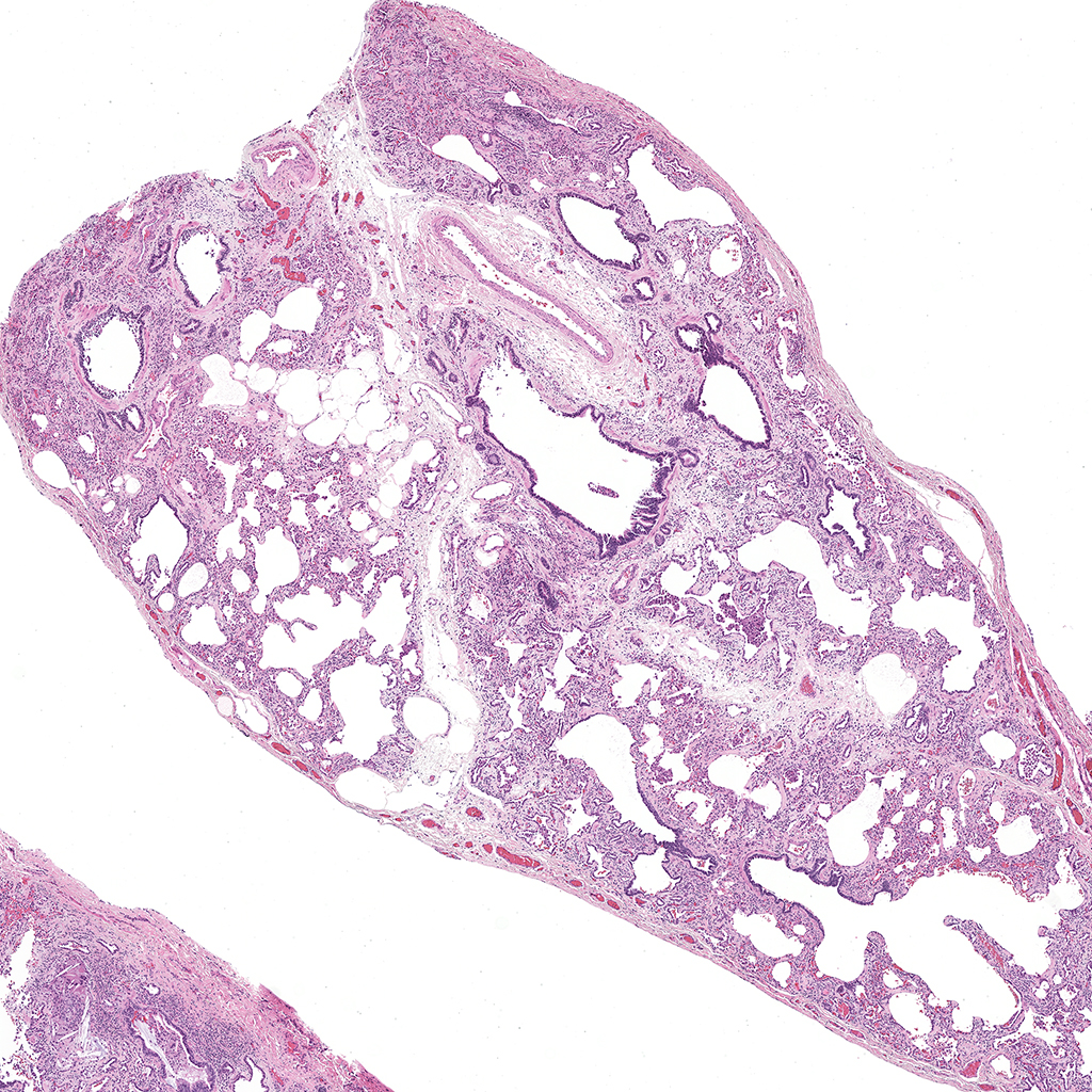
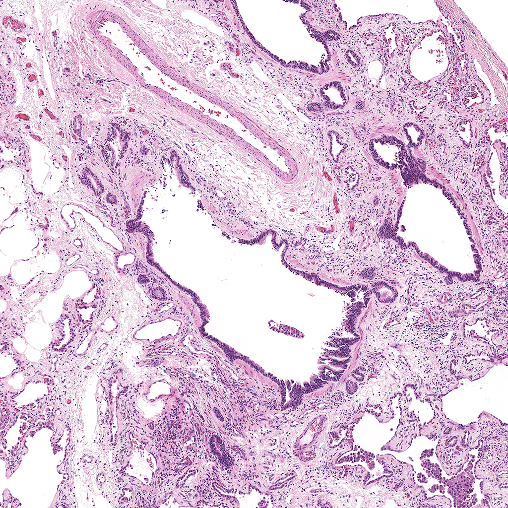
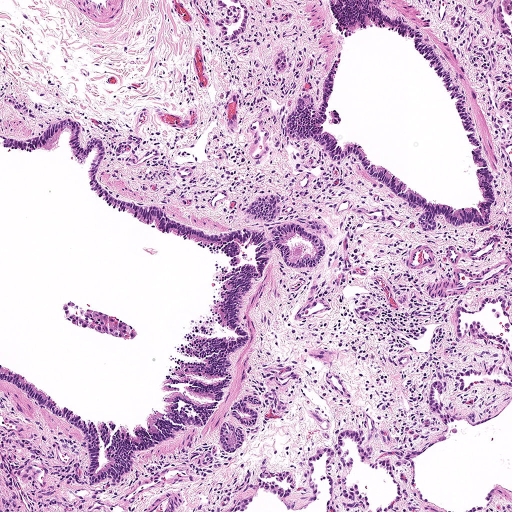
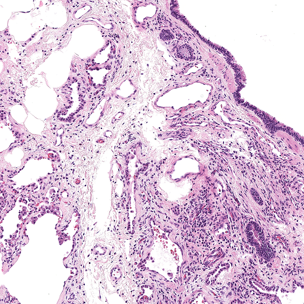
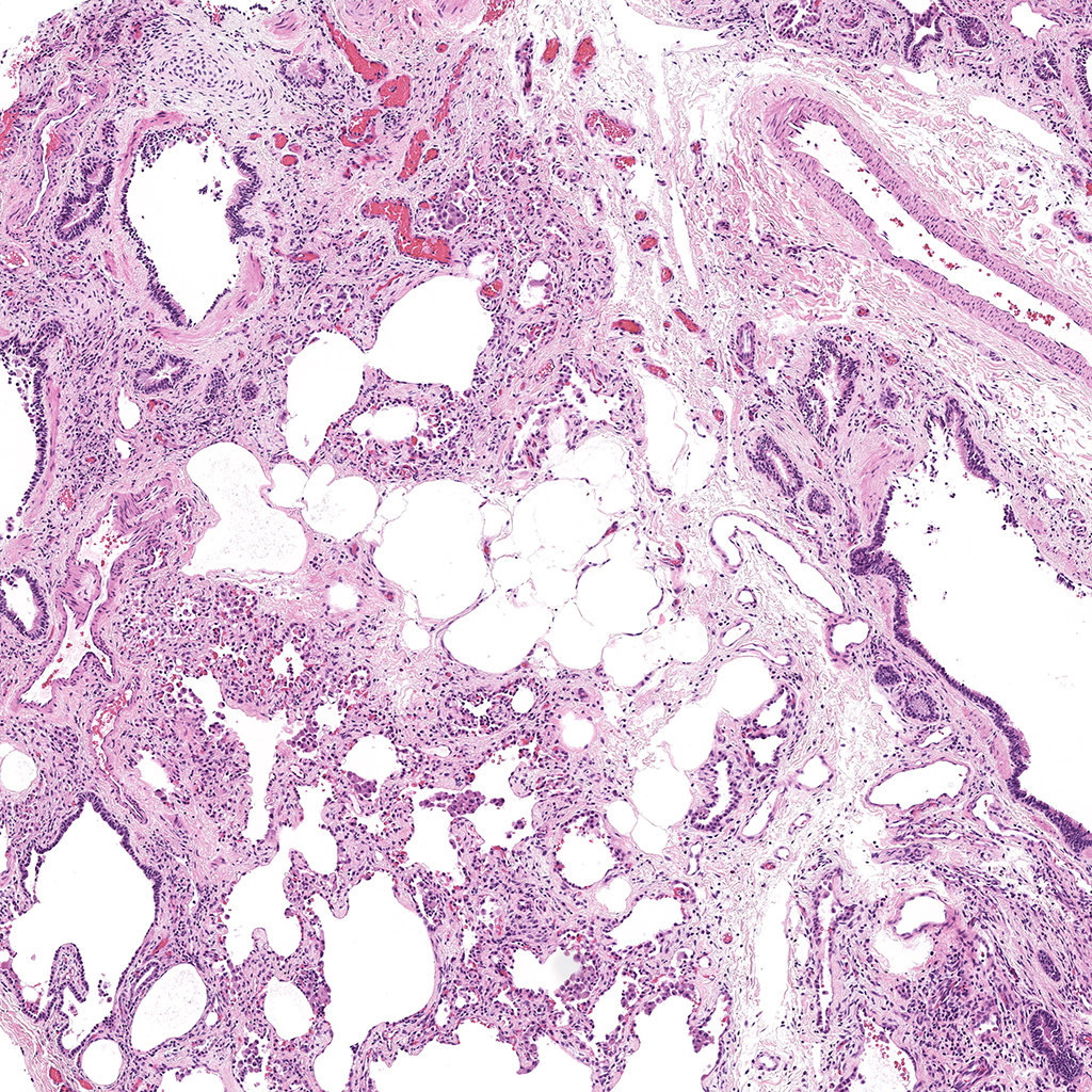
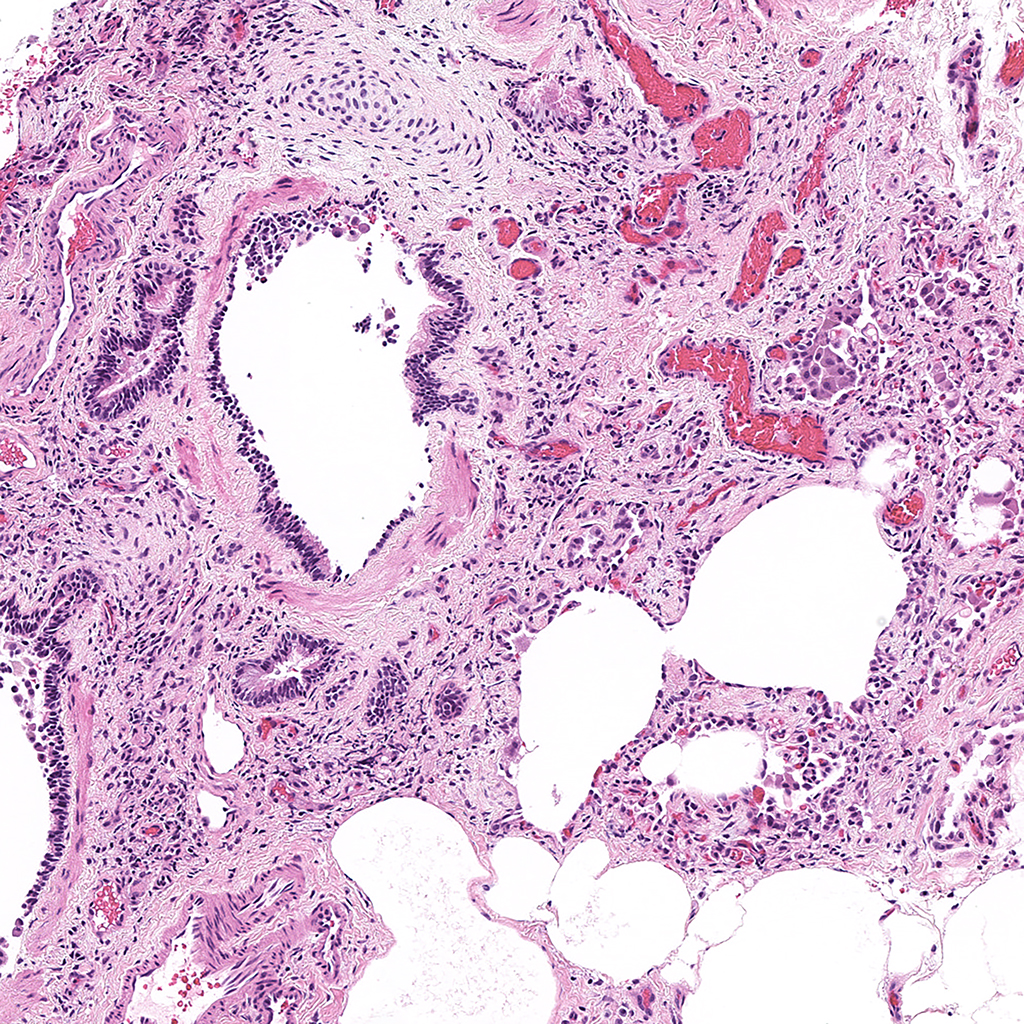
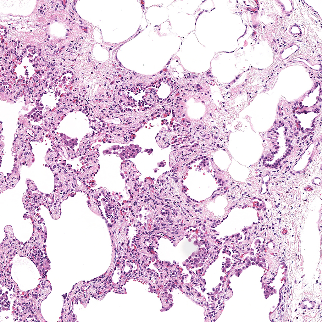
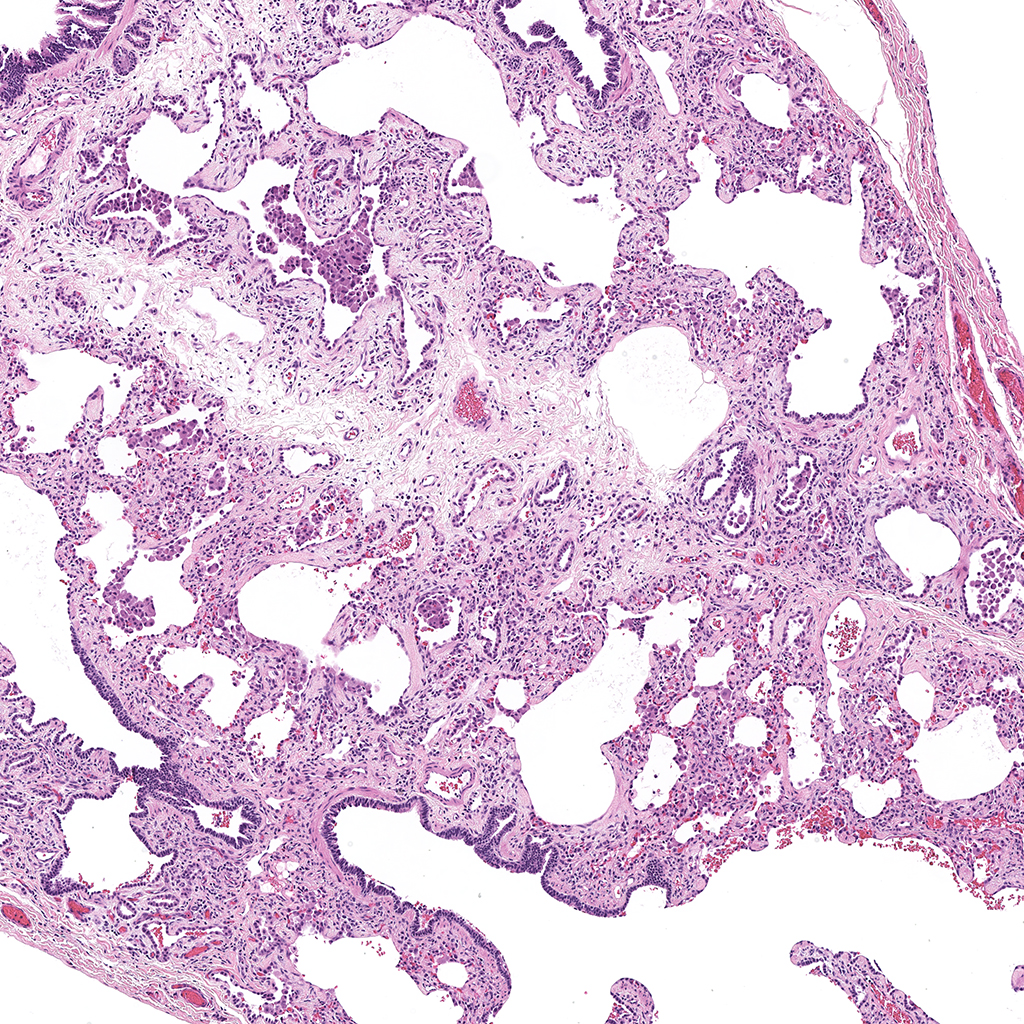
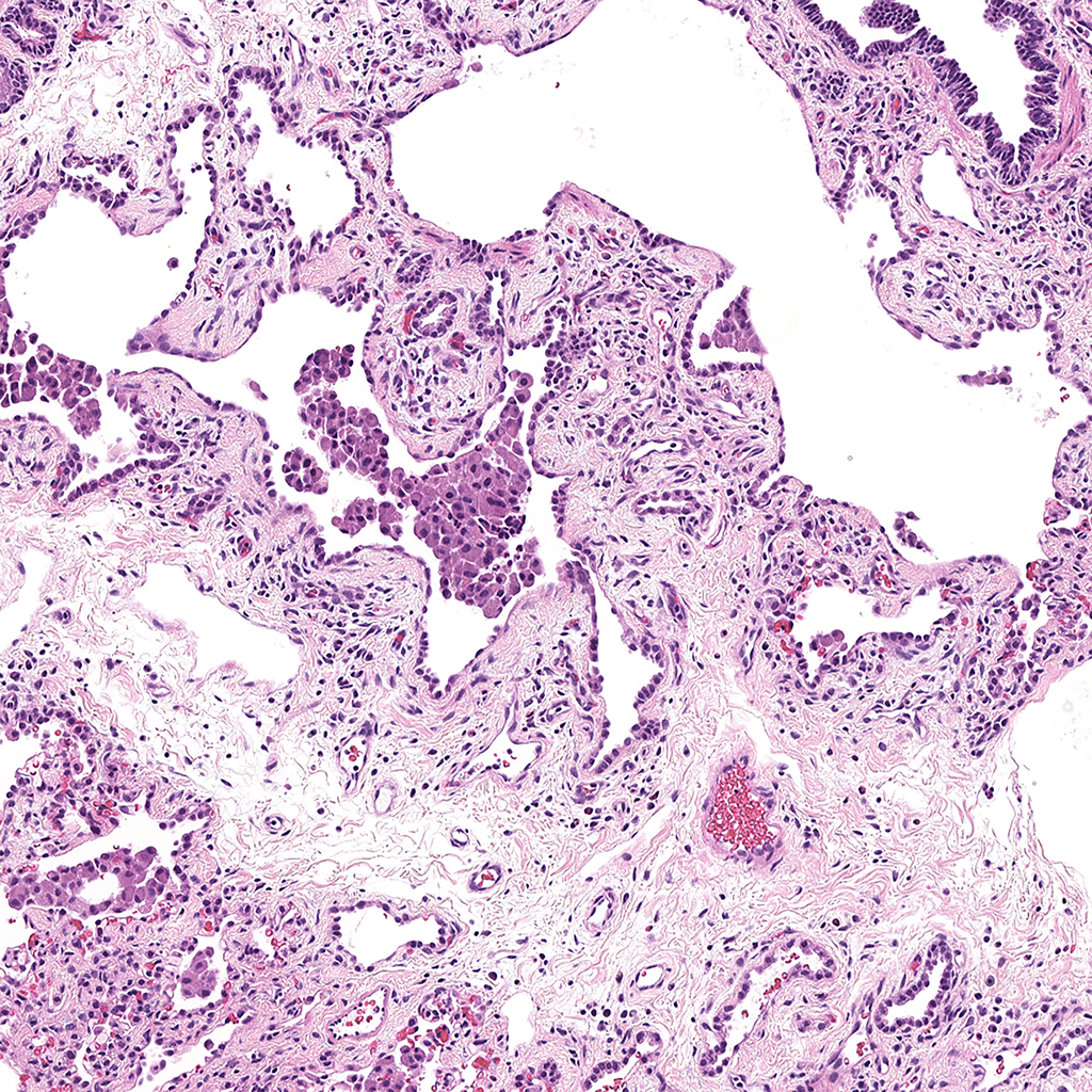
| SFTPC |
Donor tissue kindly provided by Dr. Scott Randell, University of North Carolina
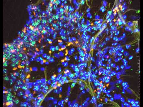
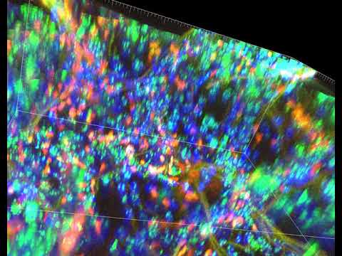
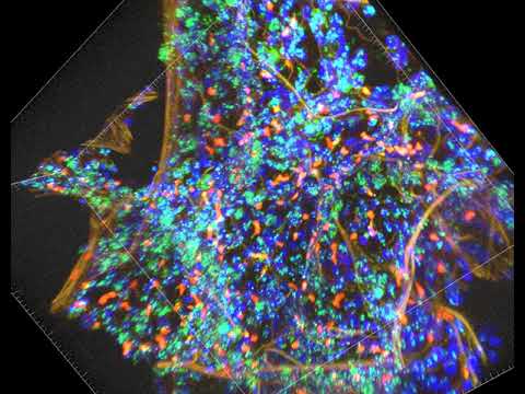
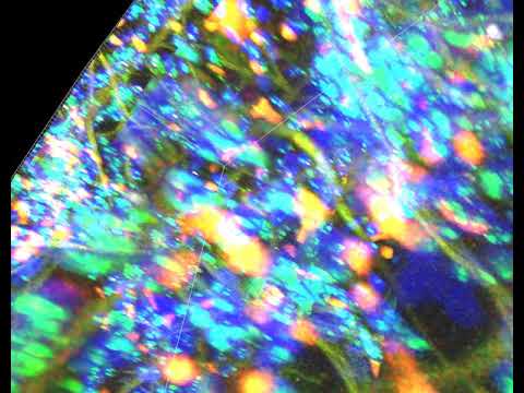
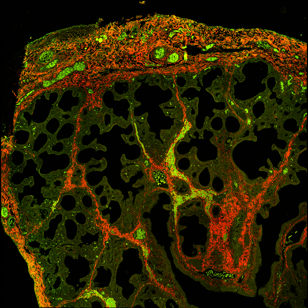
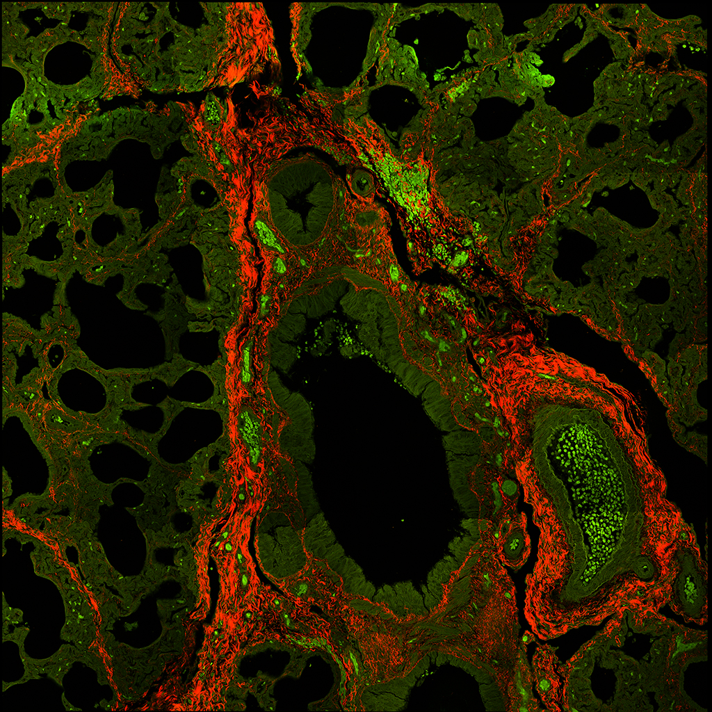
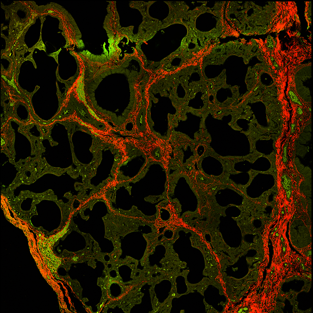
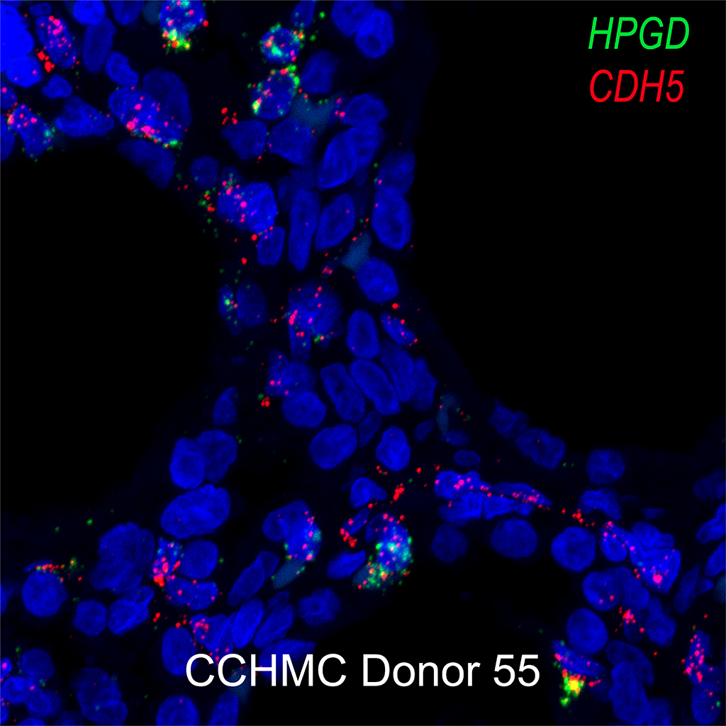
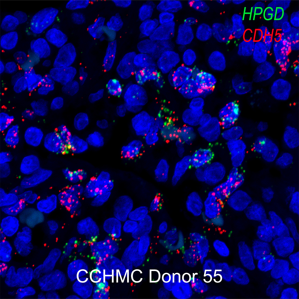
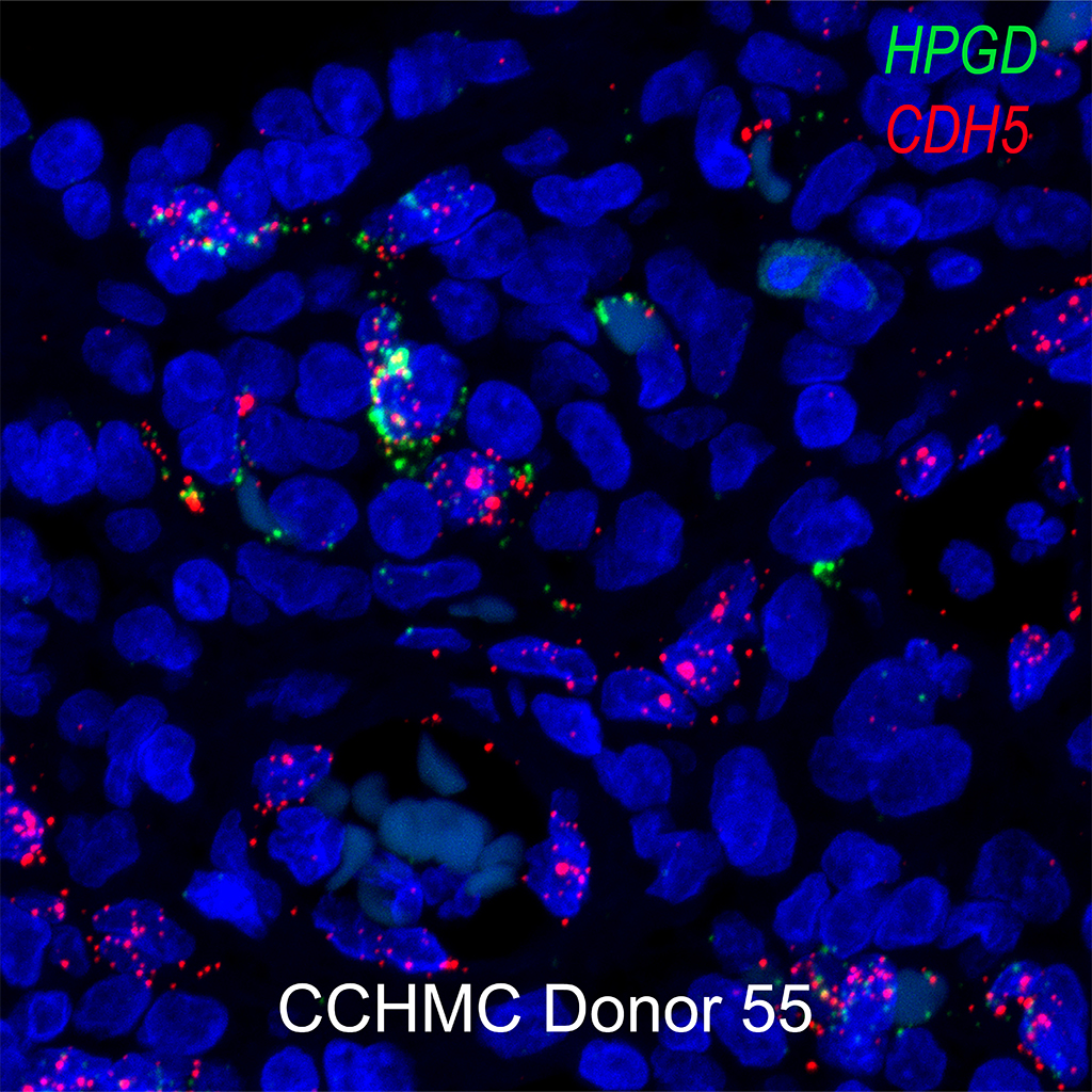
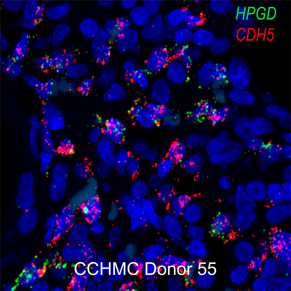
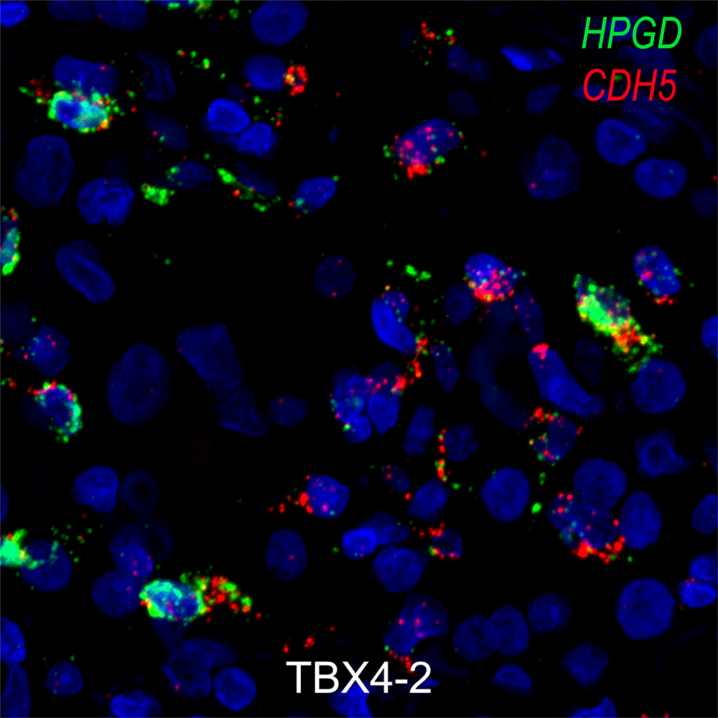
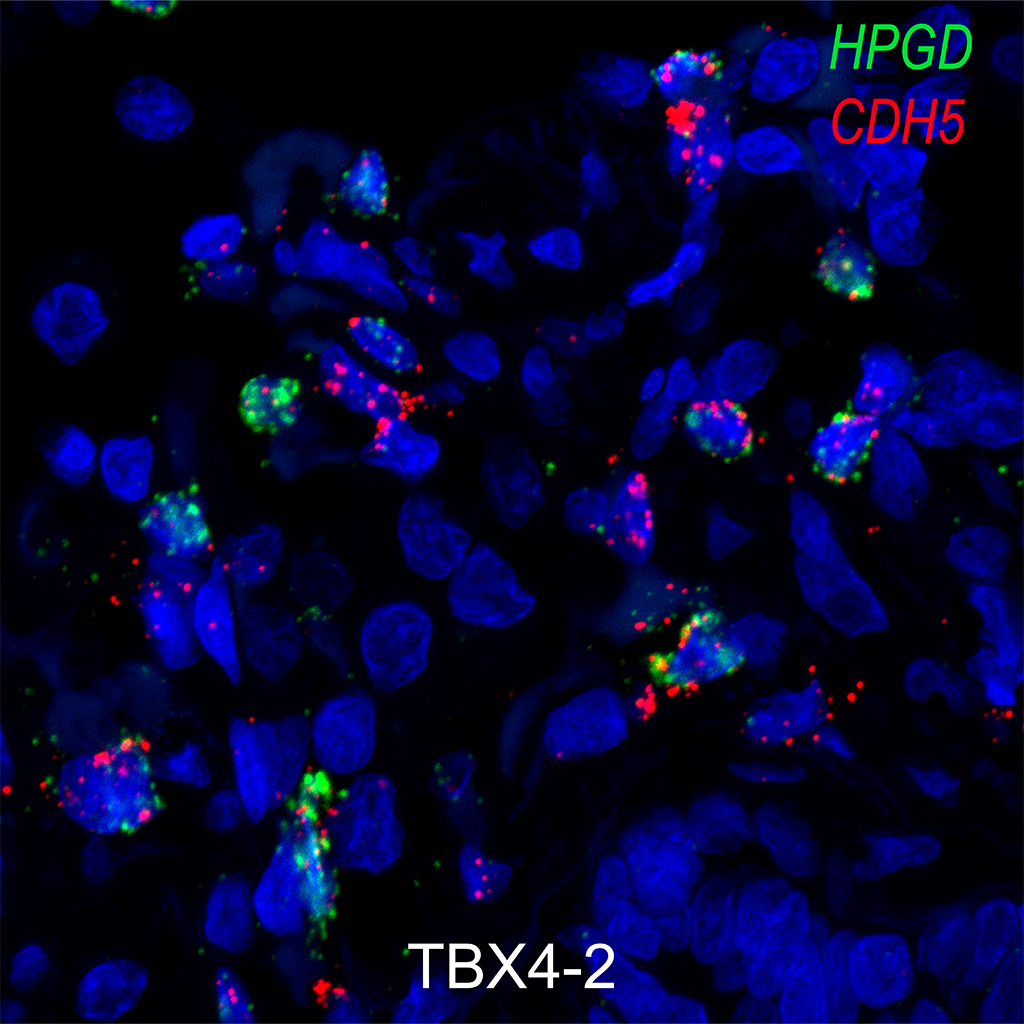
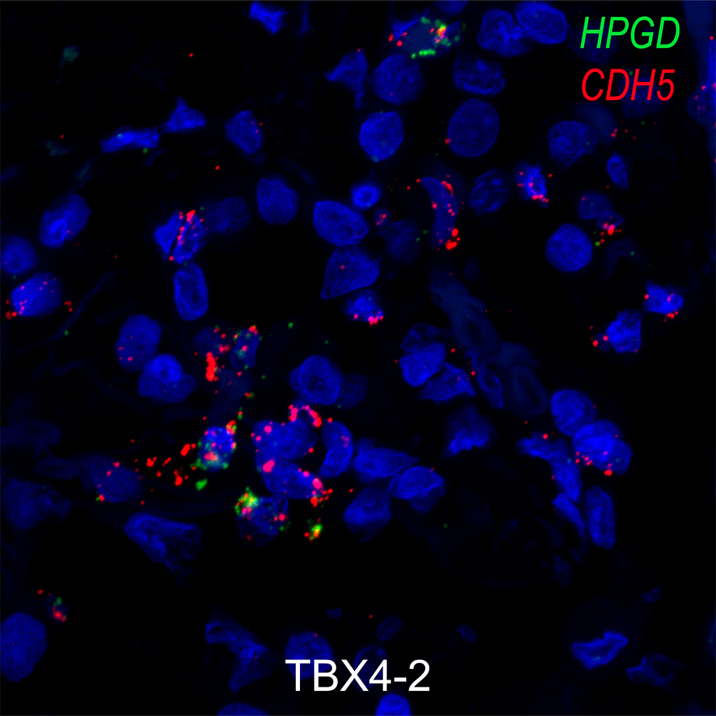
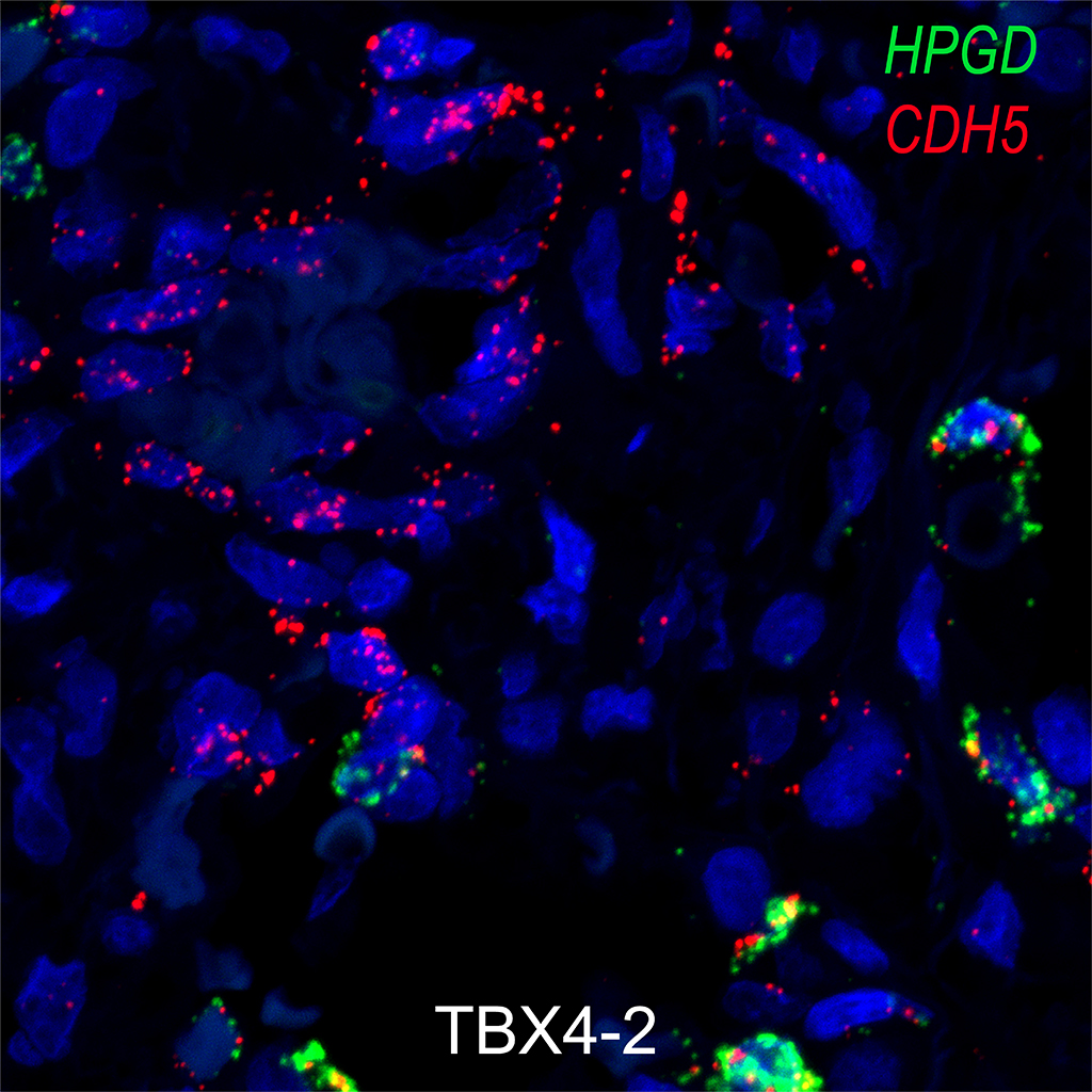
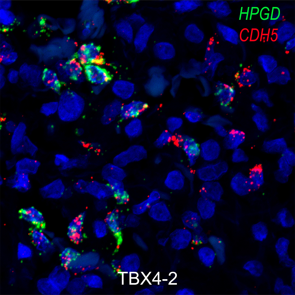
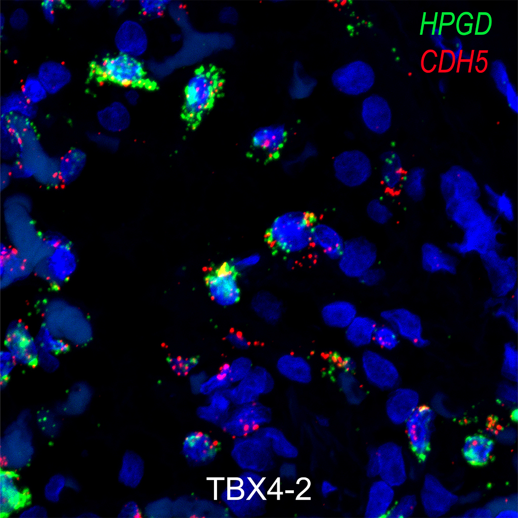
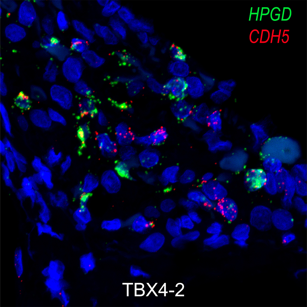
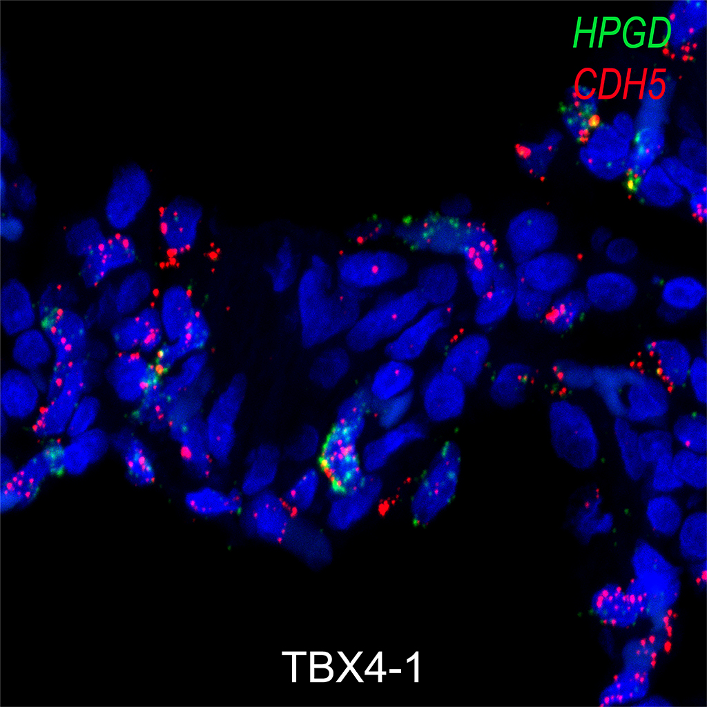
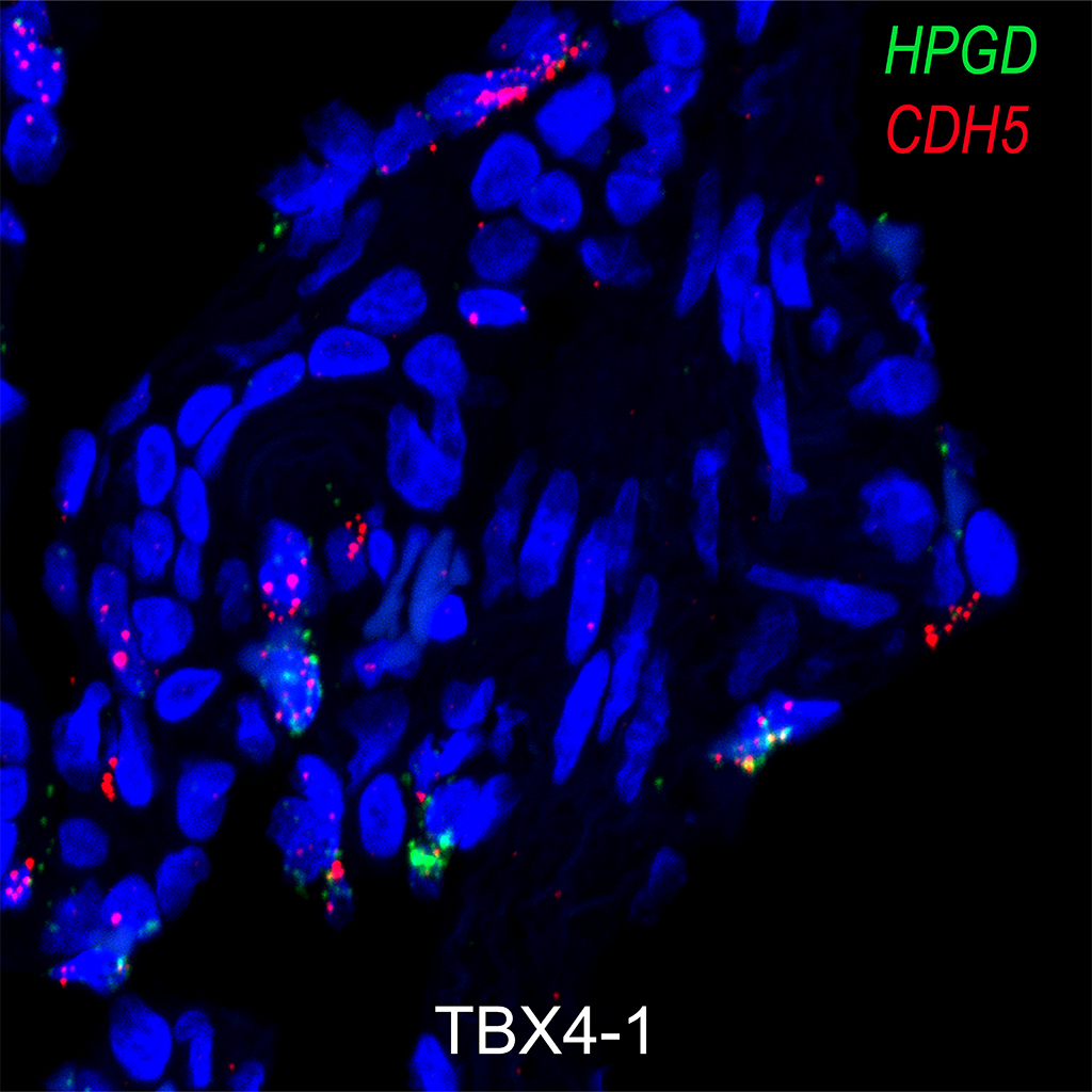
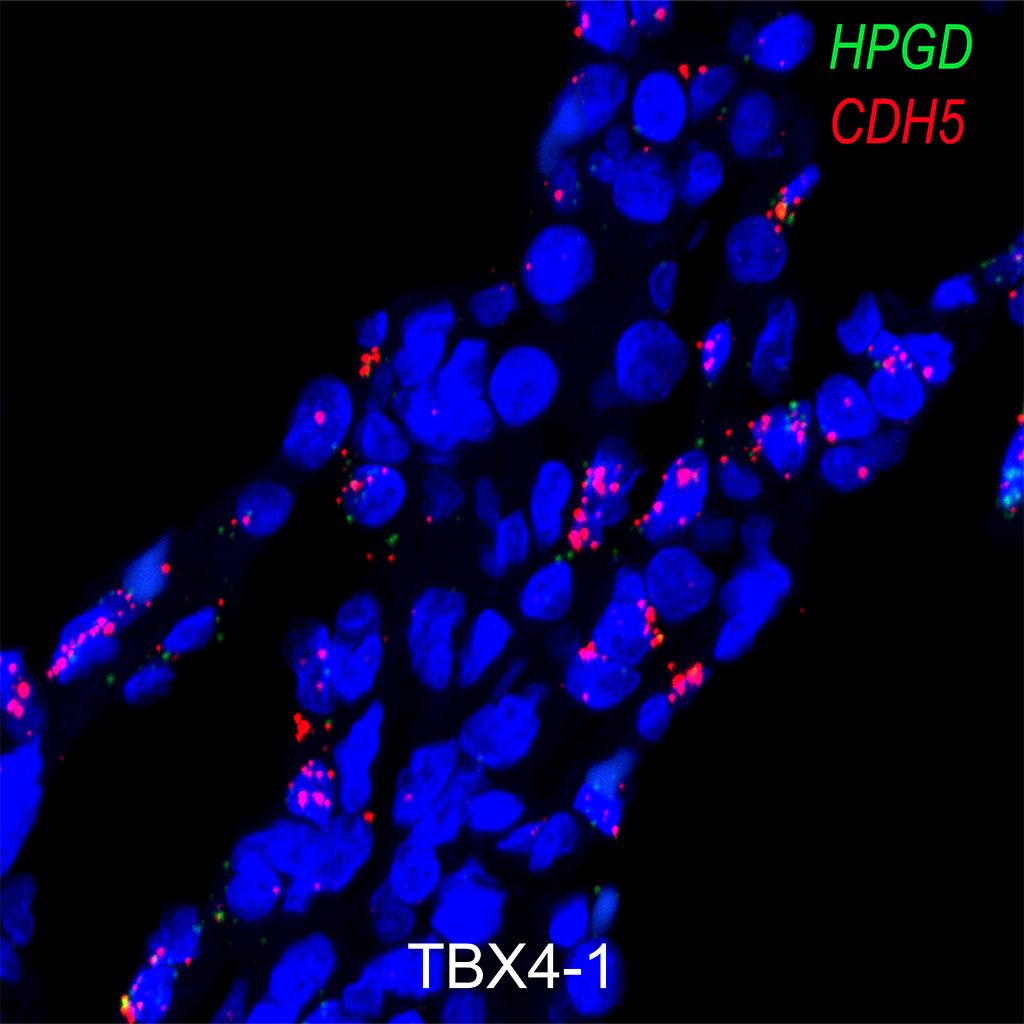
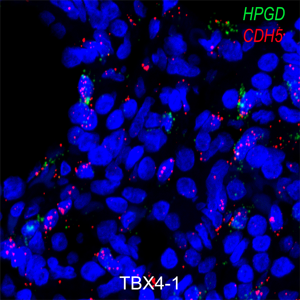
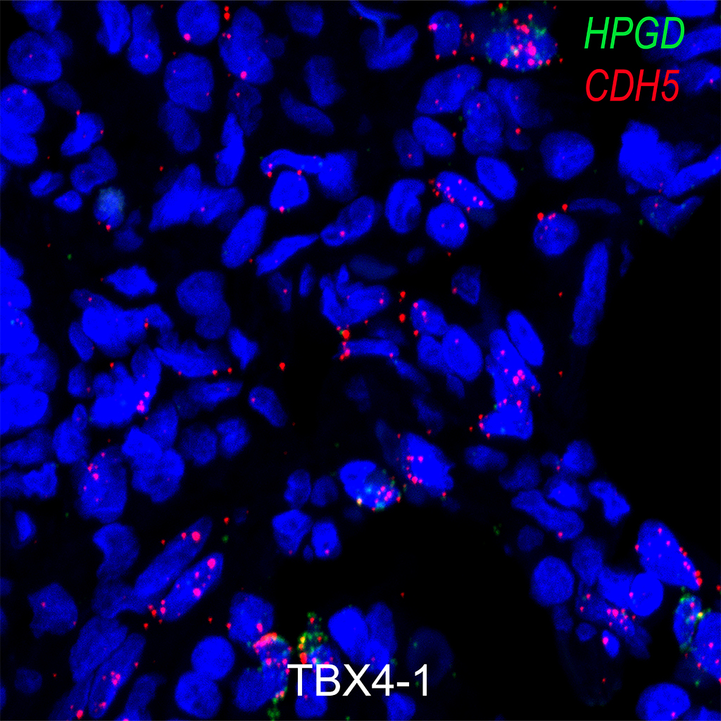
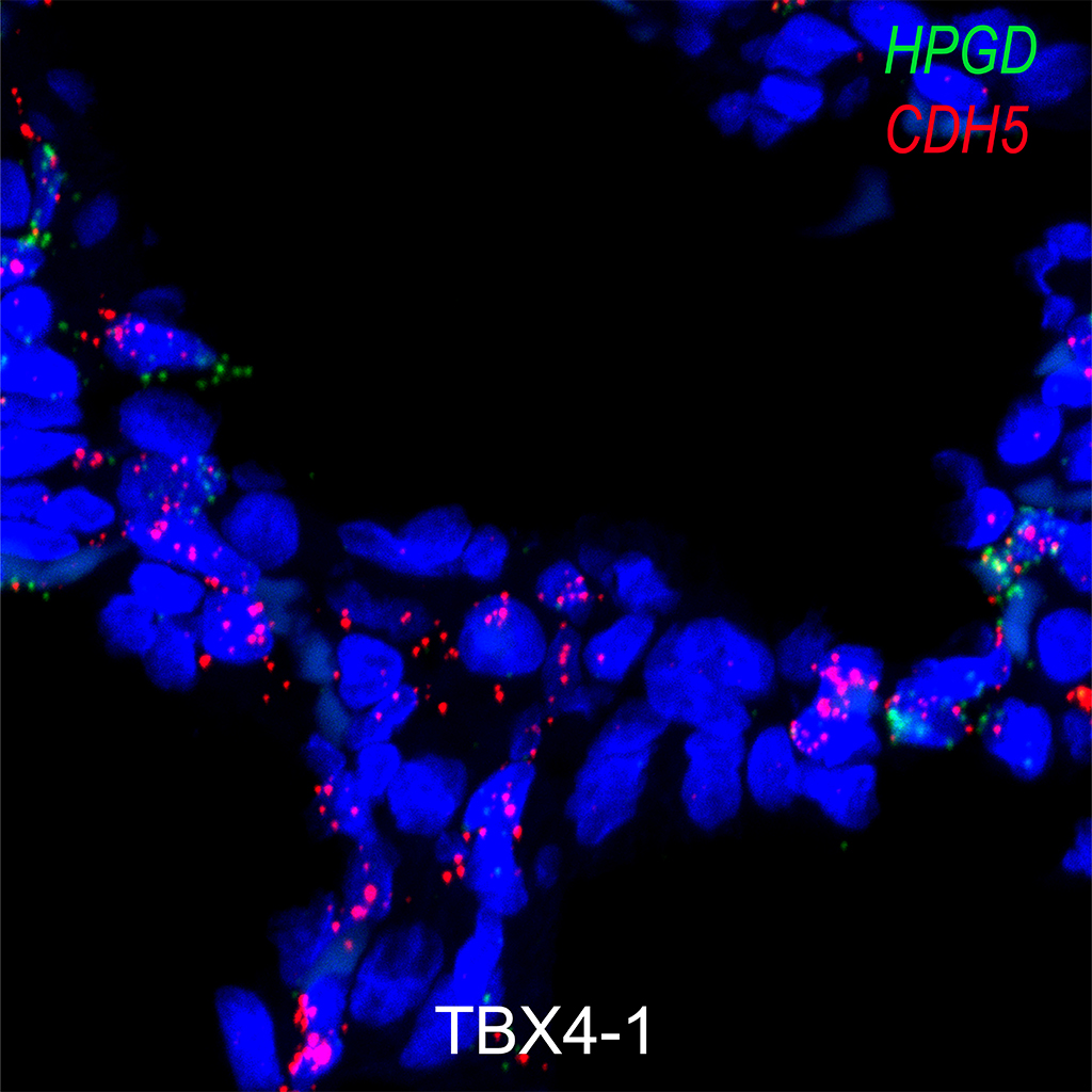
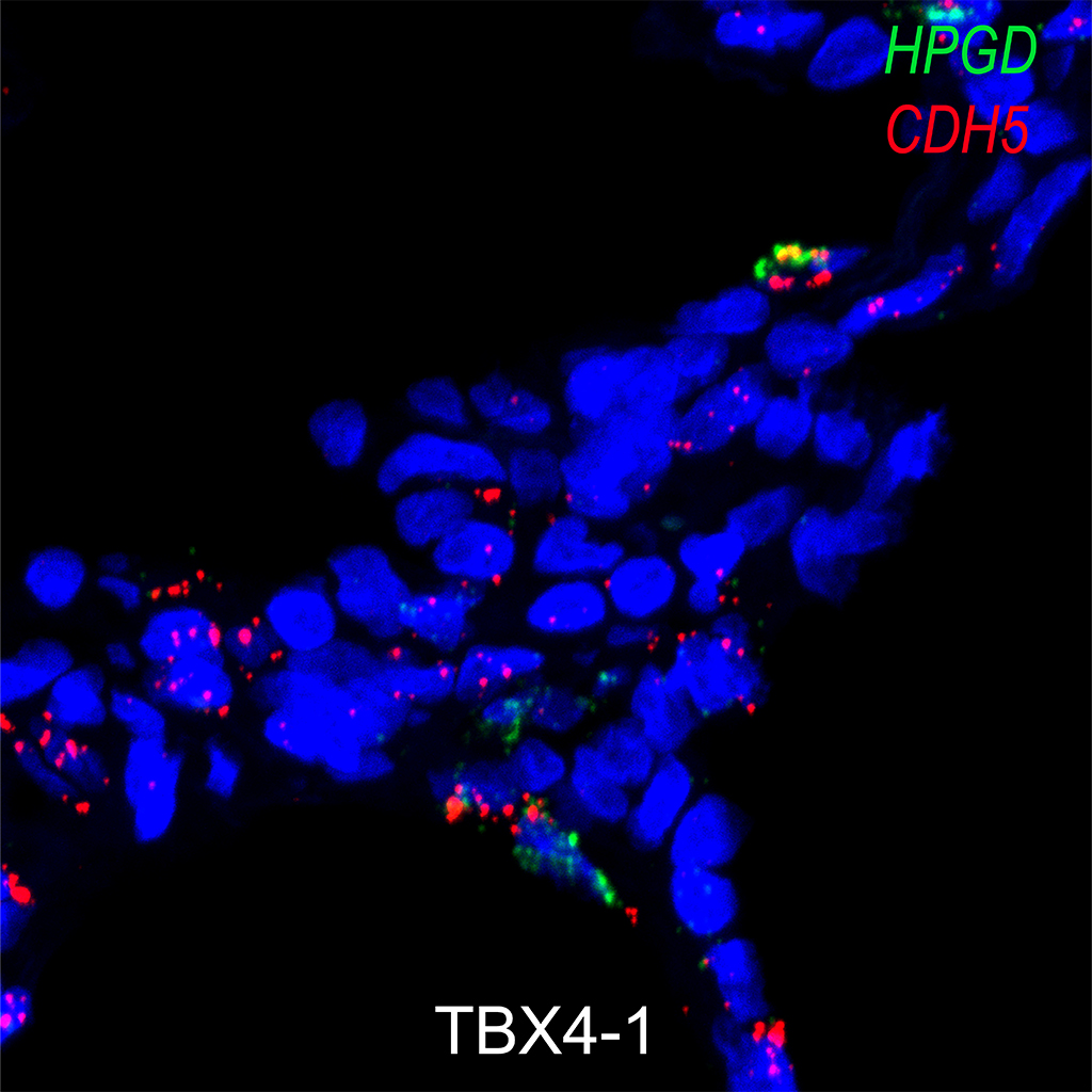
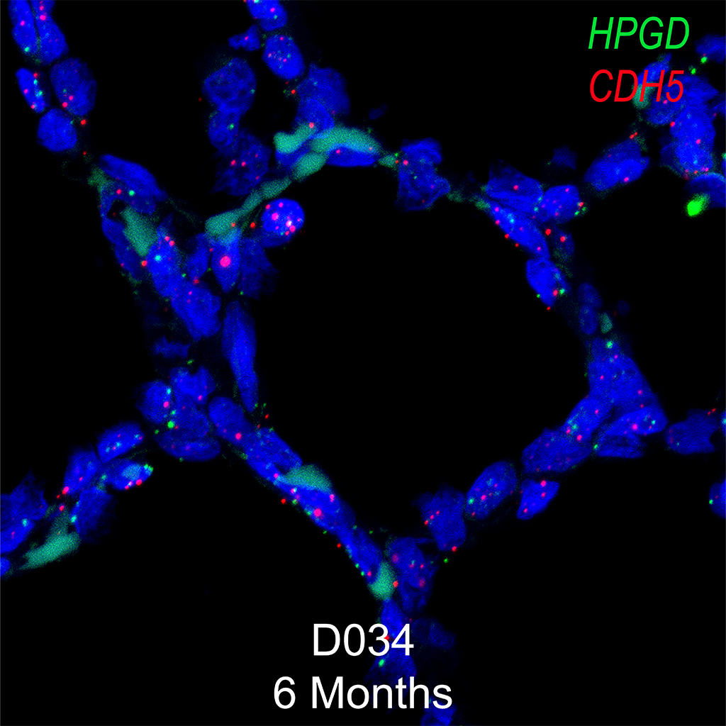
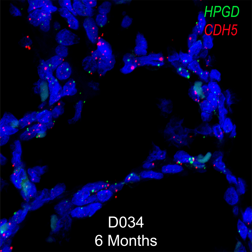
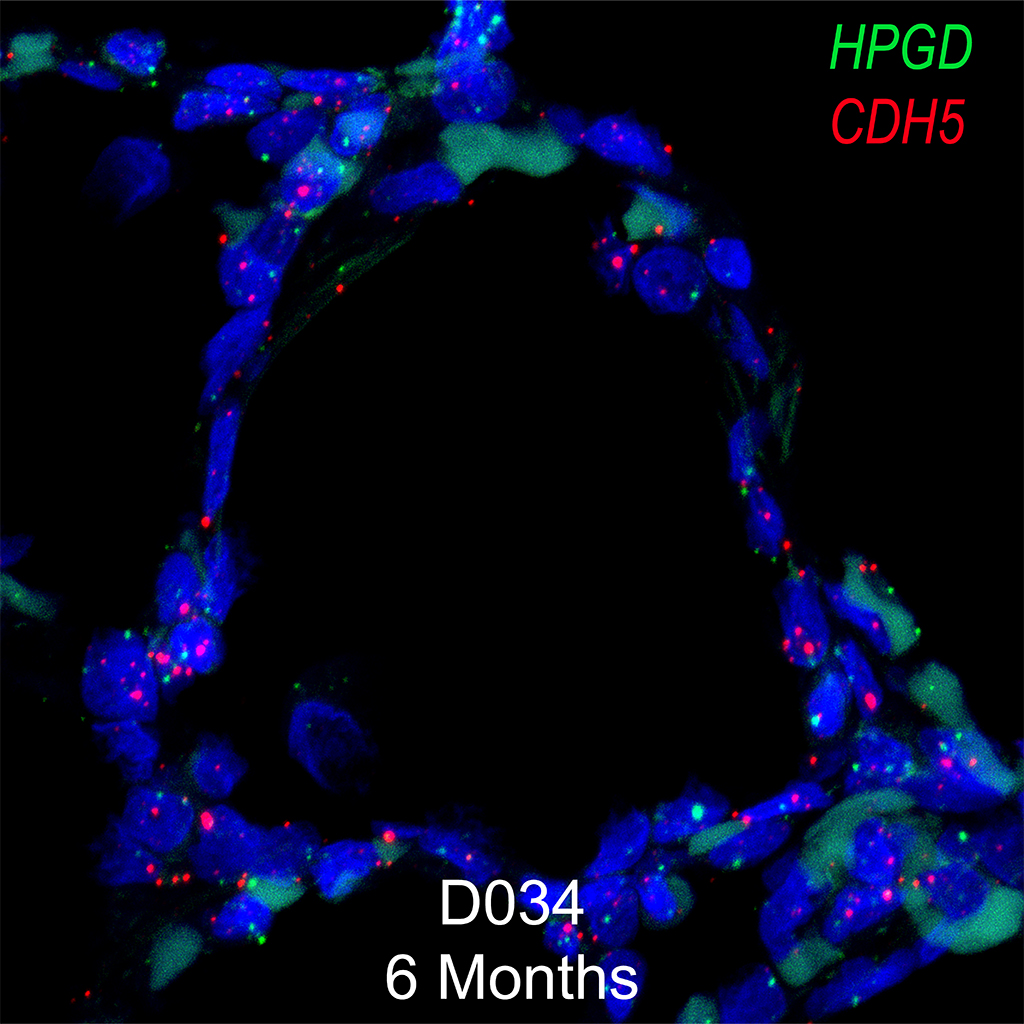
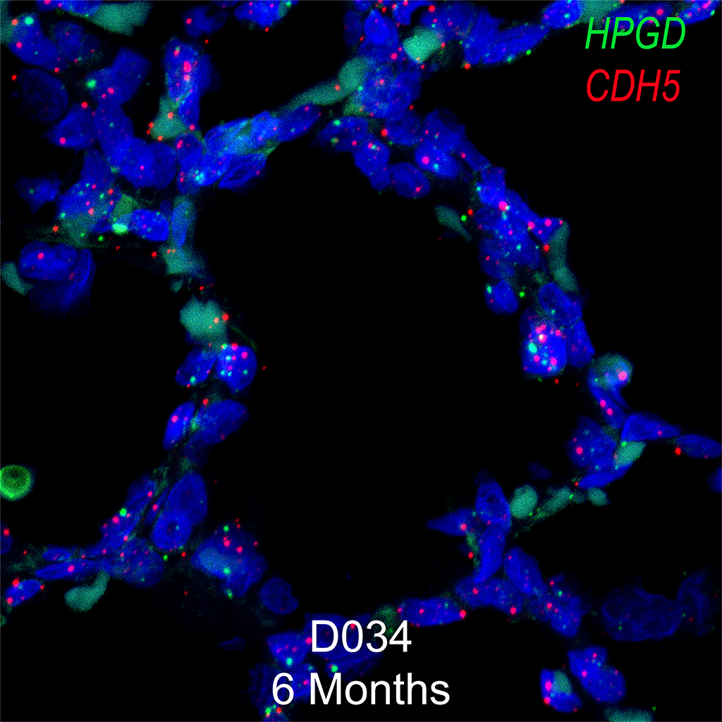
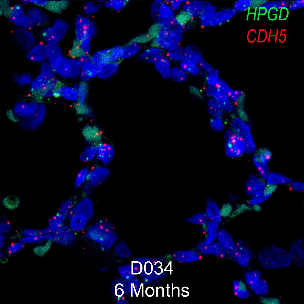
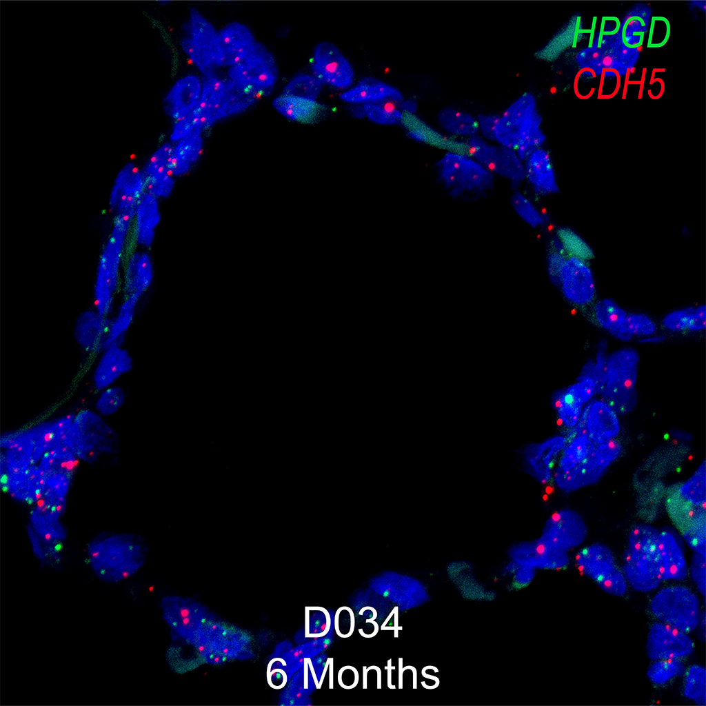
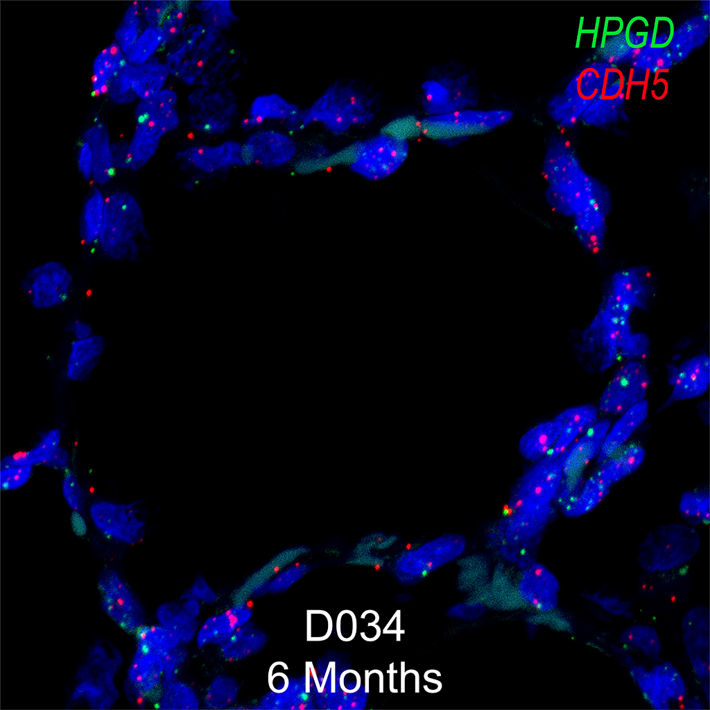
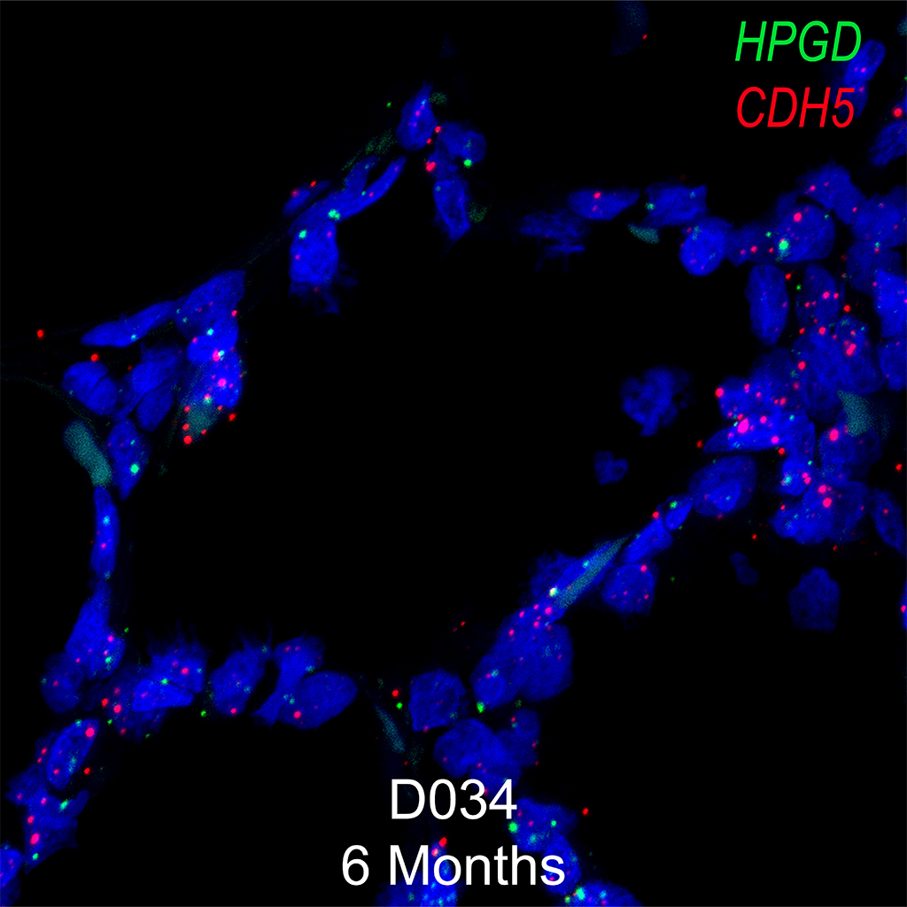
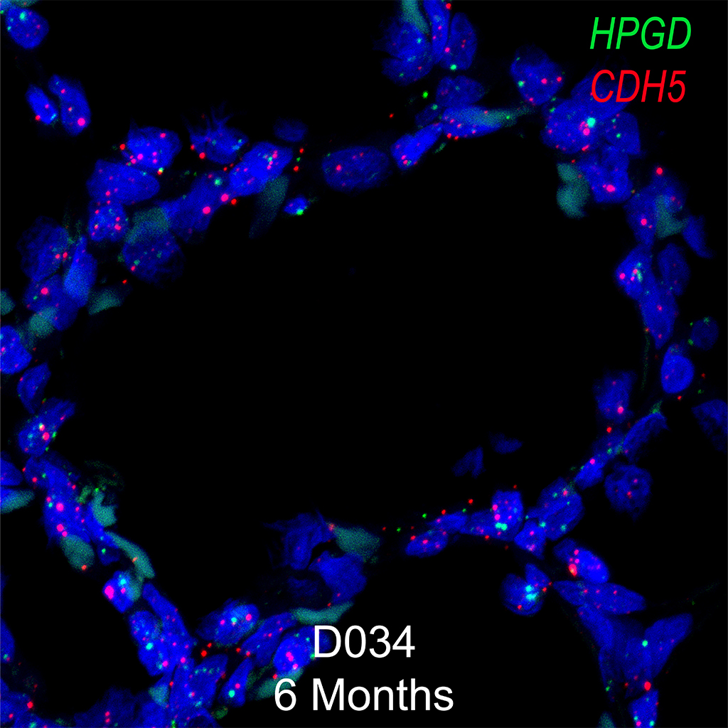
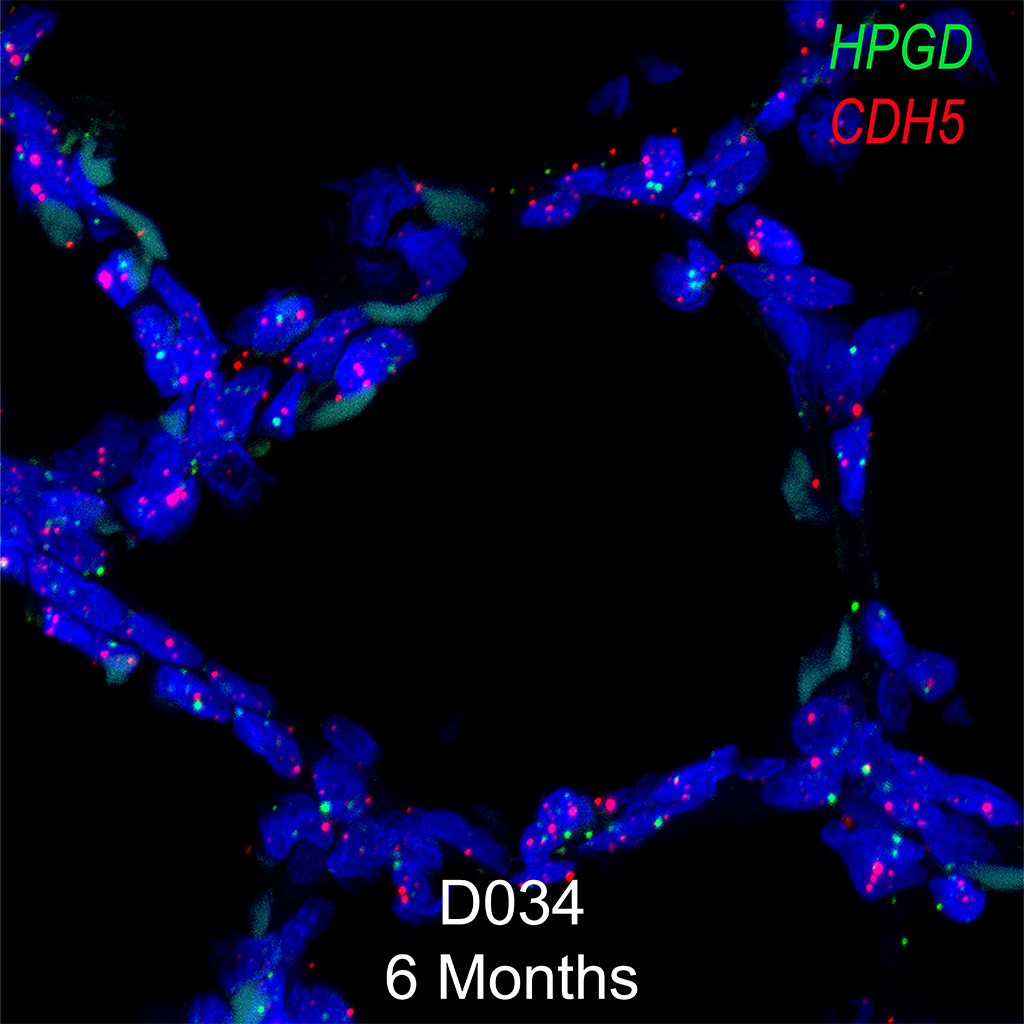
Purpose: Plish in situ hybridization on PACT cleared tissue for SFTPC, MIR29-1, and AXIN2
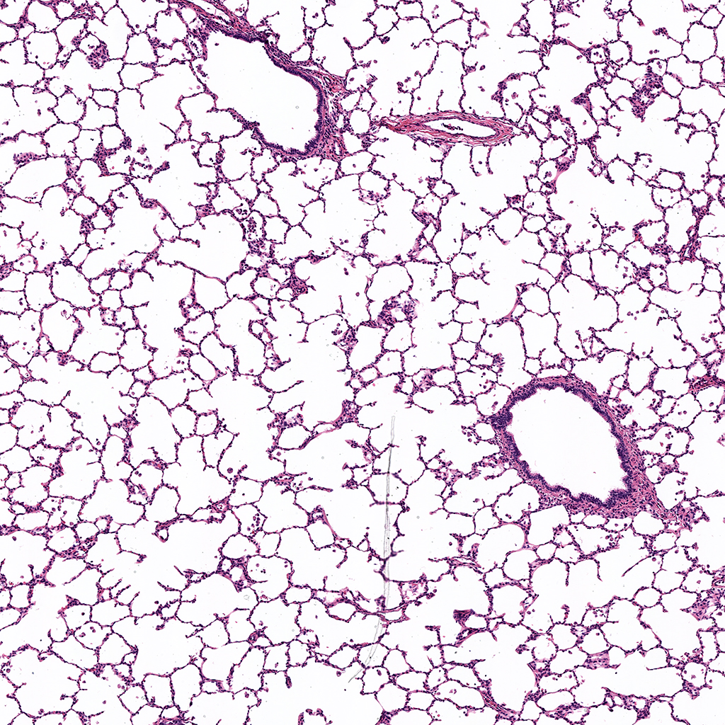
Human lung tissue (kindly provided by Dr. Scott Randell) was fixed in 10% formalin. Tissue was then washed in DEPC-treated 1X PBS and incubated in 30% Sucrose in DEPC-treated 1X PBS. 24 hours prior to cryoprotection, tissue was placed in Sucrose/OCT (2:1). Upon freezing, lung tissue was placed in OCT and frozen on dry ice and stored at -80oC. 200 µm sections of OCT protected frozen human lung were sectioned on a cryostat and stored at -20oC until clearing.
Thick sections were rehydrated in 1X PBS and washed several times to remove OCT. Lungs were incubated in 4% Acrylamide/0.25% 2,2’-Azobis[2-(2-imidazolin-2-yl)propane]dihydrochloride photoinitiator (VA-044, Wako) in 1X PBS overnight at 4oC in a 50ml conical tube. Thick lung sections were then polymerized for 3 hours at 37oC. Following polymerization, excess hydrogel was washed away with several washes of 1X PBS. PBS was removed, and the thick sections were incubated in 8% SDS in 1X PBS overnight in a shaking incubator at 37oC. Thick lung sections were washed several times in 1X PBS to completely remove SDS.
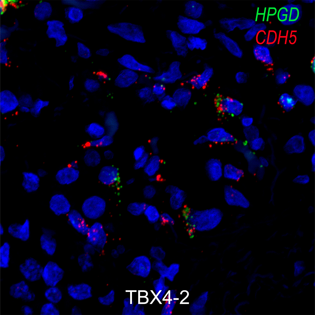
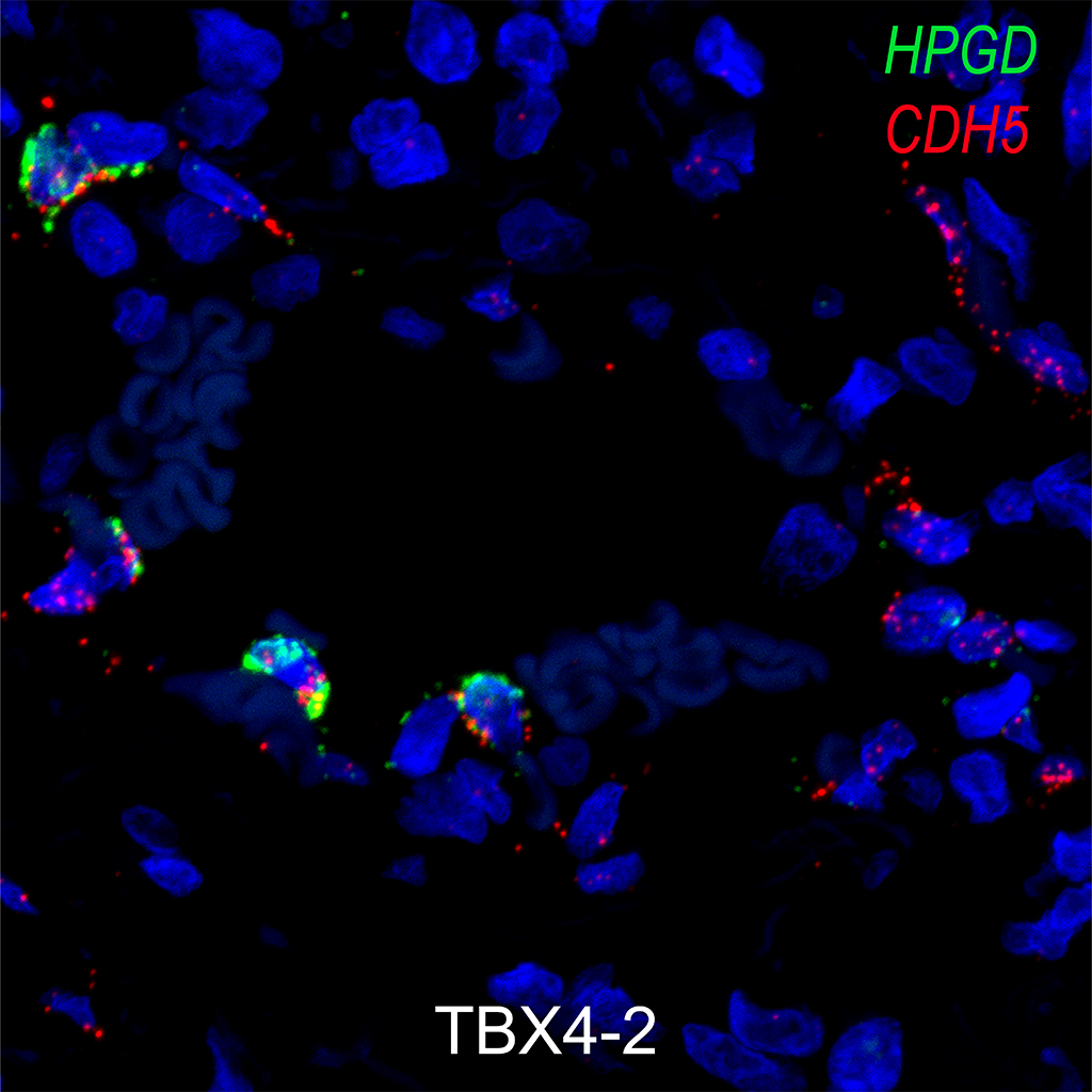
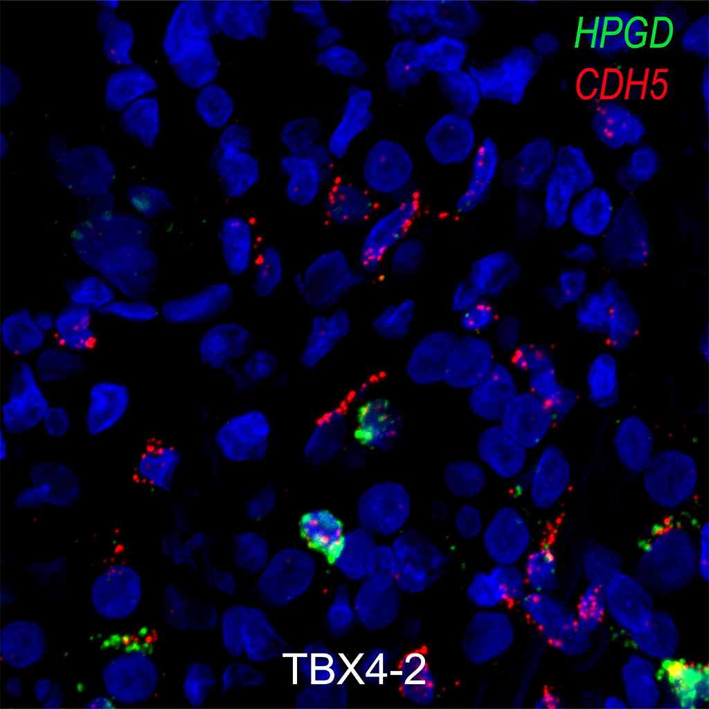
Hybridization probes were incubated at concentrations of 100 nM each in hybridization buffer (1M NaTCA, 5mM EDTA, 50mM Tris pH7.4, 0.2mg.mL Heparin in DEPC water) for two hours at 37ºC and 100% humidity. The sections were then incubated with ligase buffer and phosphorylated common bridge and connector circle oligos at 10 nM for 60 minutes and then with ligase buffer, T4 ligase, and ligase buffer for two hours under the same conditions. DNA amplification was then accomplished using Phi-29 polymerase (Lucigen-30221) in polymerase buffer at the same conditions overnight. After the reaction was complete, slides were washed with label buffer (2X SCC/20% Formamide in DEPC water) and then incubated with 100nM Tye705 label probe in label probe buffer for one hour in the same conditions. Immunofluorescent co-staining and imaging were obtained and analyzed as follows. Negative controls of secondary antibody alone and reactions performed with identically labeled probes for the bacteria Bacillus subtilis gene mgsA were imaged and used for thresholding for quantitative image analysis. All probes were ordered from Integrated DNA Technologies, target probes were ordered with standard desalting and all probes were stored as 100μM stocks.
Single-Cell Phenotyping within Transparent Intact Tissue through Whole-Body Clearing
Yang, Bin et al. Cell , Volume 158 , Issue 4 , 945 – 958
http://dx.doi.org/10.1016/j.cell.2014.07.017
Automated cell-type classification in intact tissues by single-cell molecular profiling
Elife. 2018 Jan 10;7. pii: e30510. doi: 10.7554/eLife.30510
https://elifesciences.org/articles/30510
Single-cell Wnt signaling niches maintain stemness of alveolar type 2 cells
Science. 2018 Feb 1. pii: eaam6603. doi: 10.1126/science.aam6603
http://science.sciencemag.org/content/early/2018/01/31/science.aam6603.long
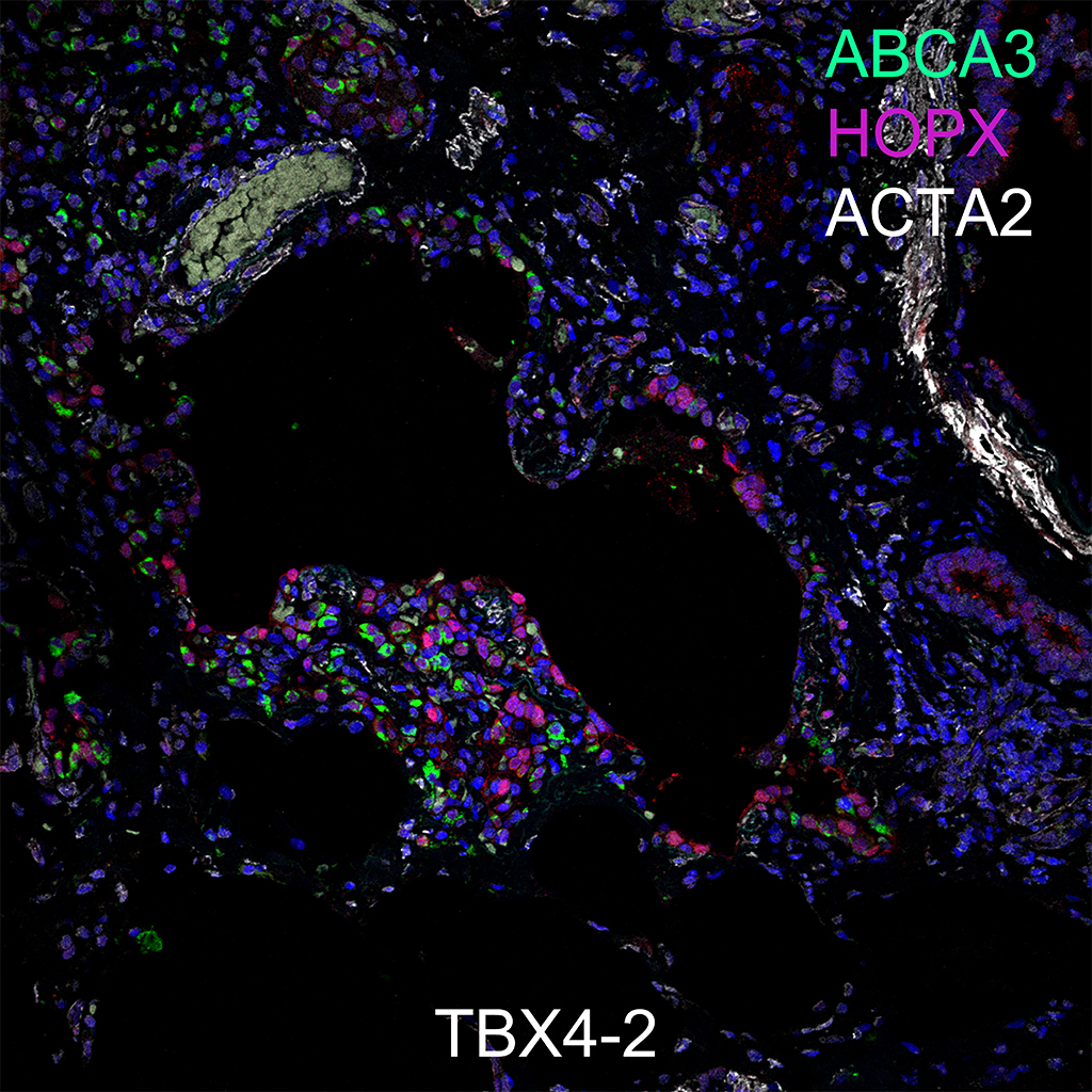
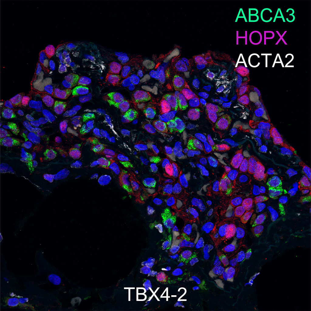
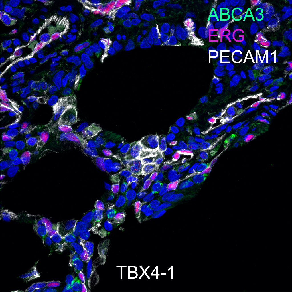
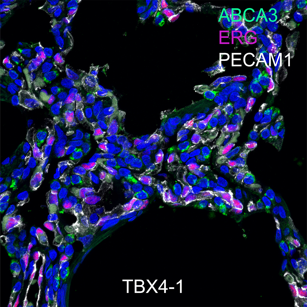
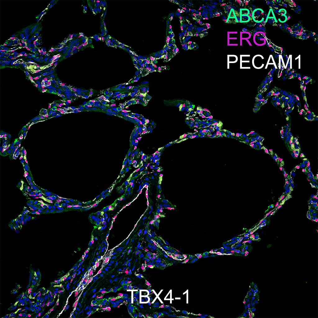
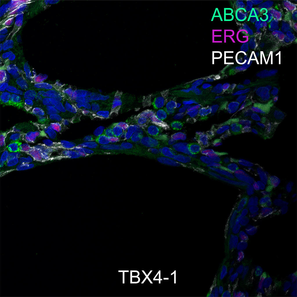
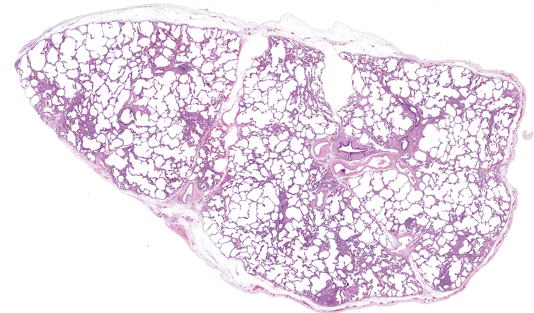
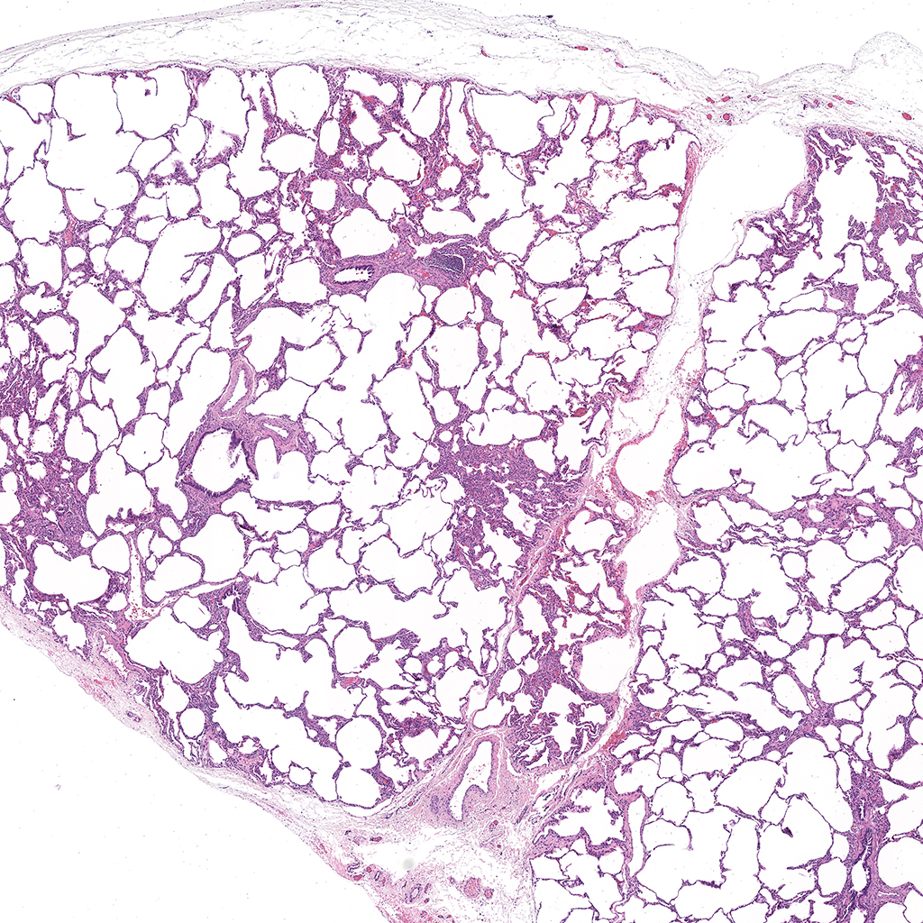
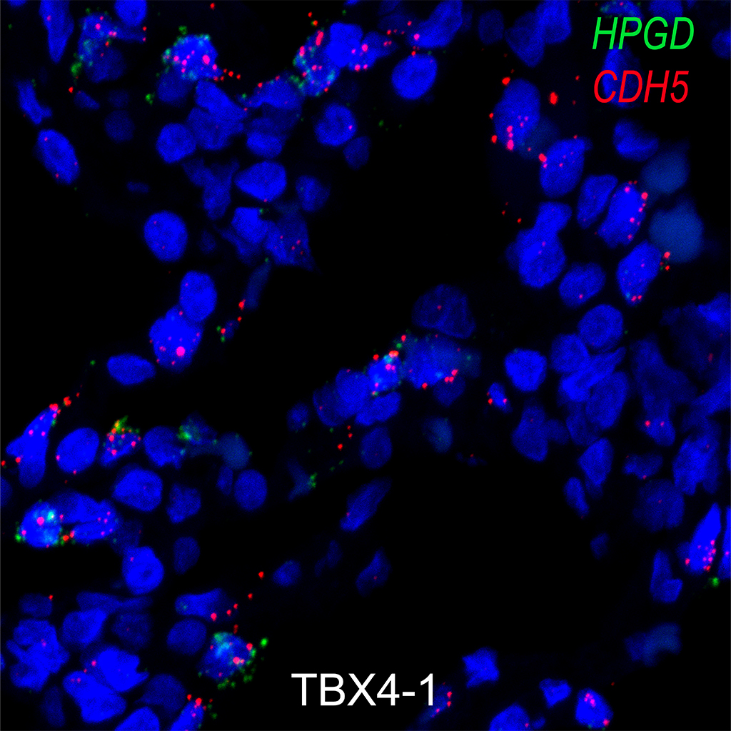
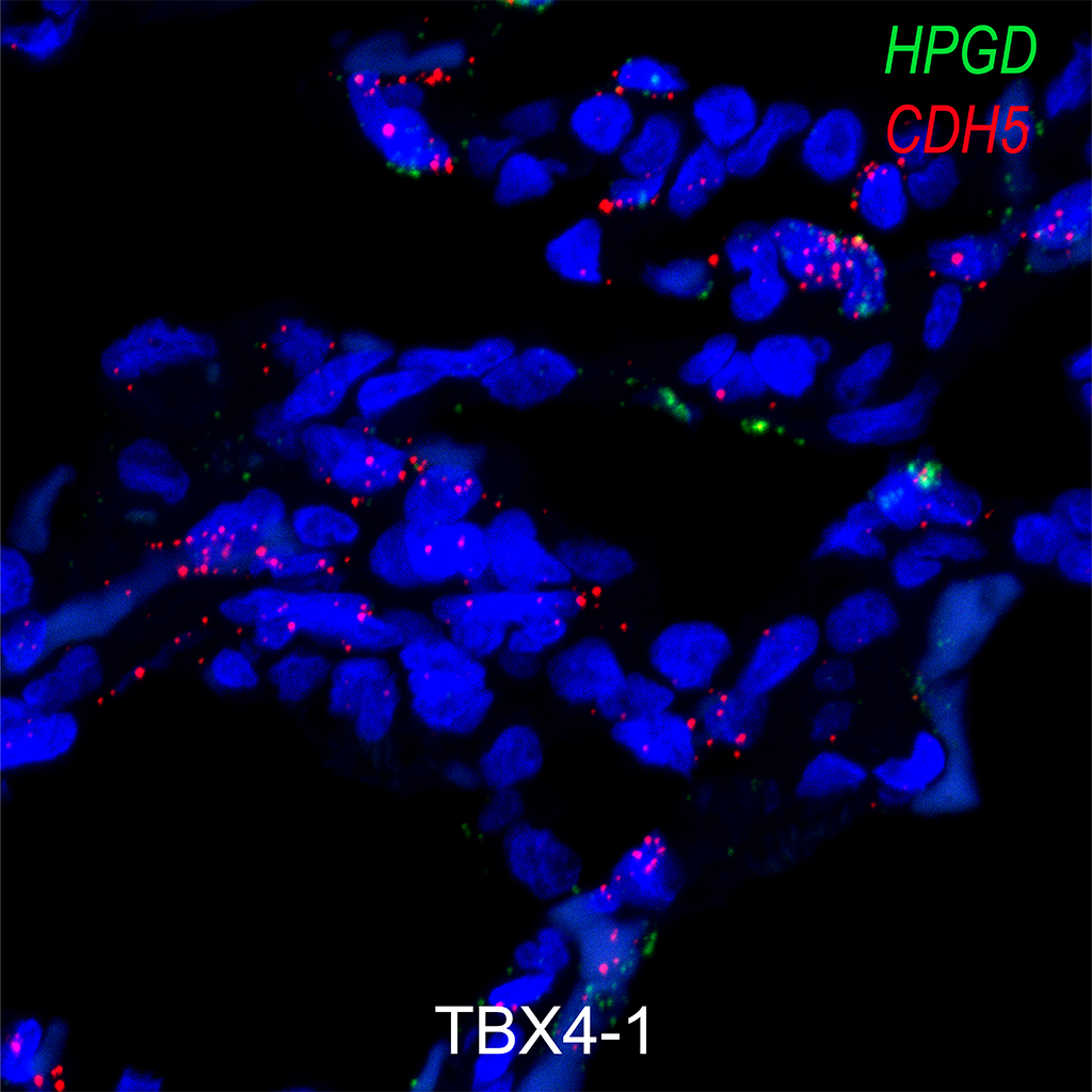
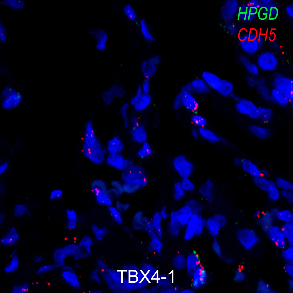
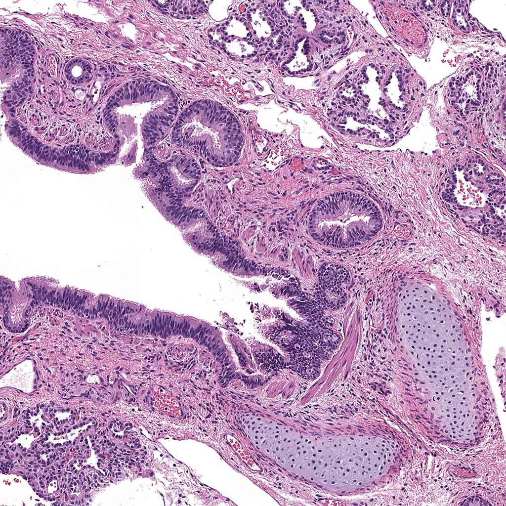
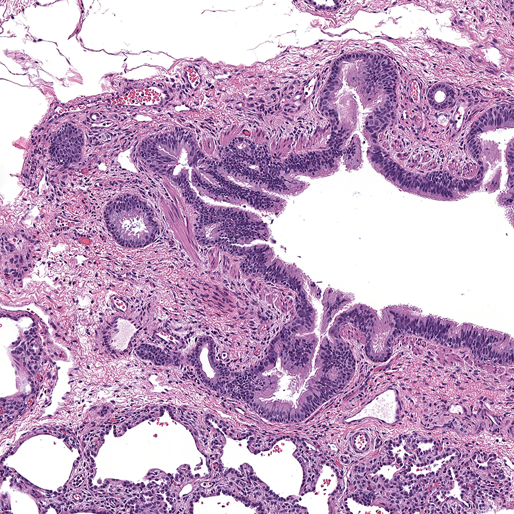
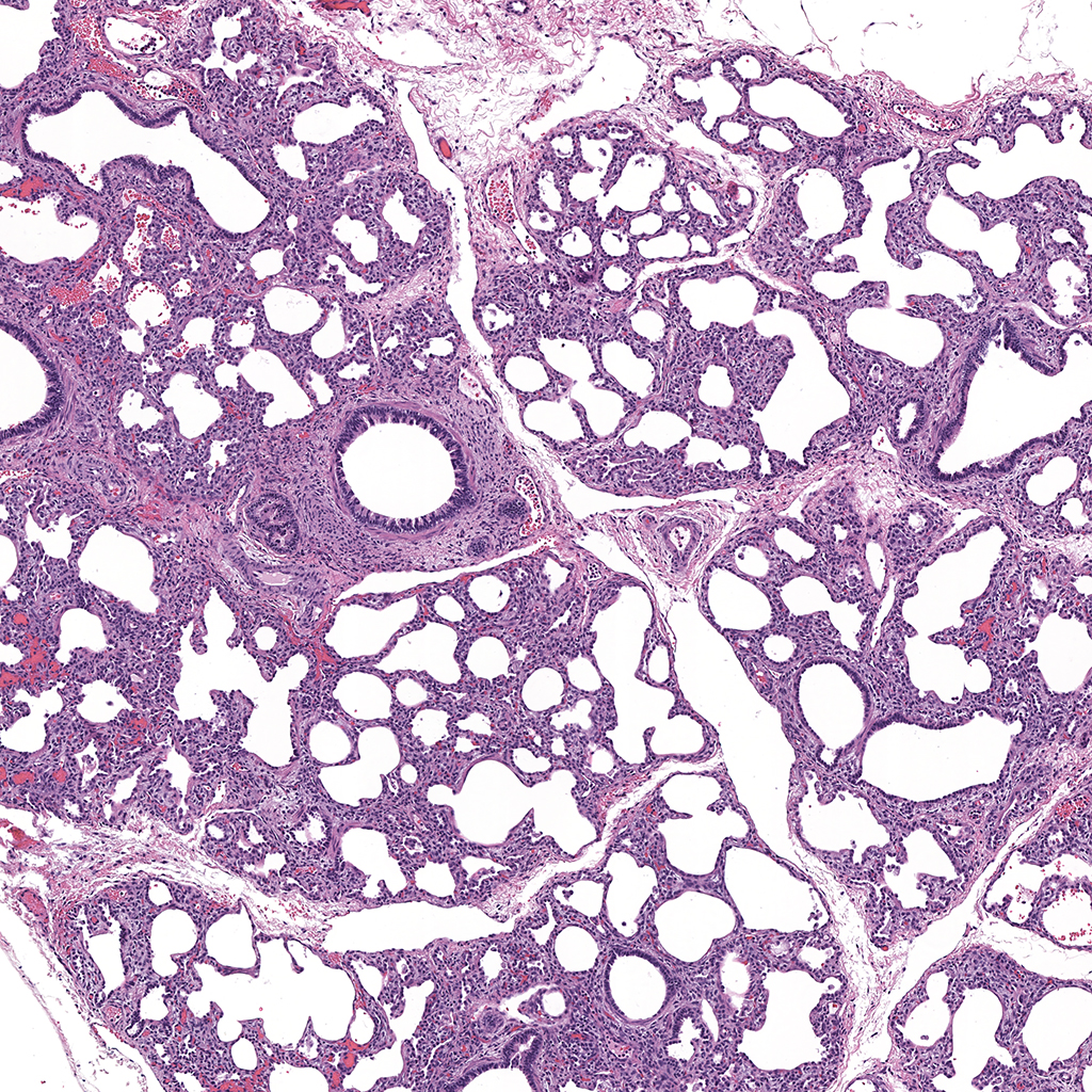
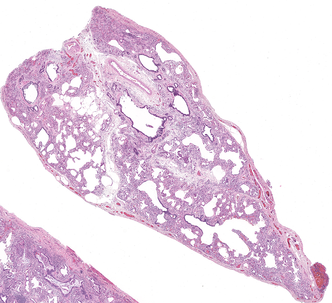
LMHA-15-UNC-DD036L_MC15-04H.4.1
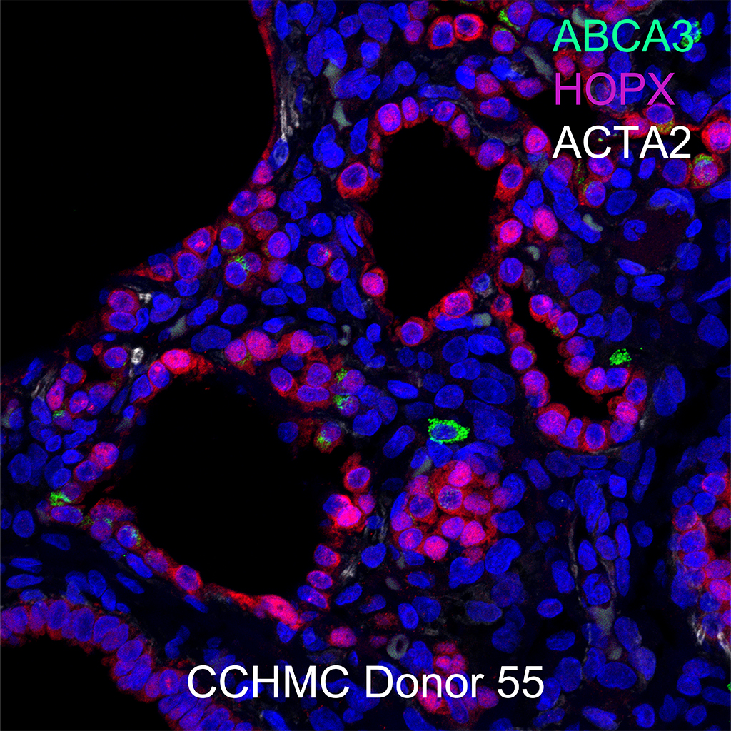
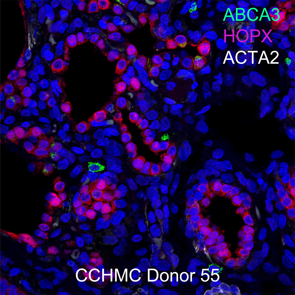
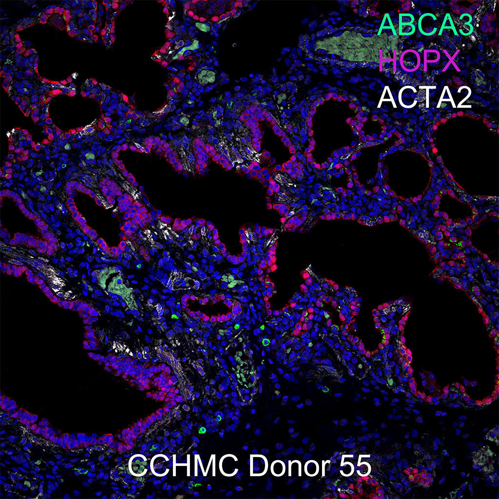
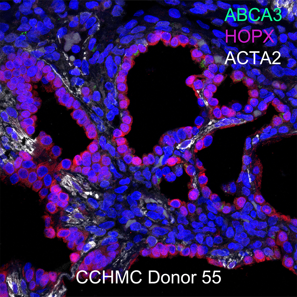
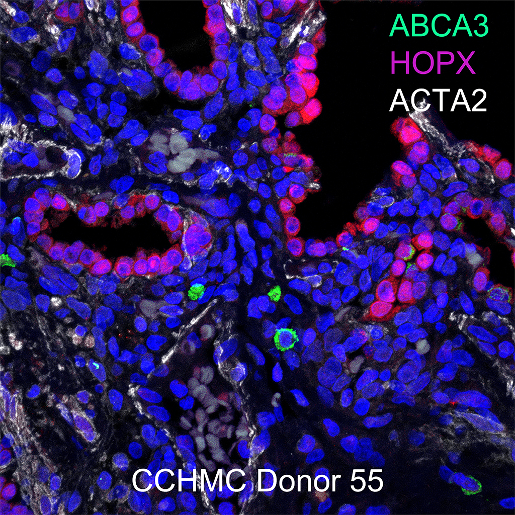
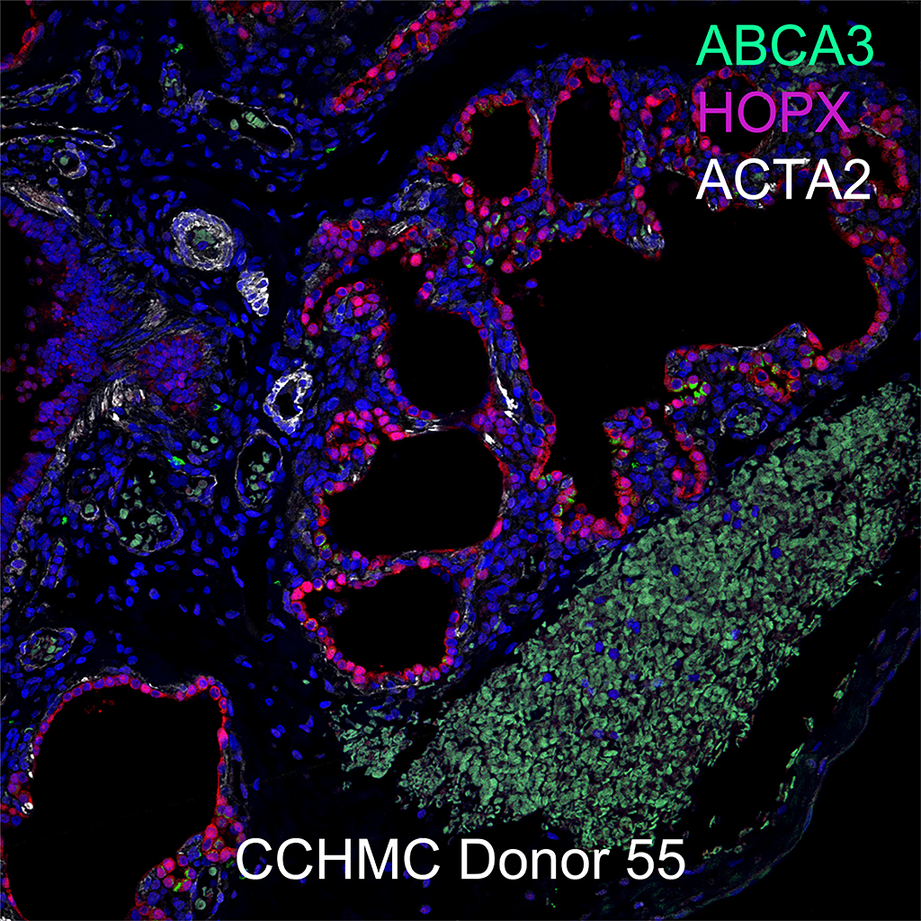
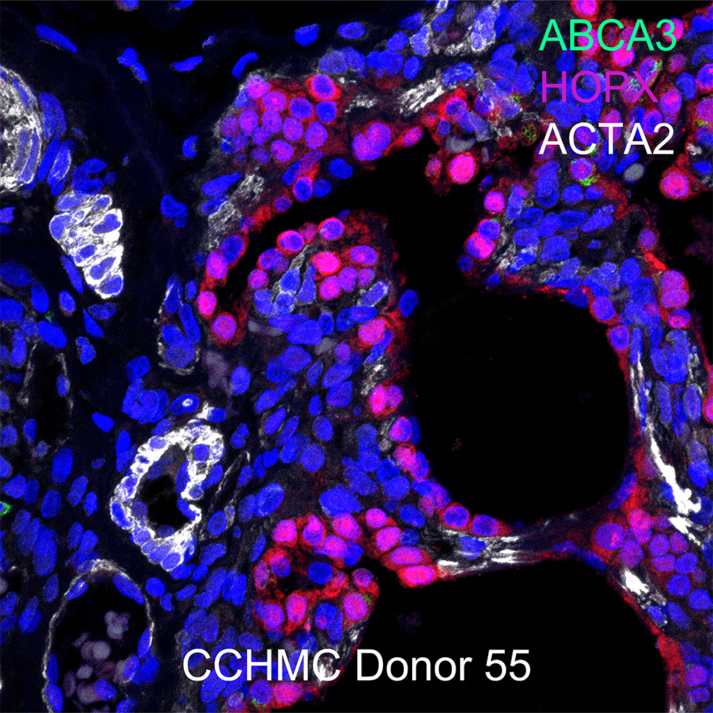
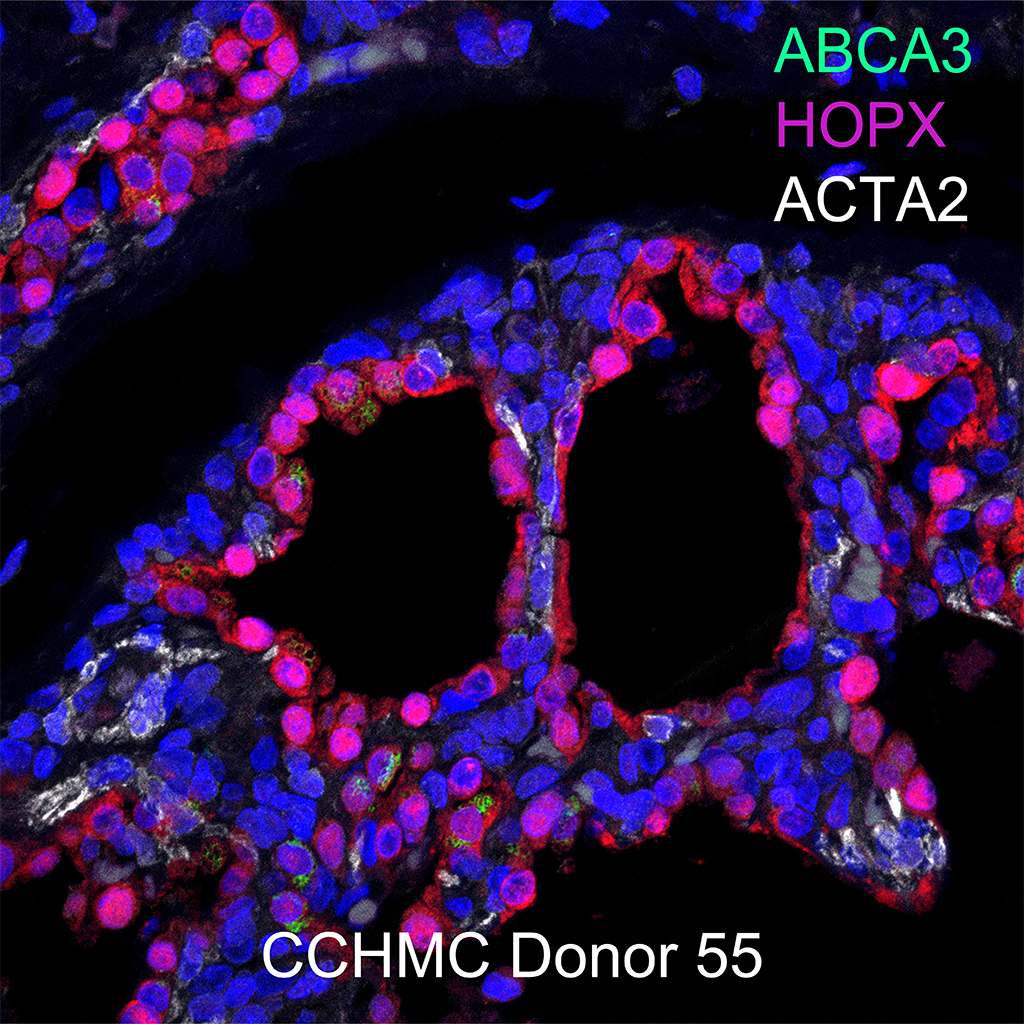
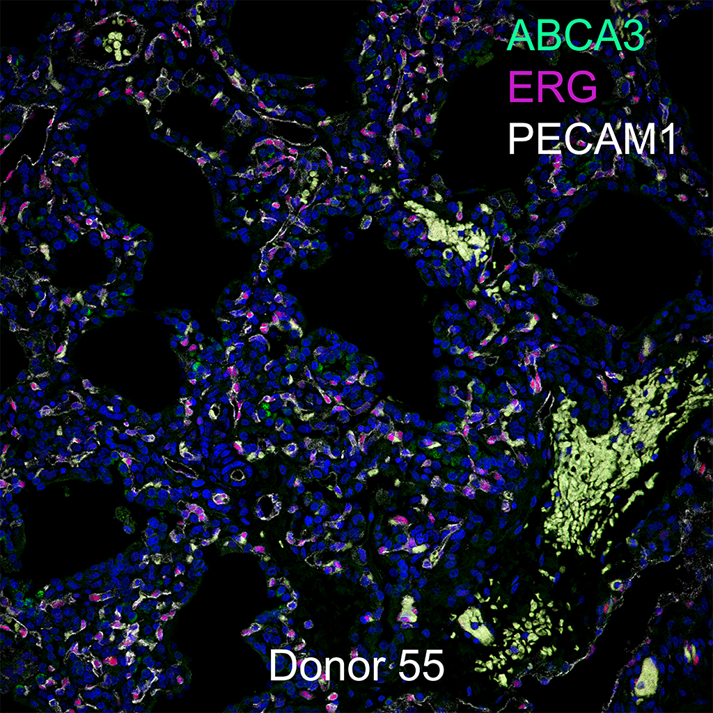
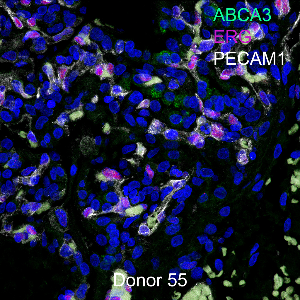
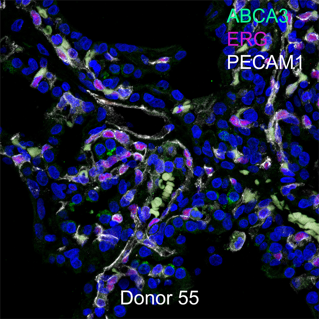
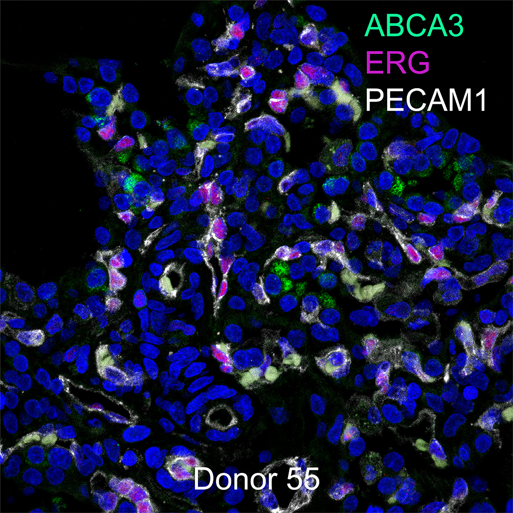
Gender: Male
Age: 31 Year Old
Race: Hispanic
Non-Smoker