
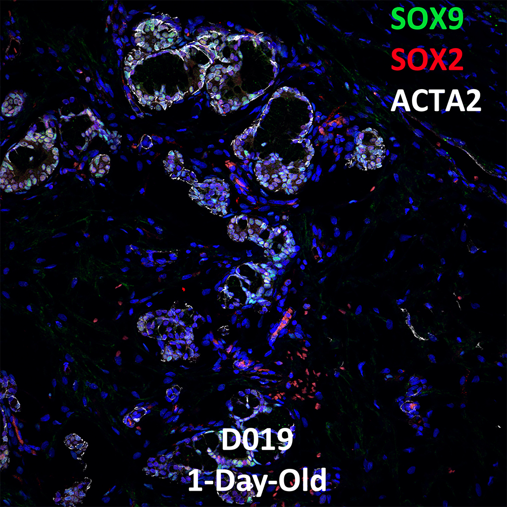
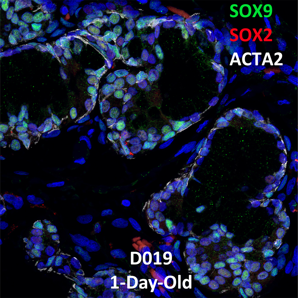
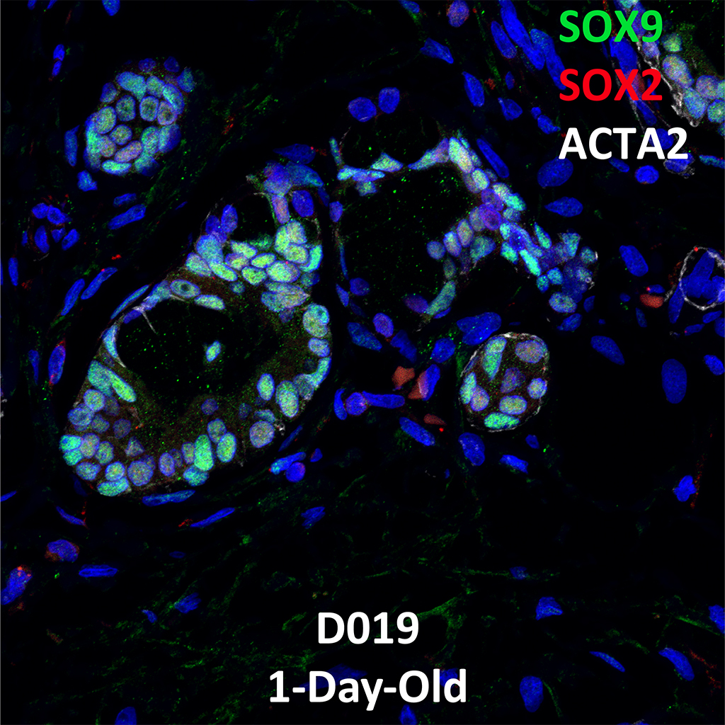
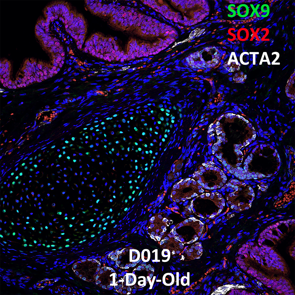
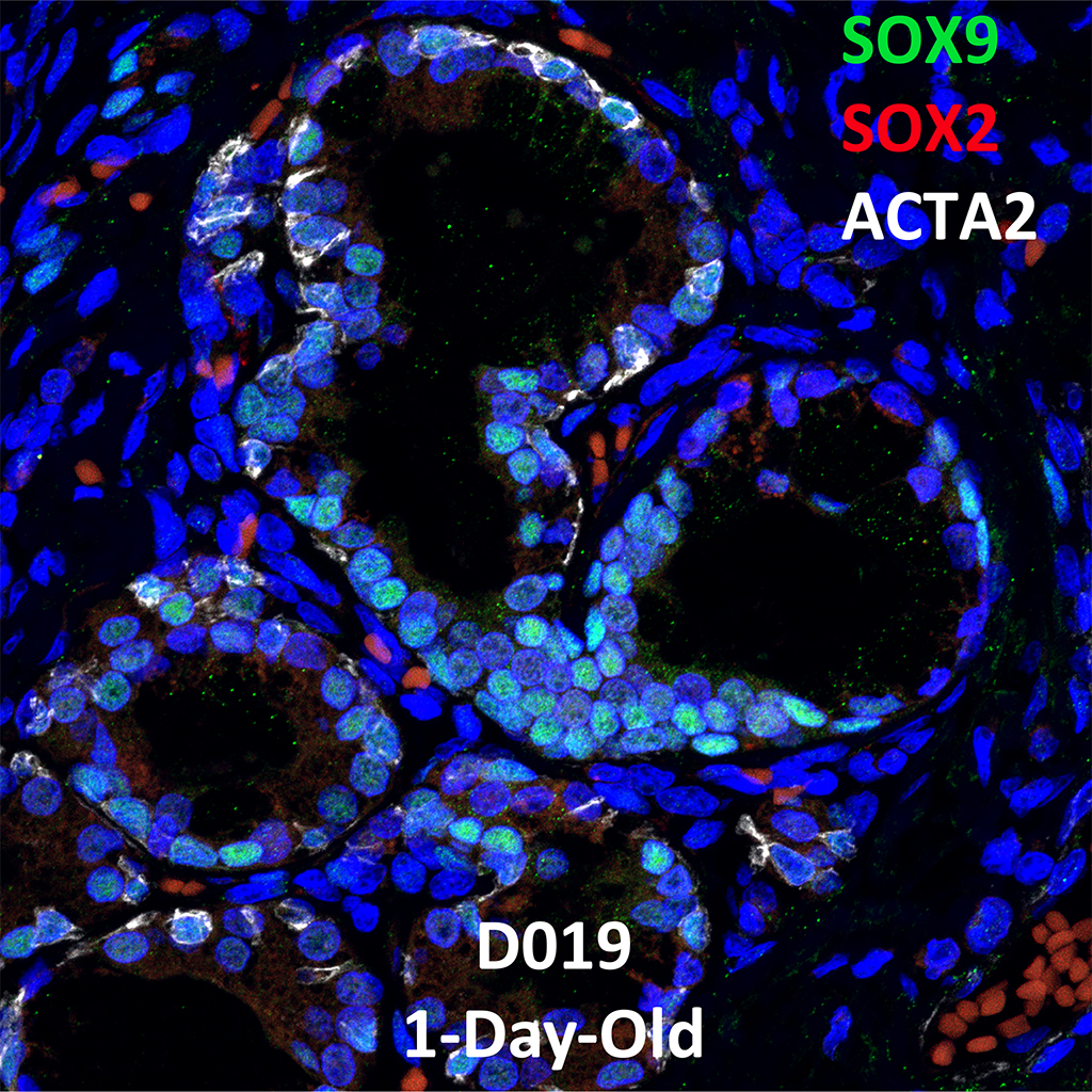
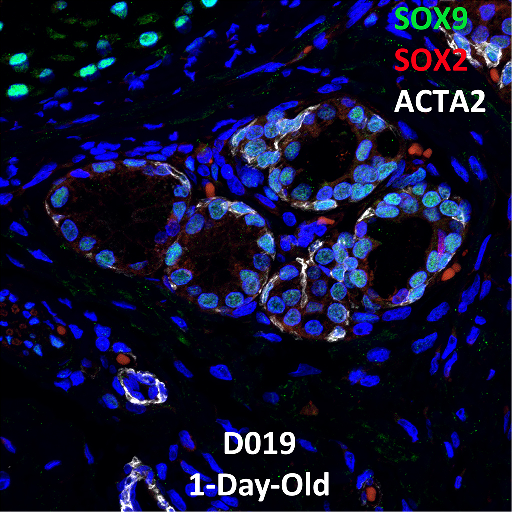
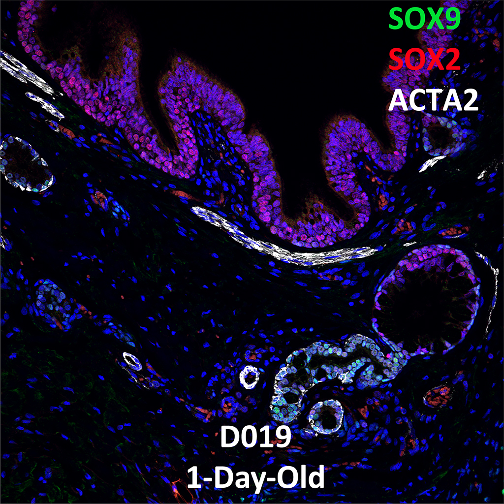
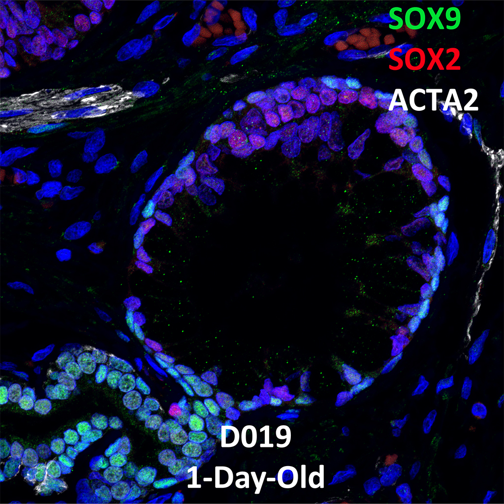
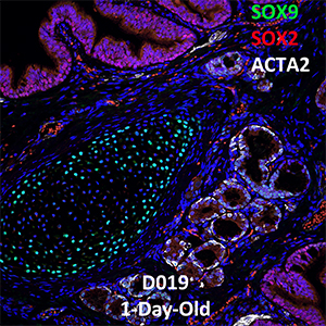
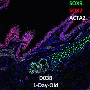
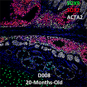
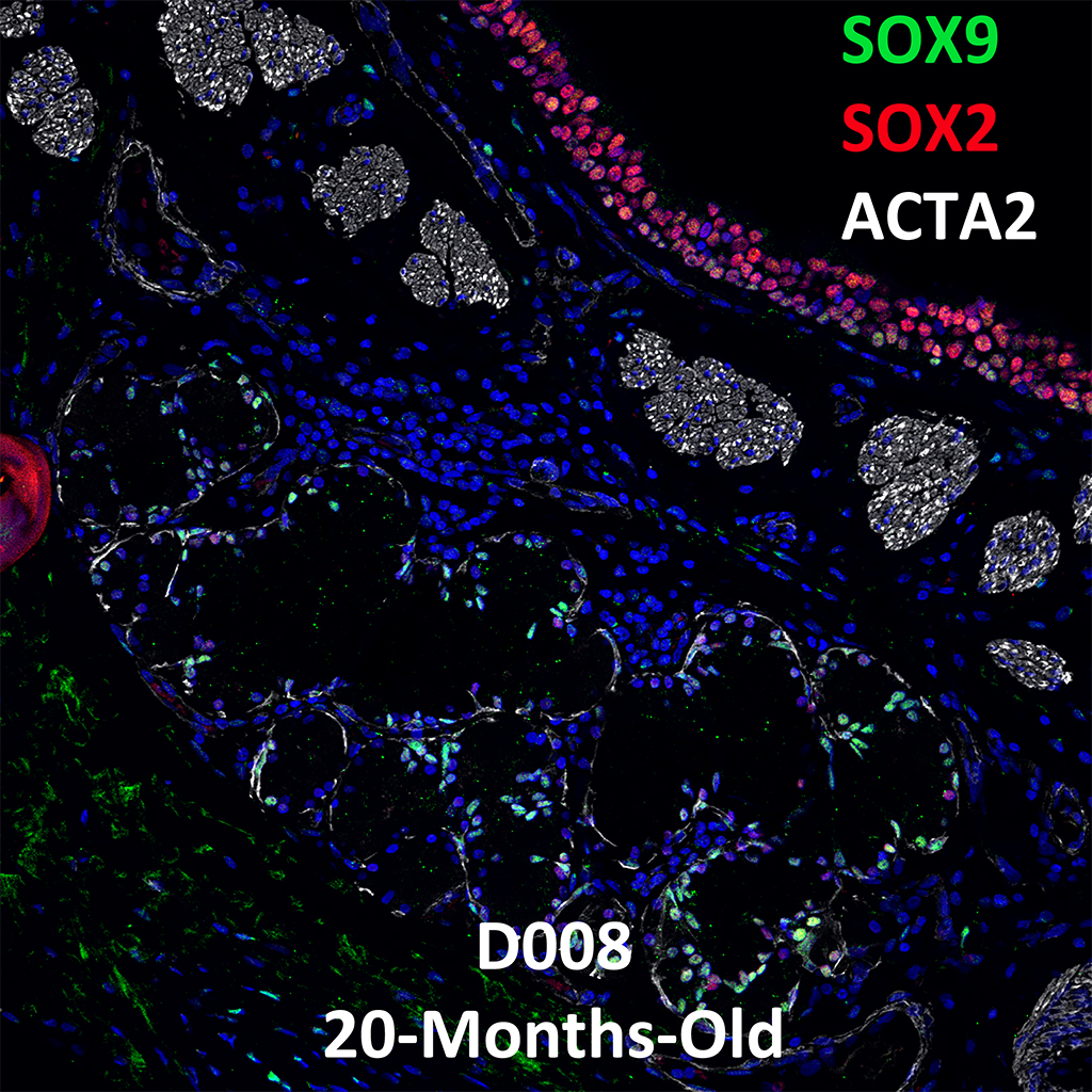
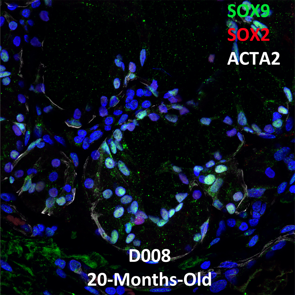
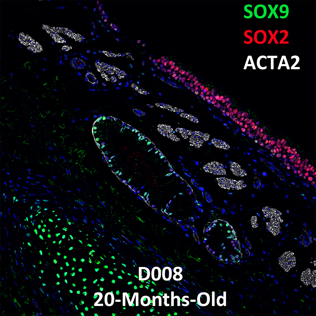
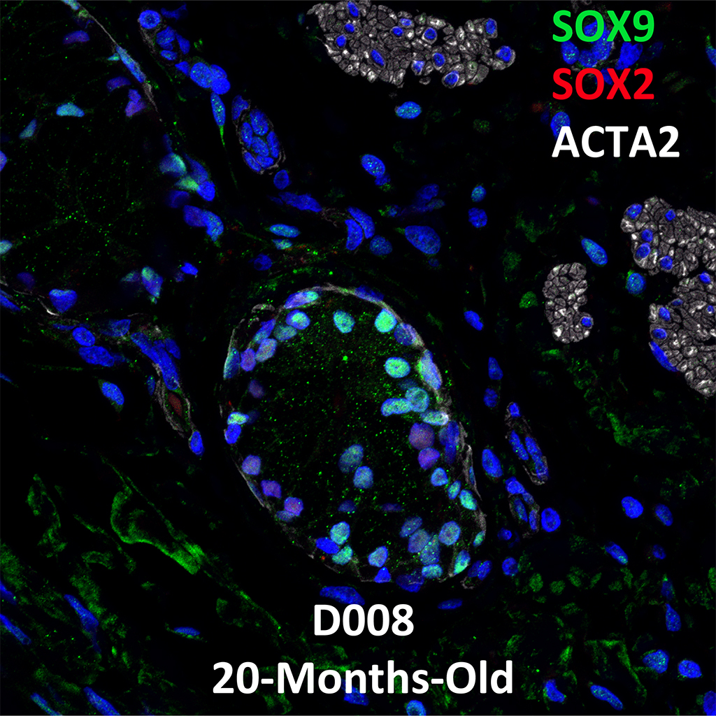
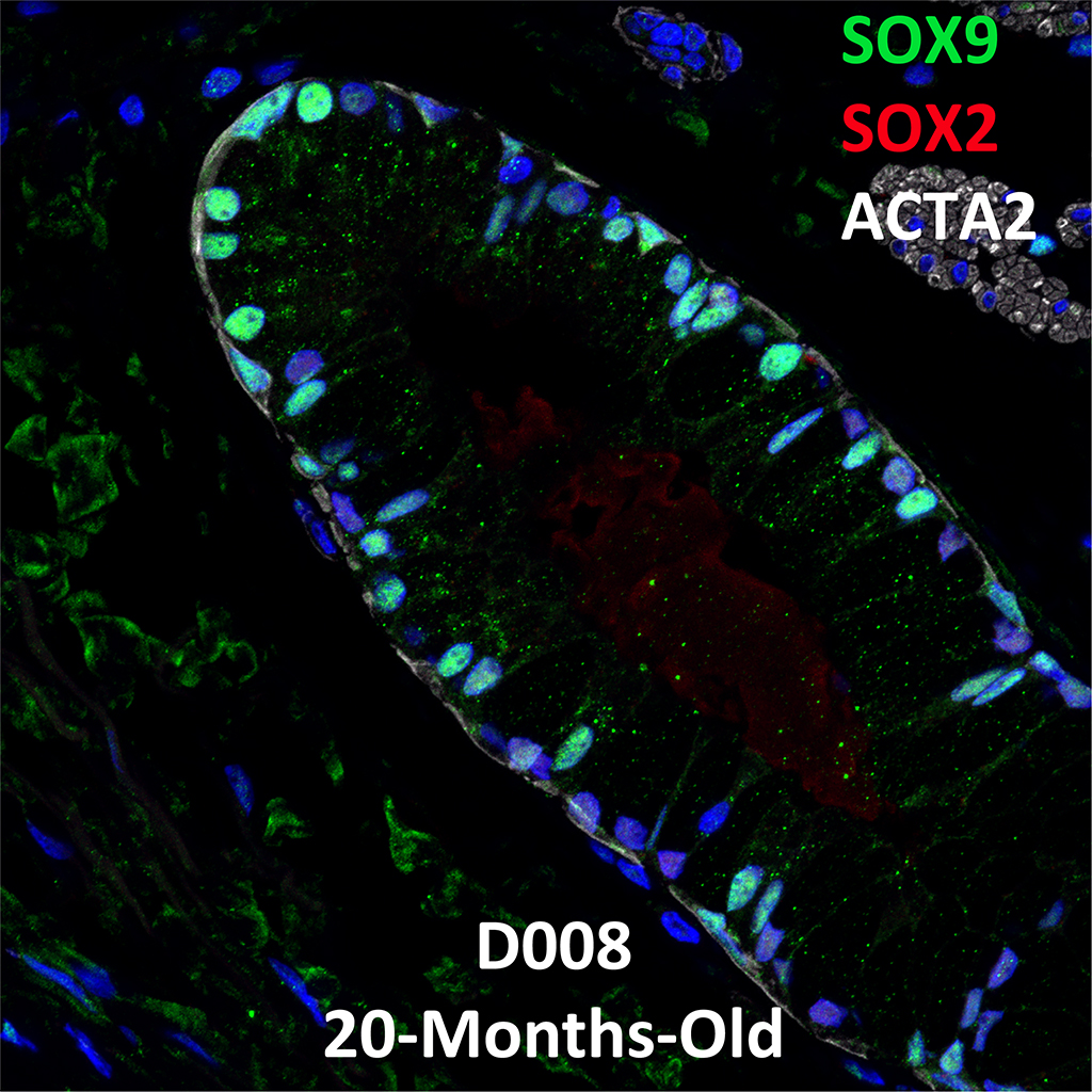
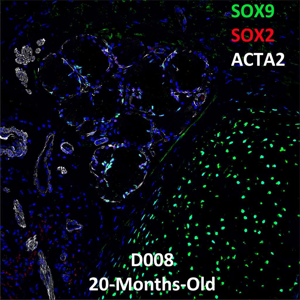
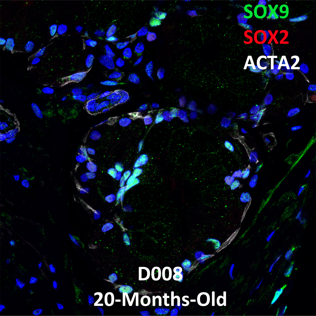
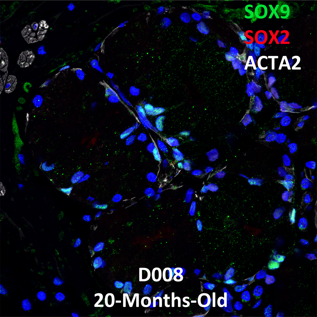
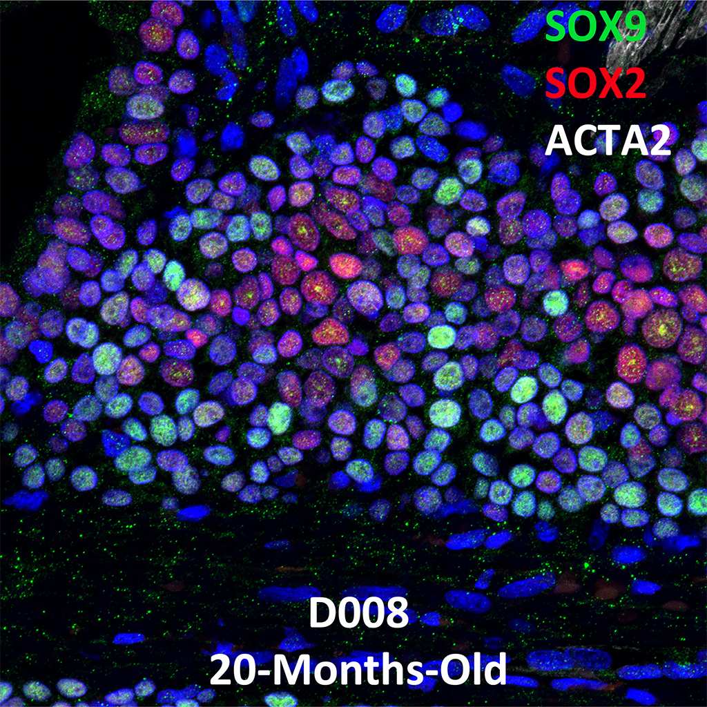
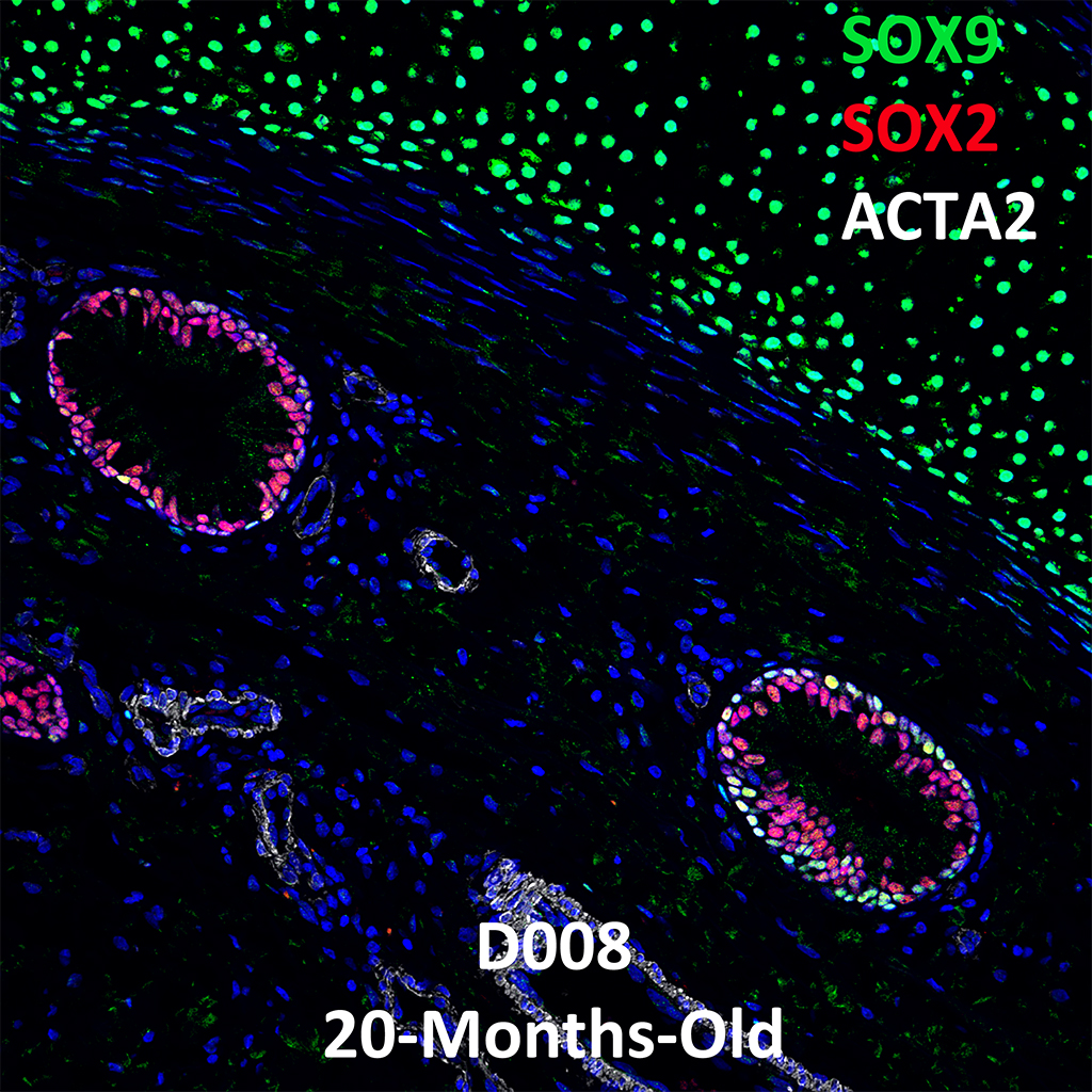
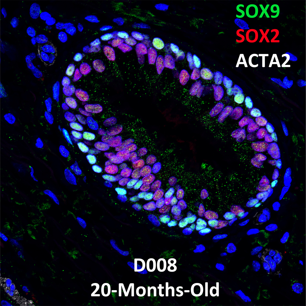
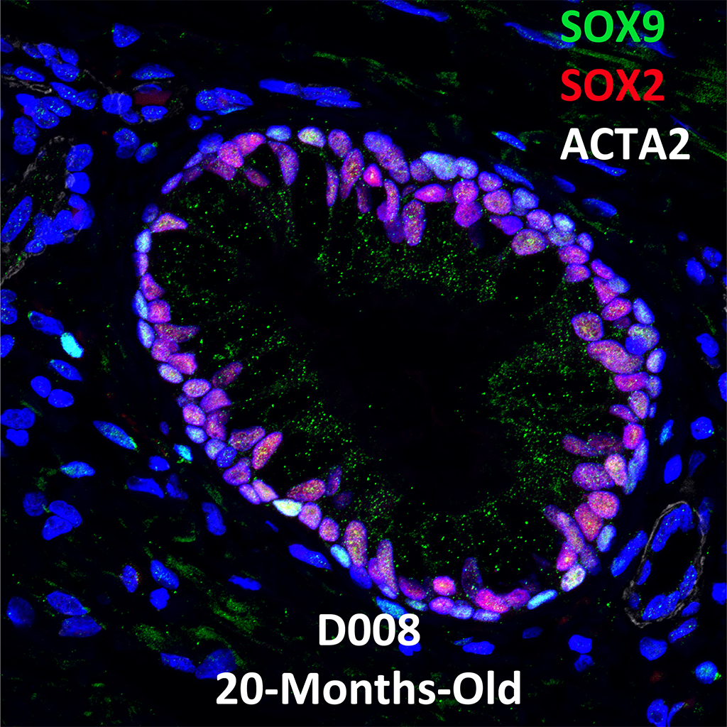
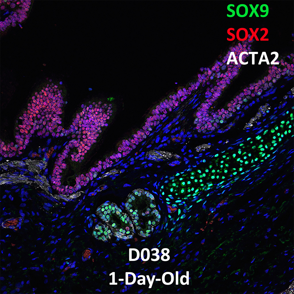
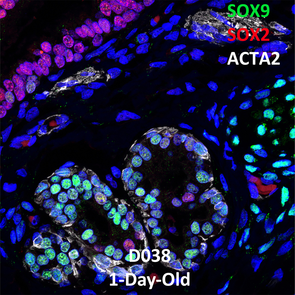
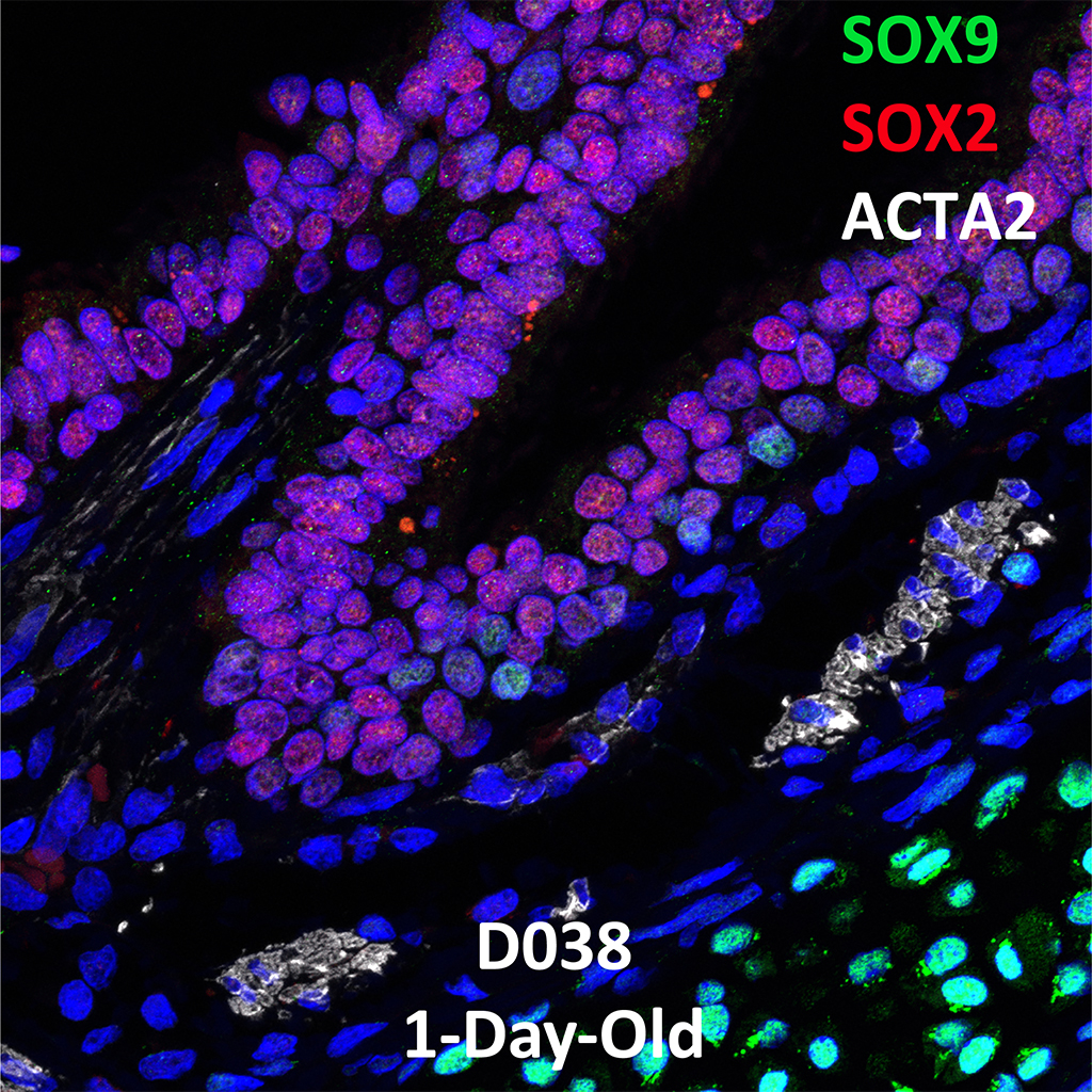
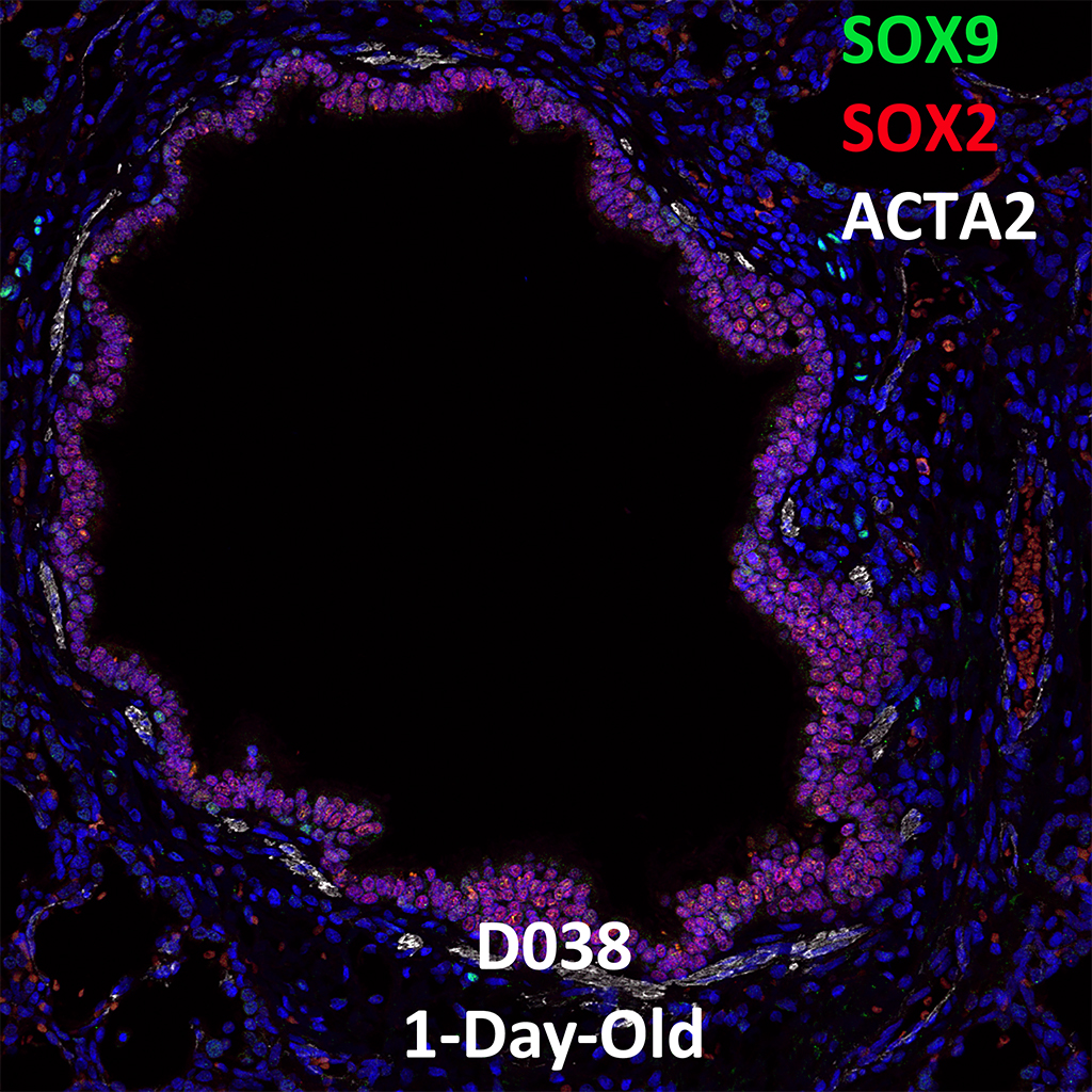
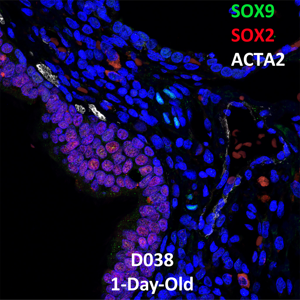
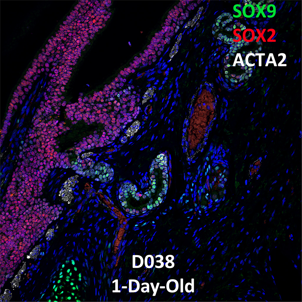
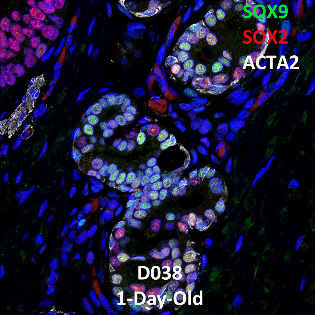
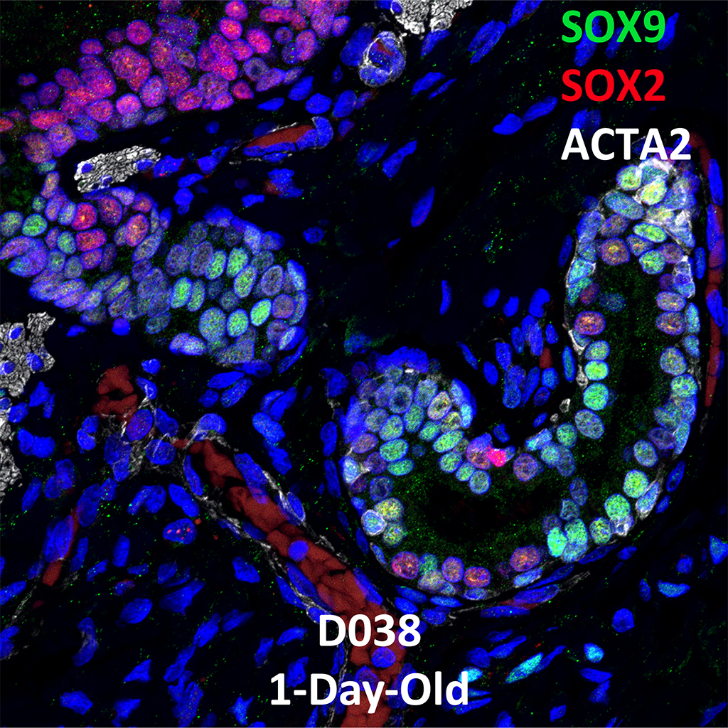
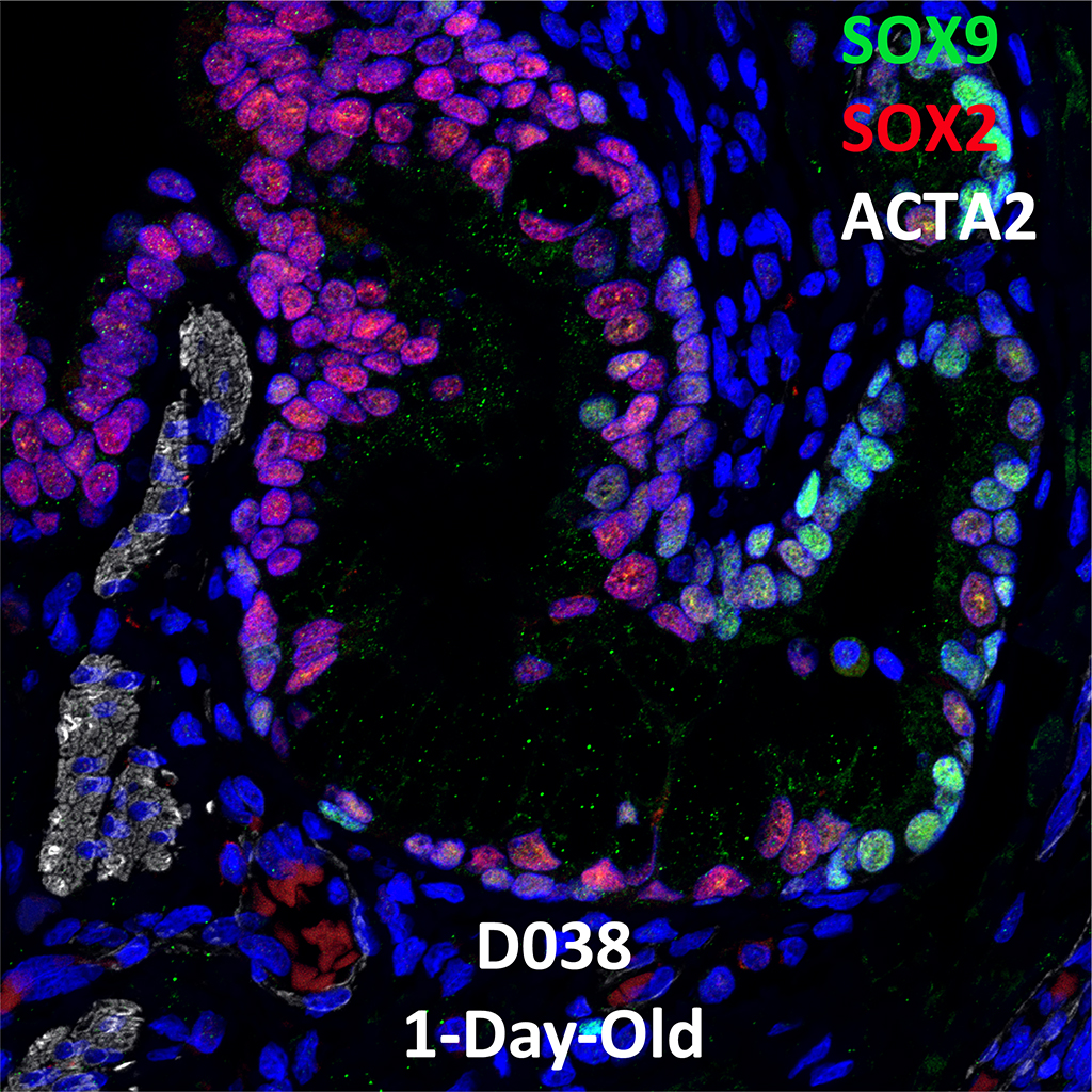
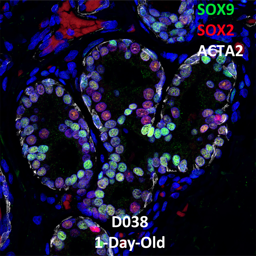
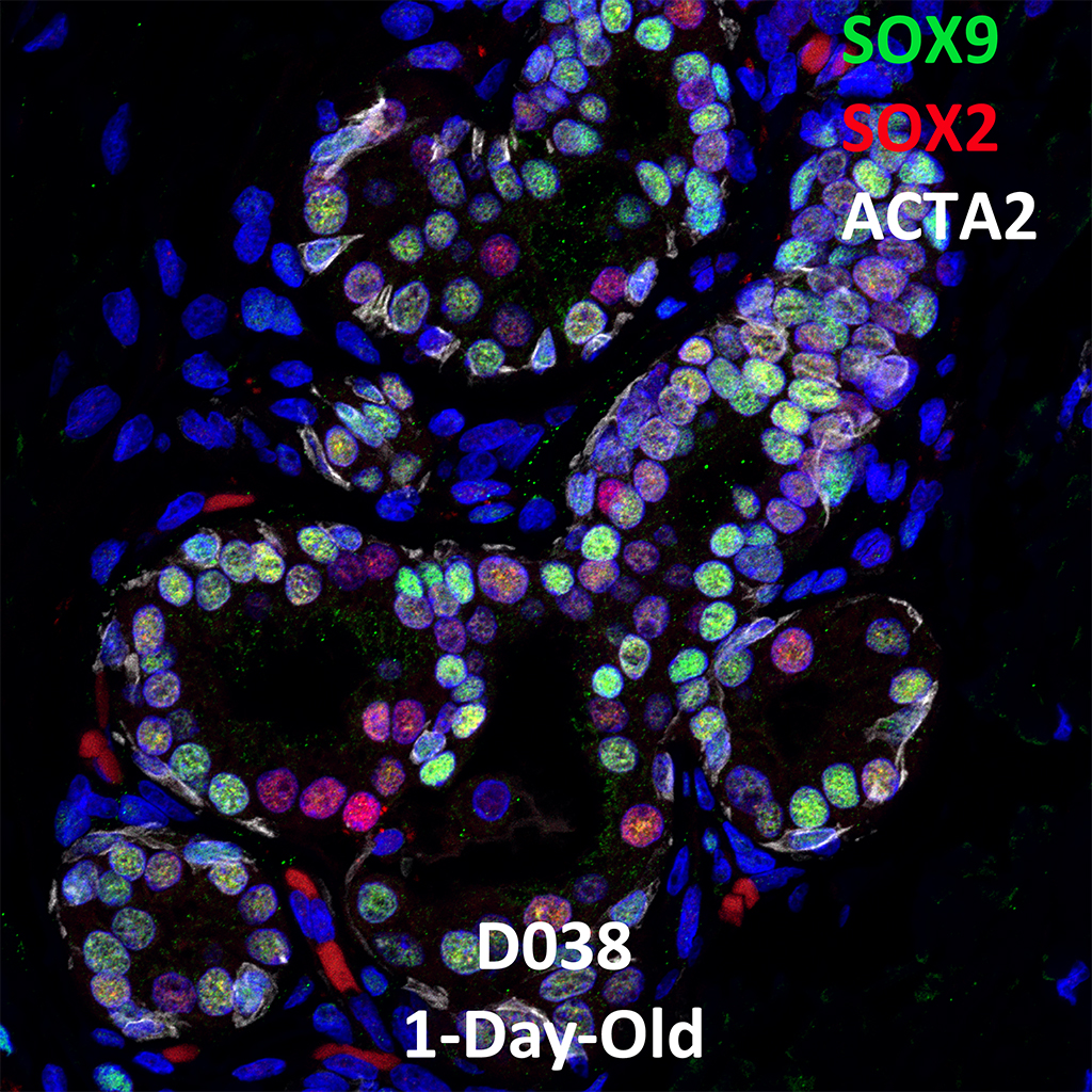
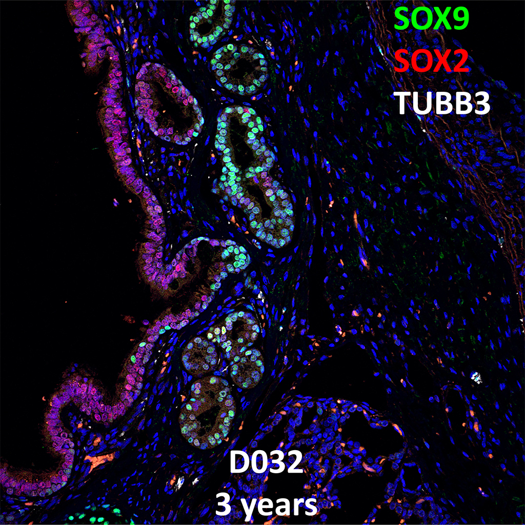
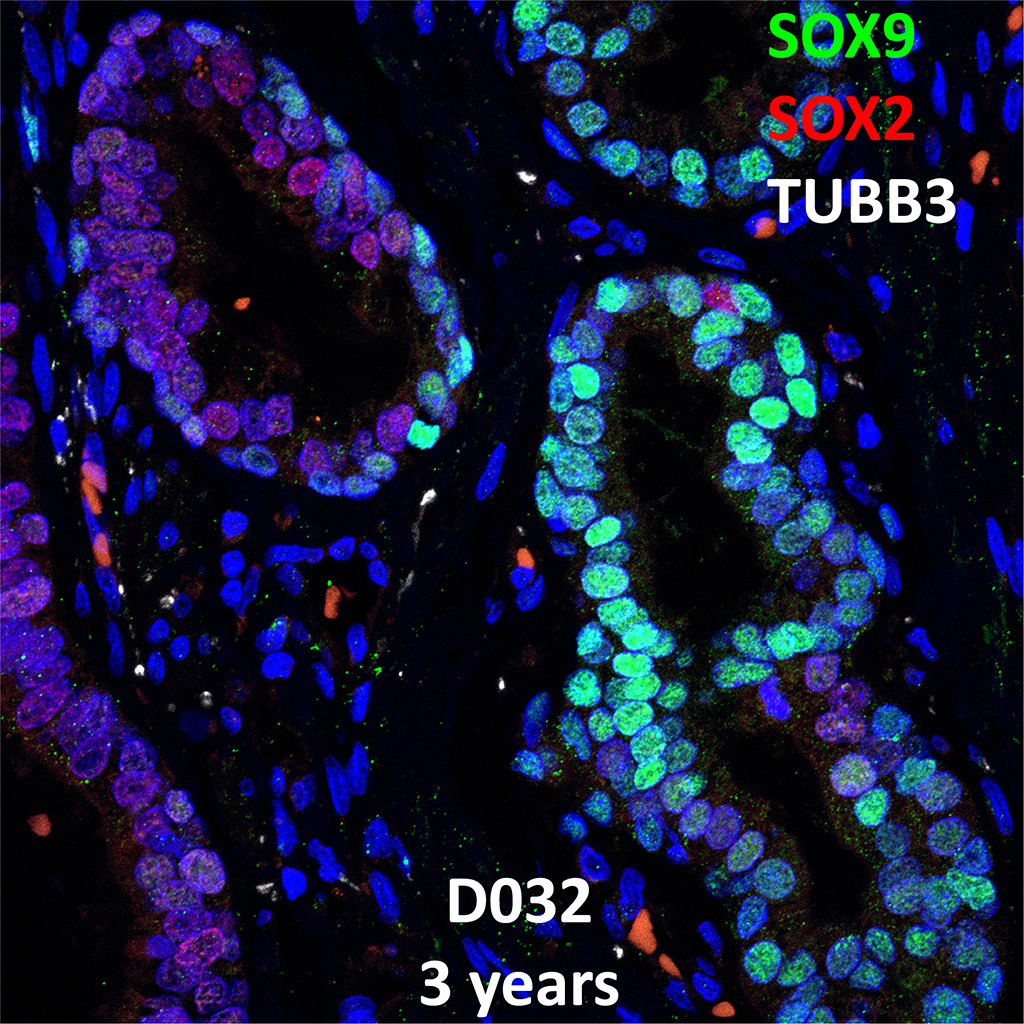
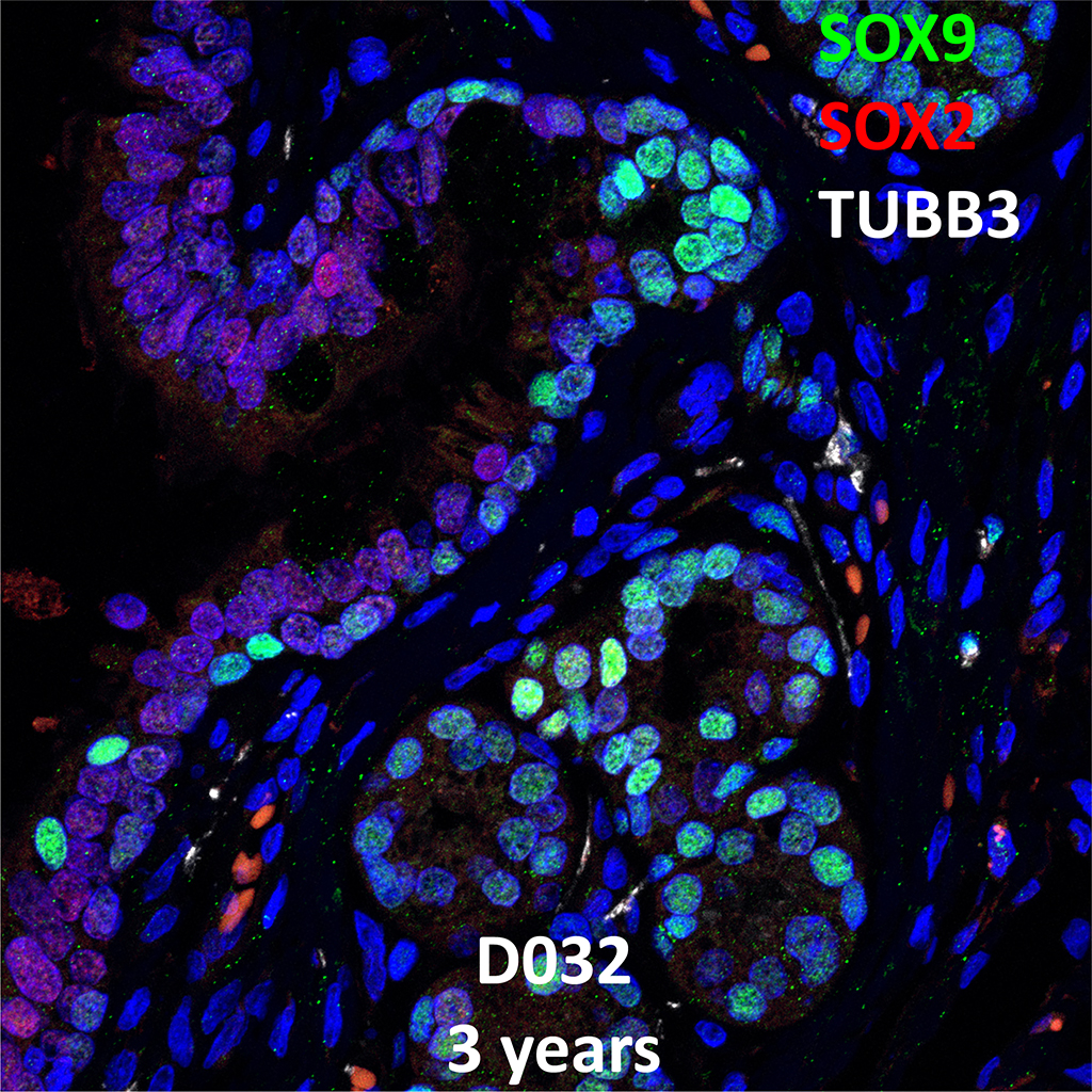
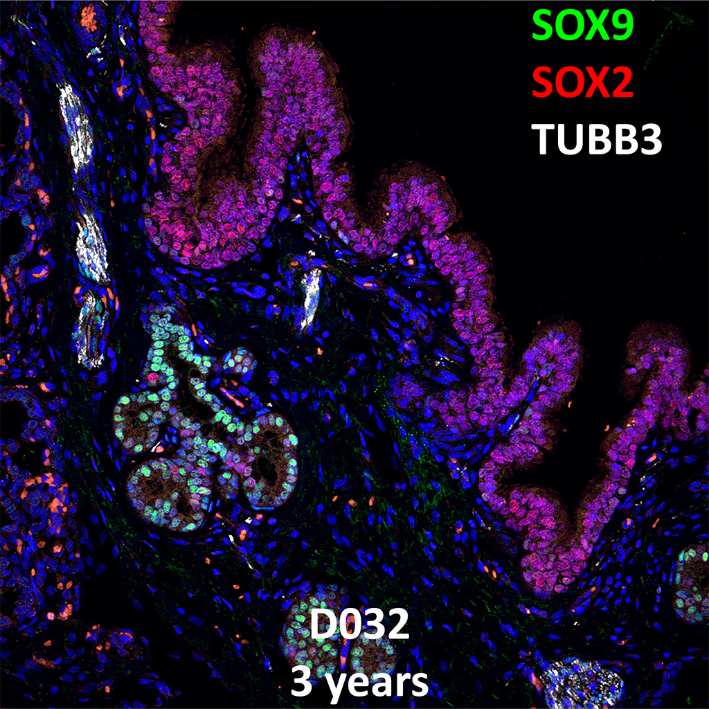
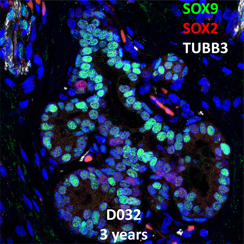
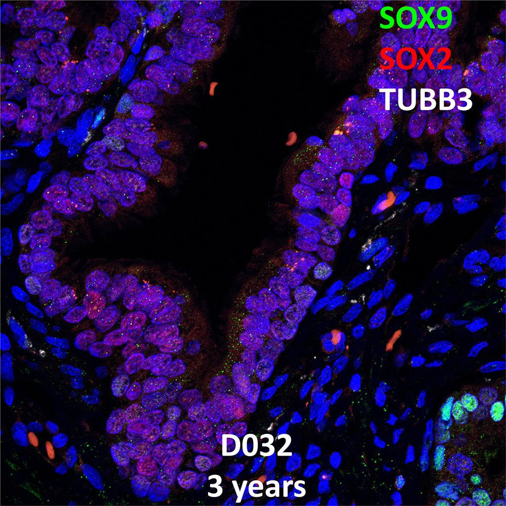
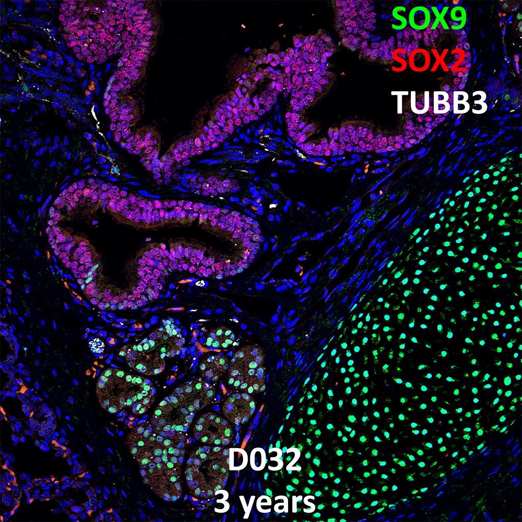
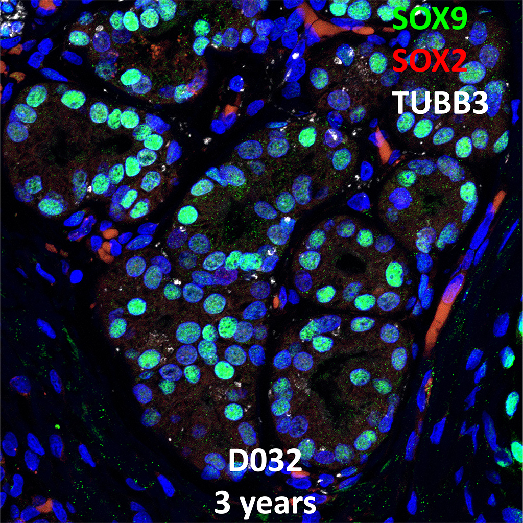
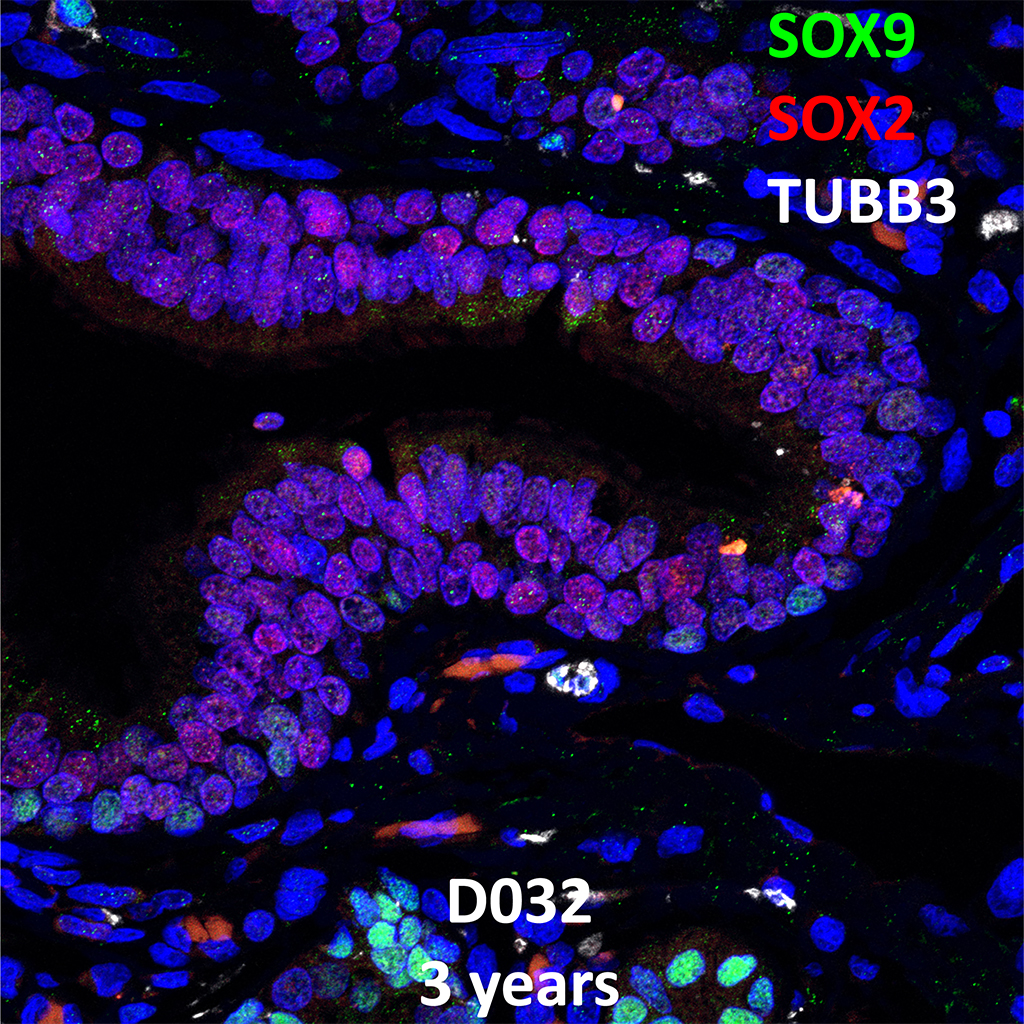
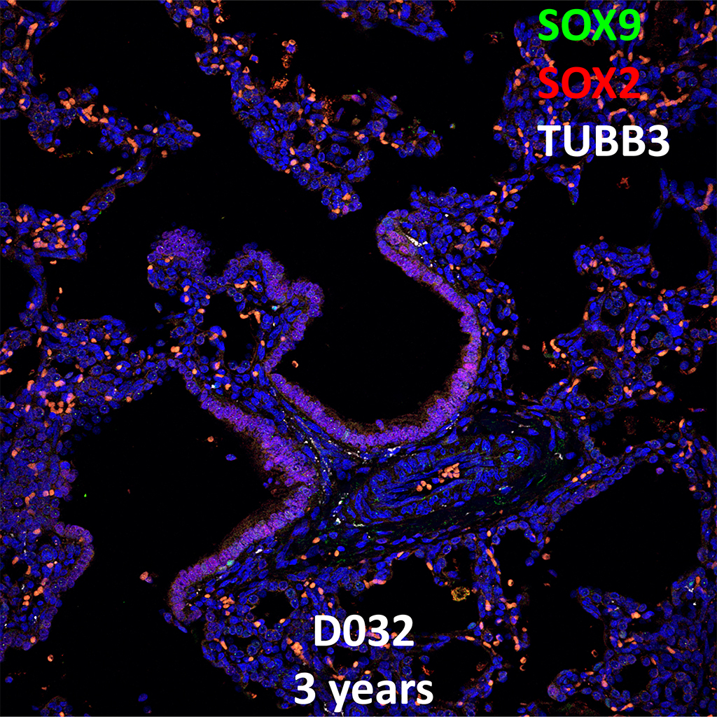
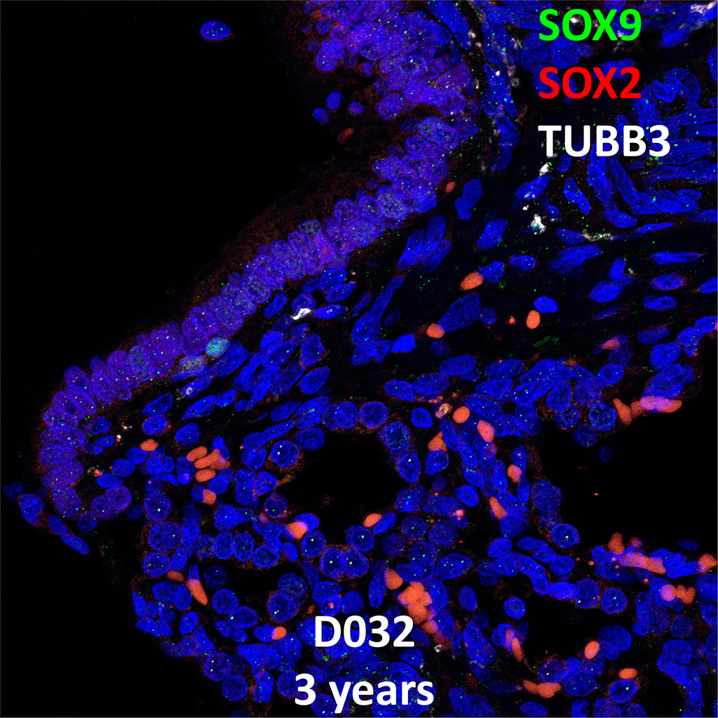
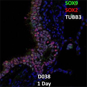
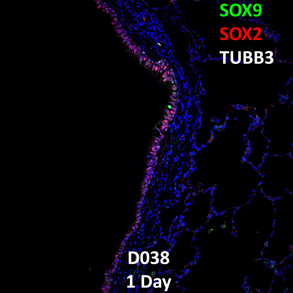
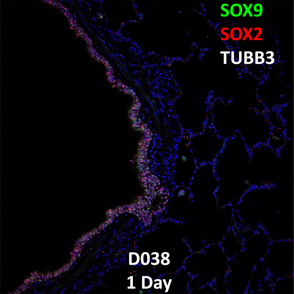
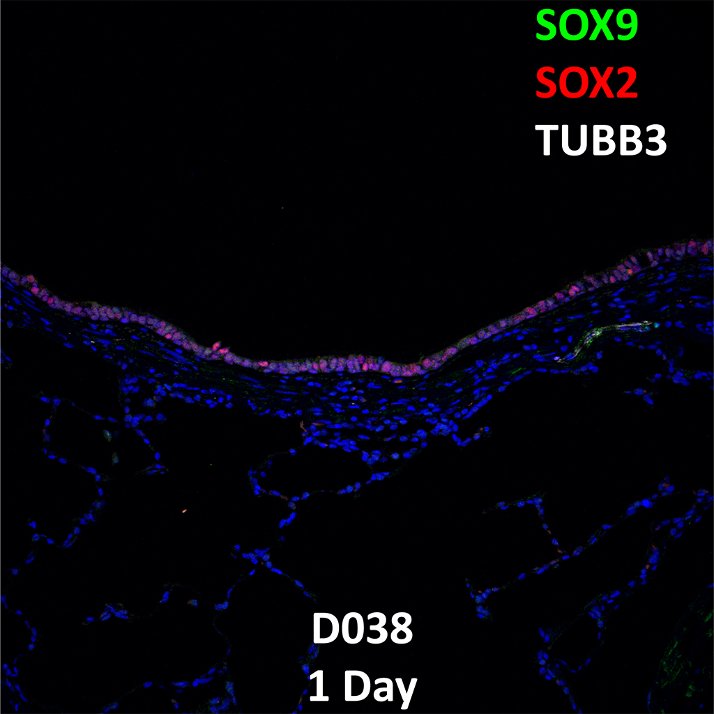
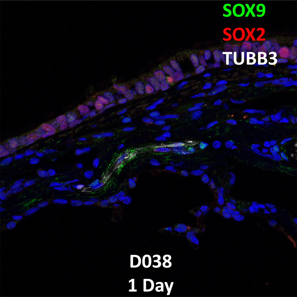
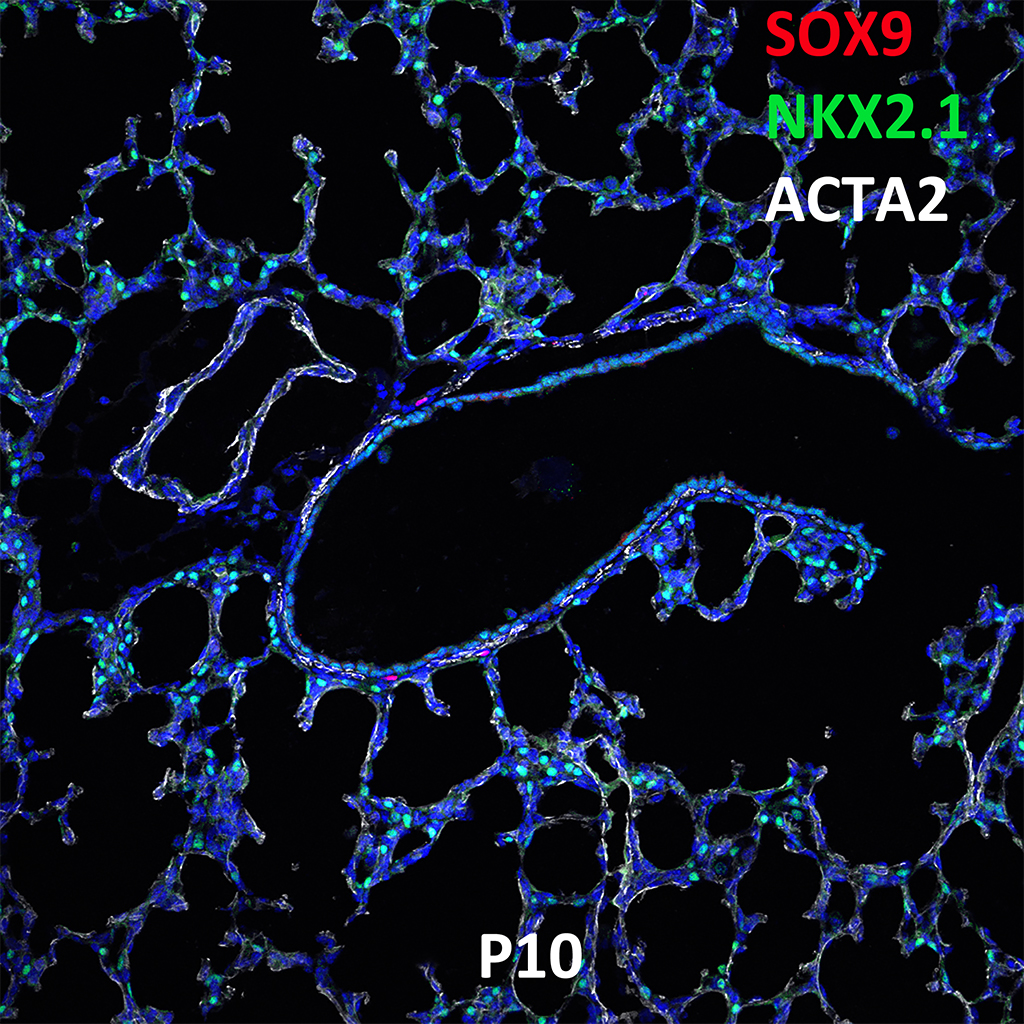
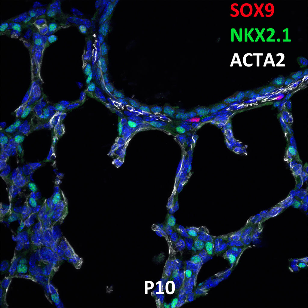
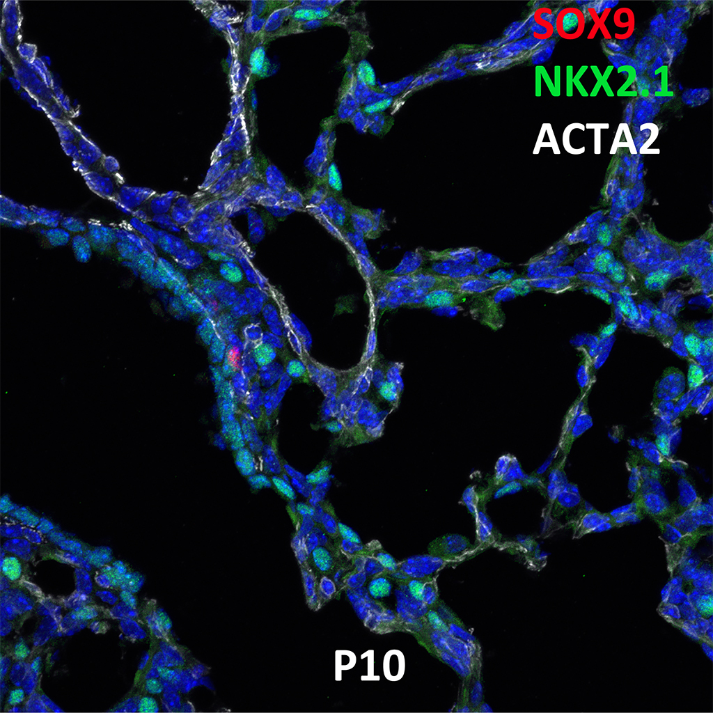
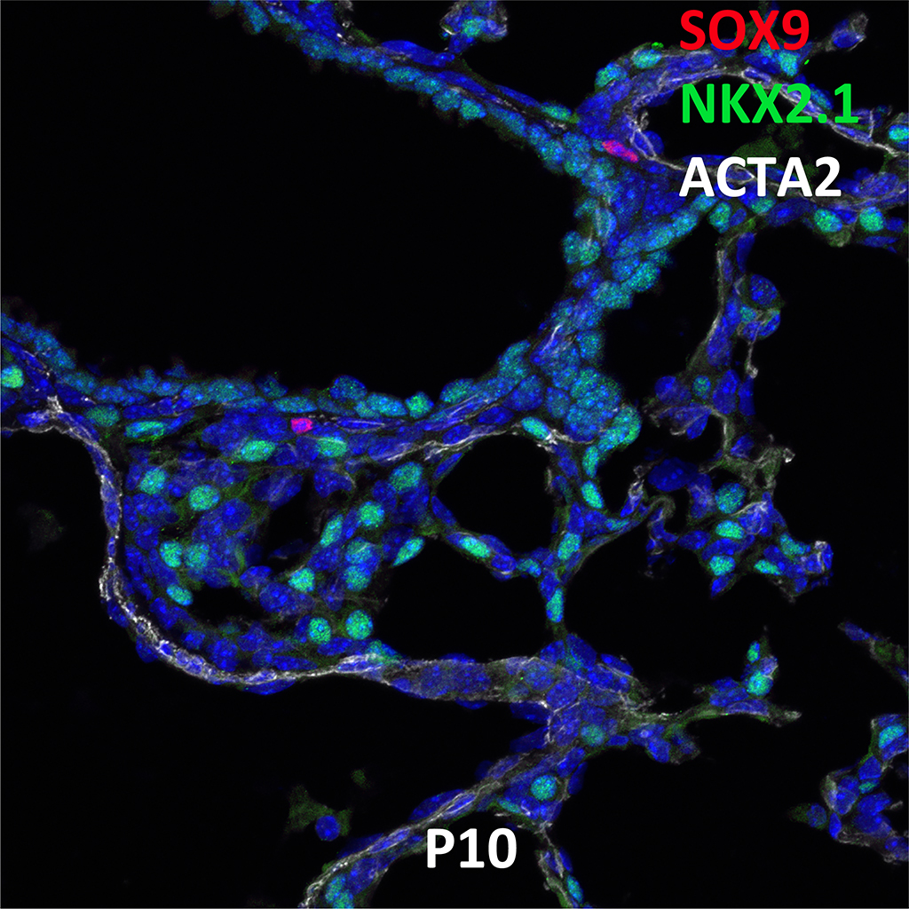
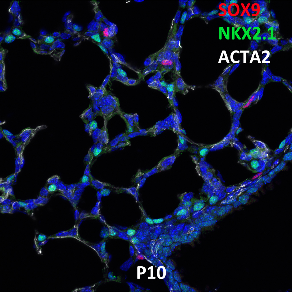
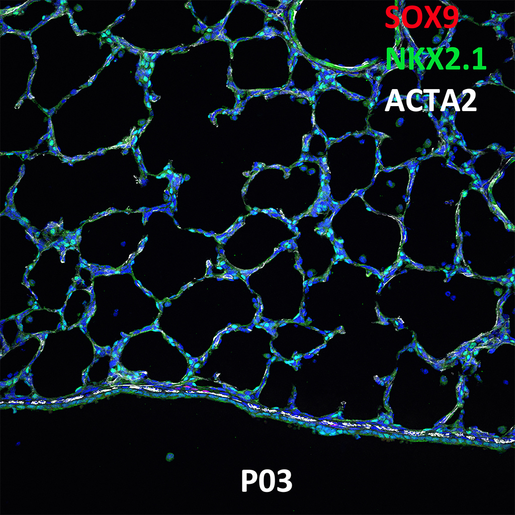
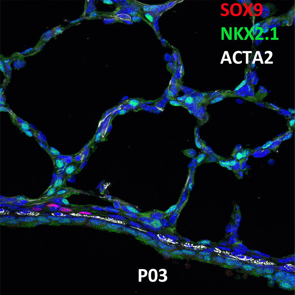
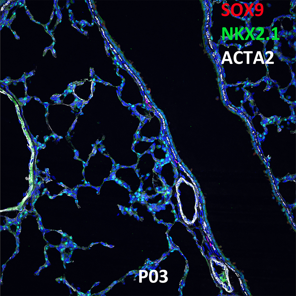
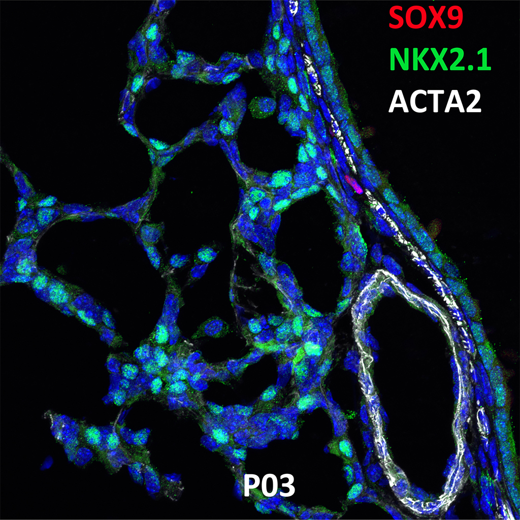
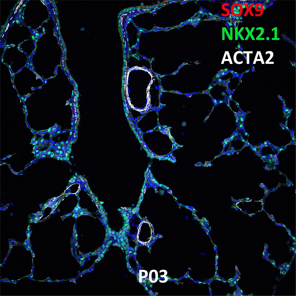
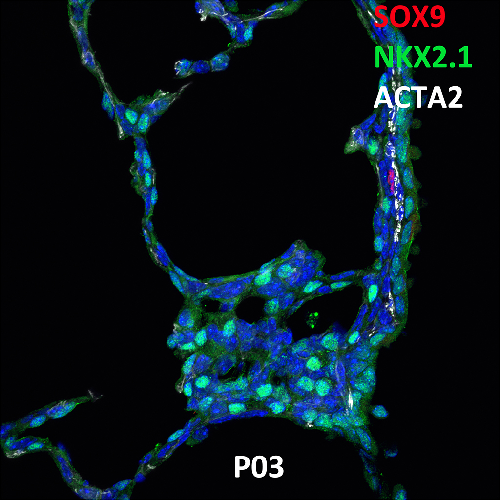
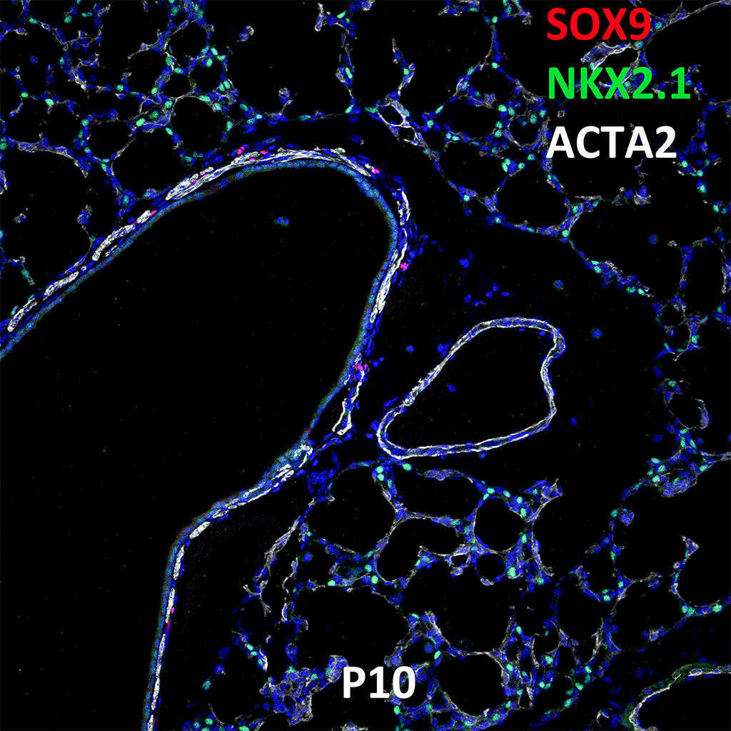
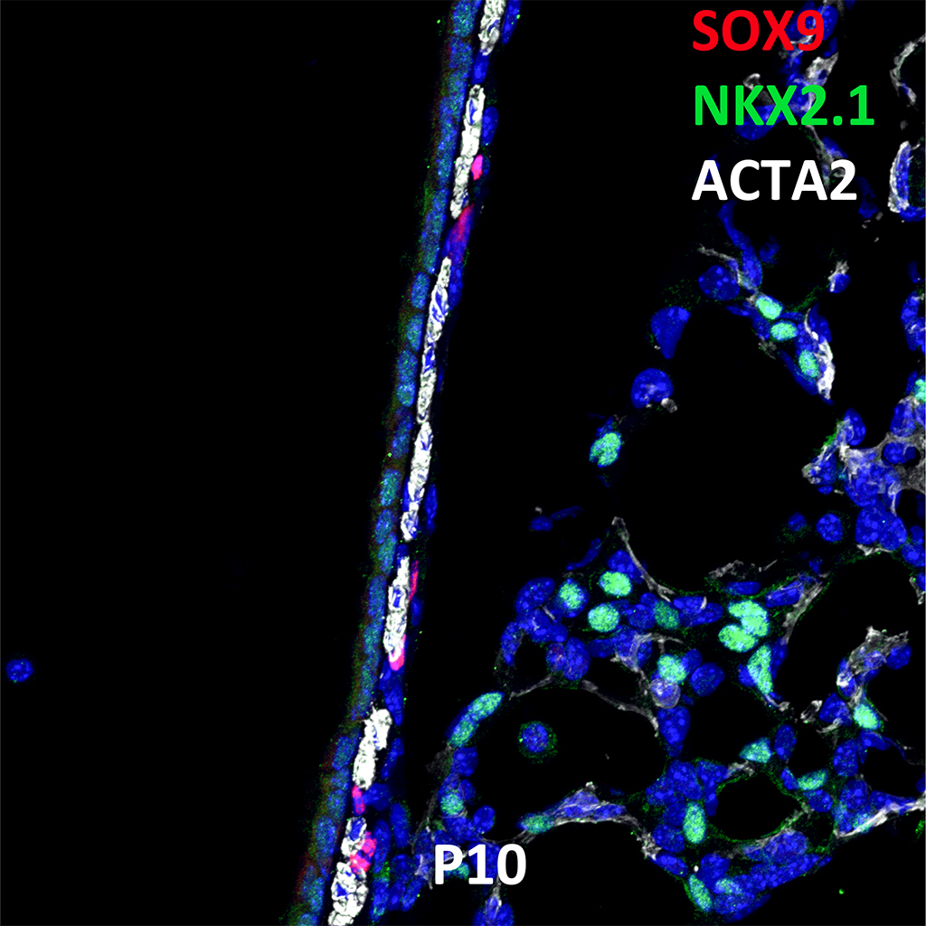
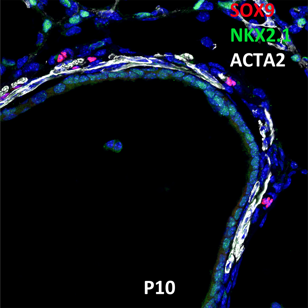
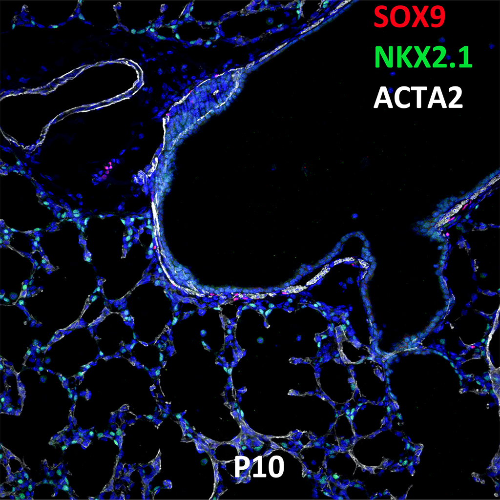
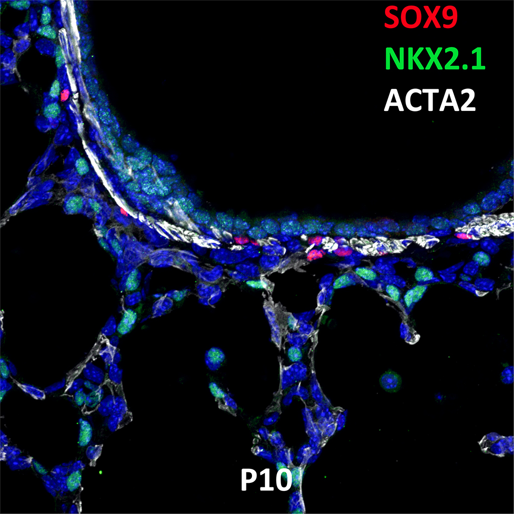
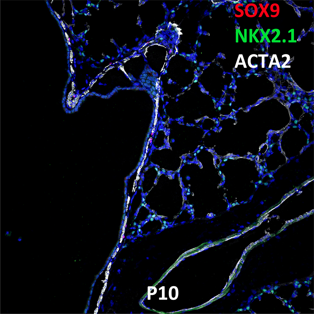
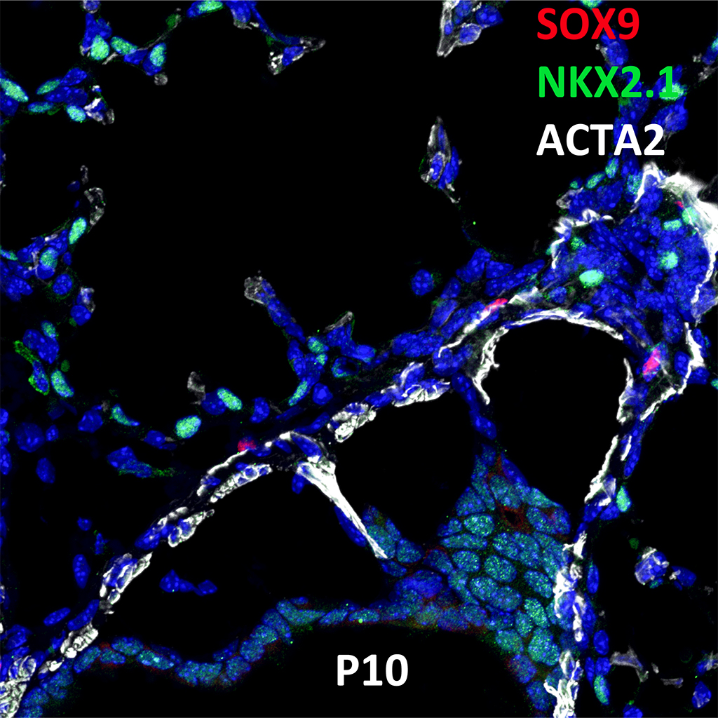
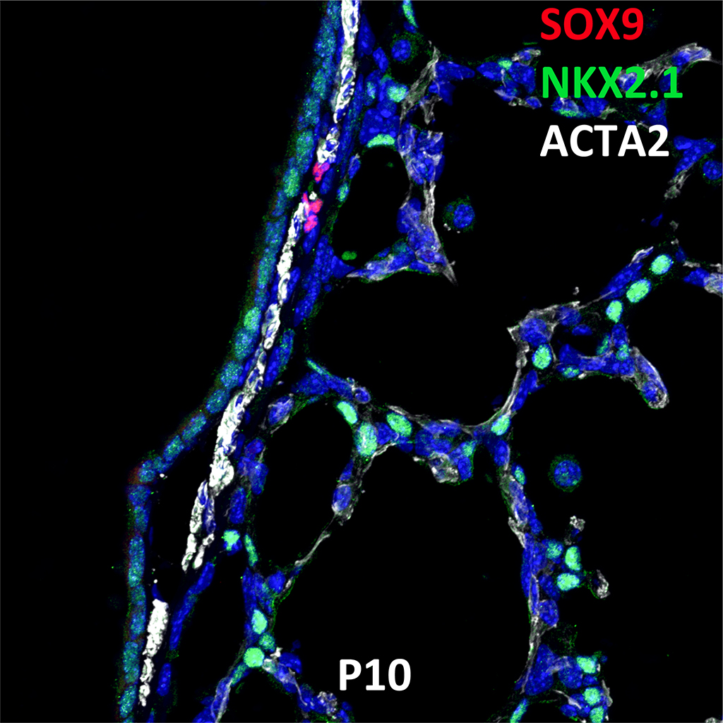
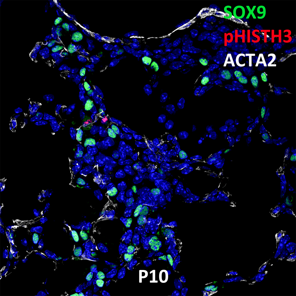
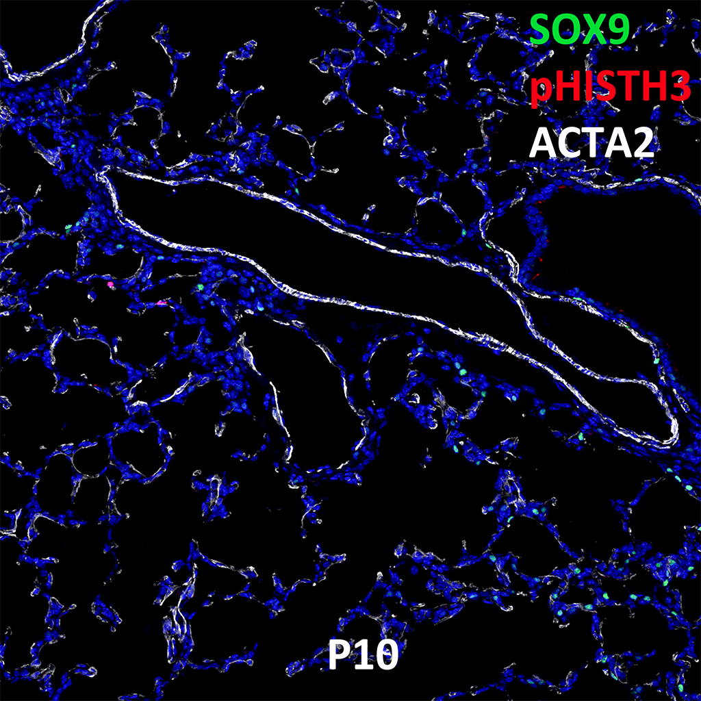
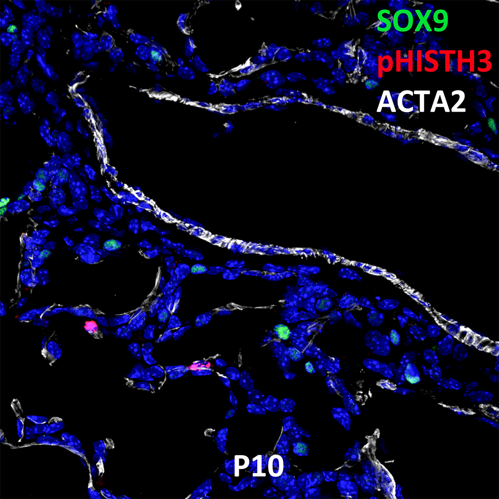
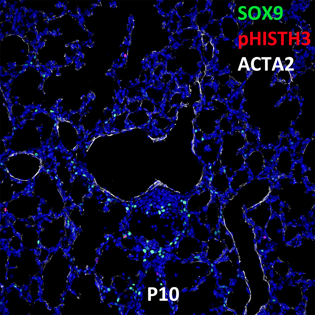
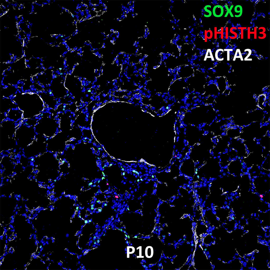
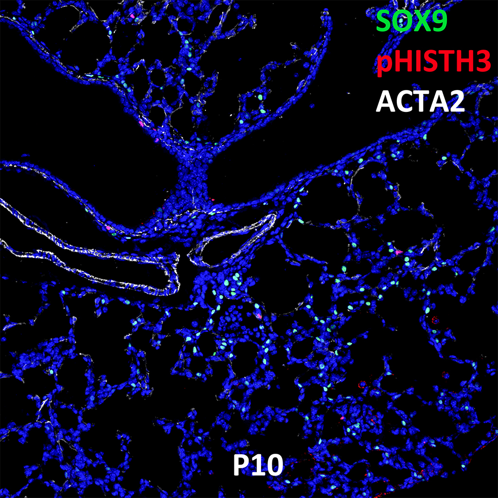
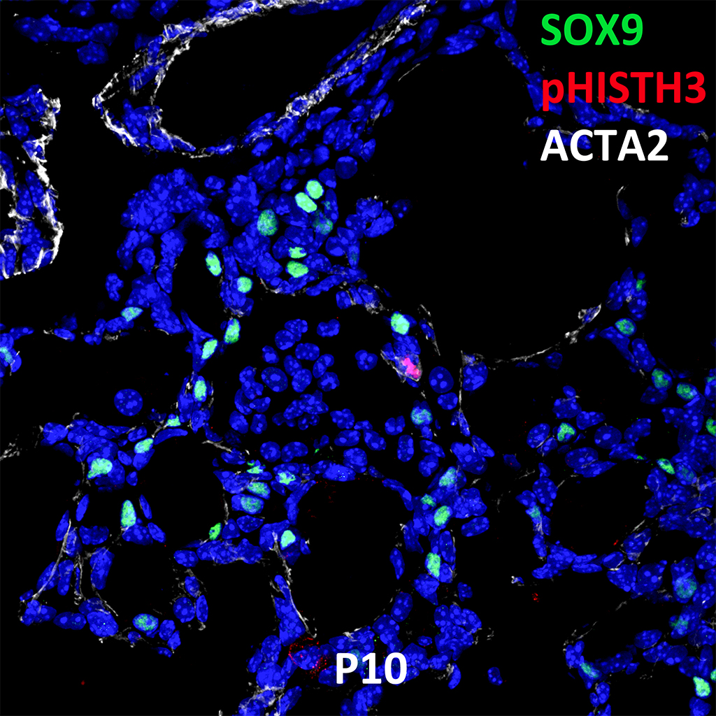
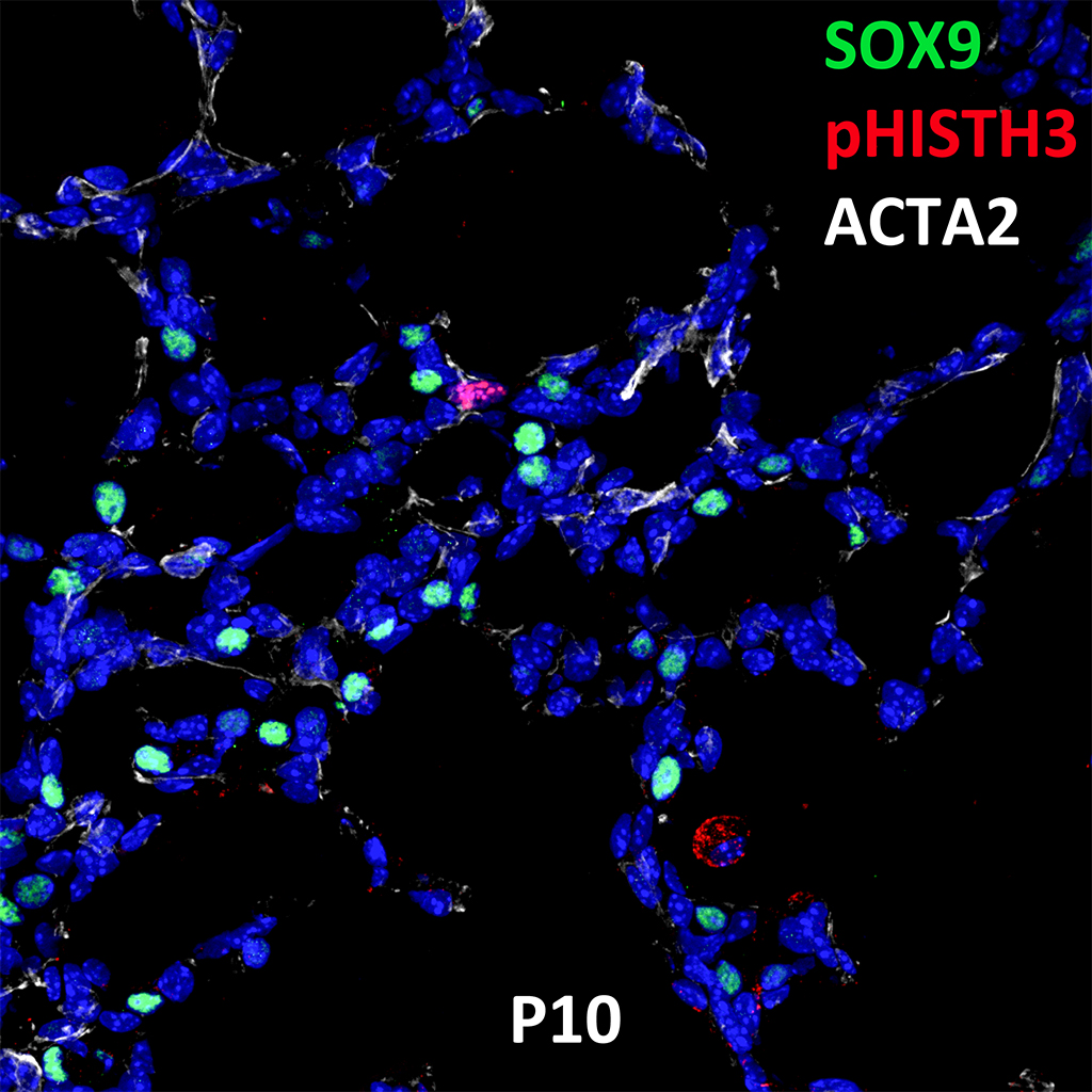
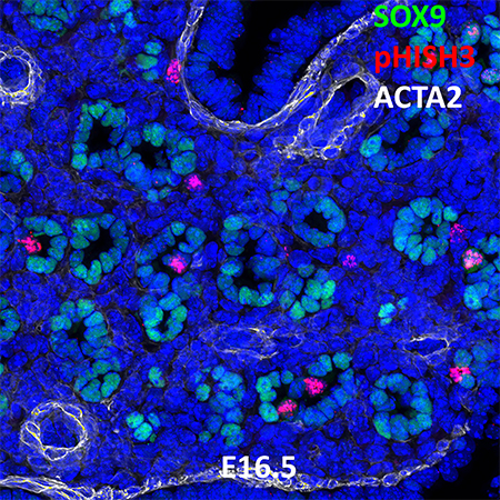
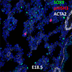
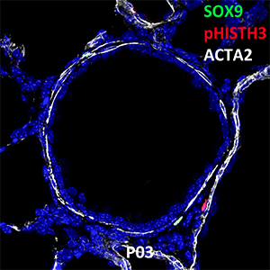
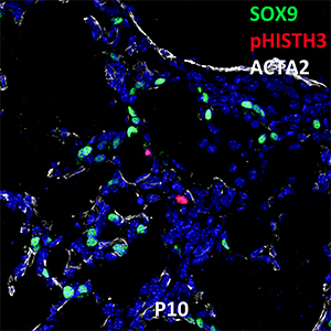
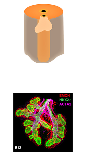
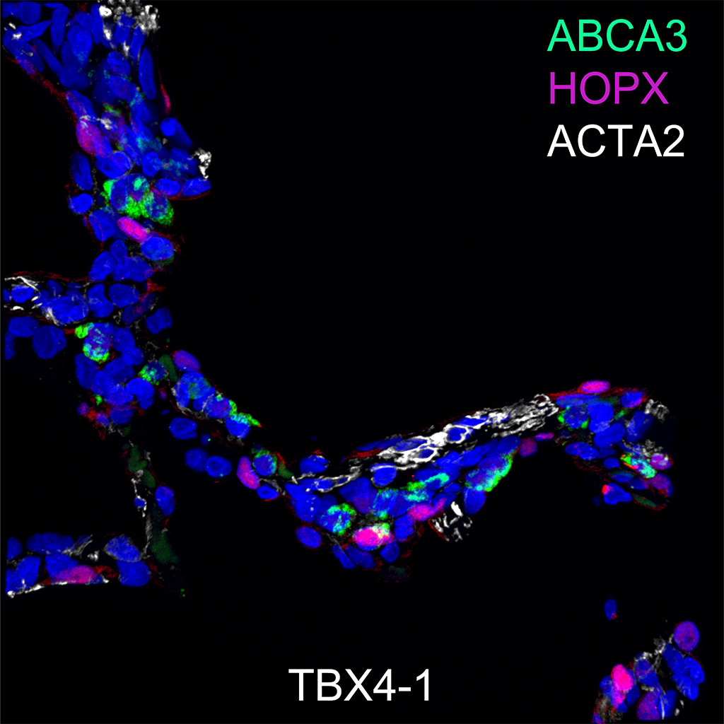
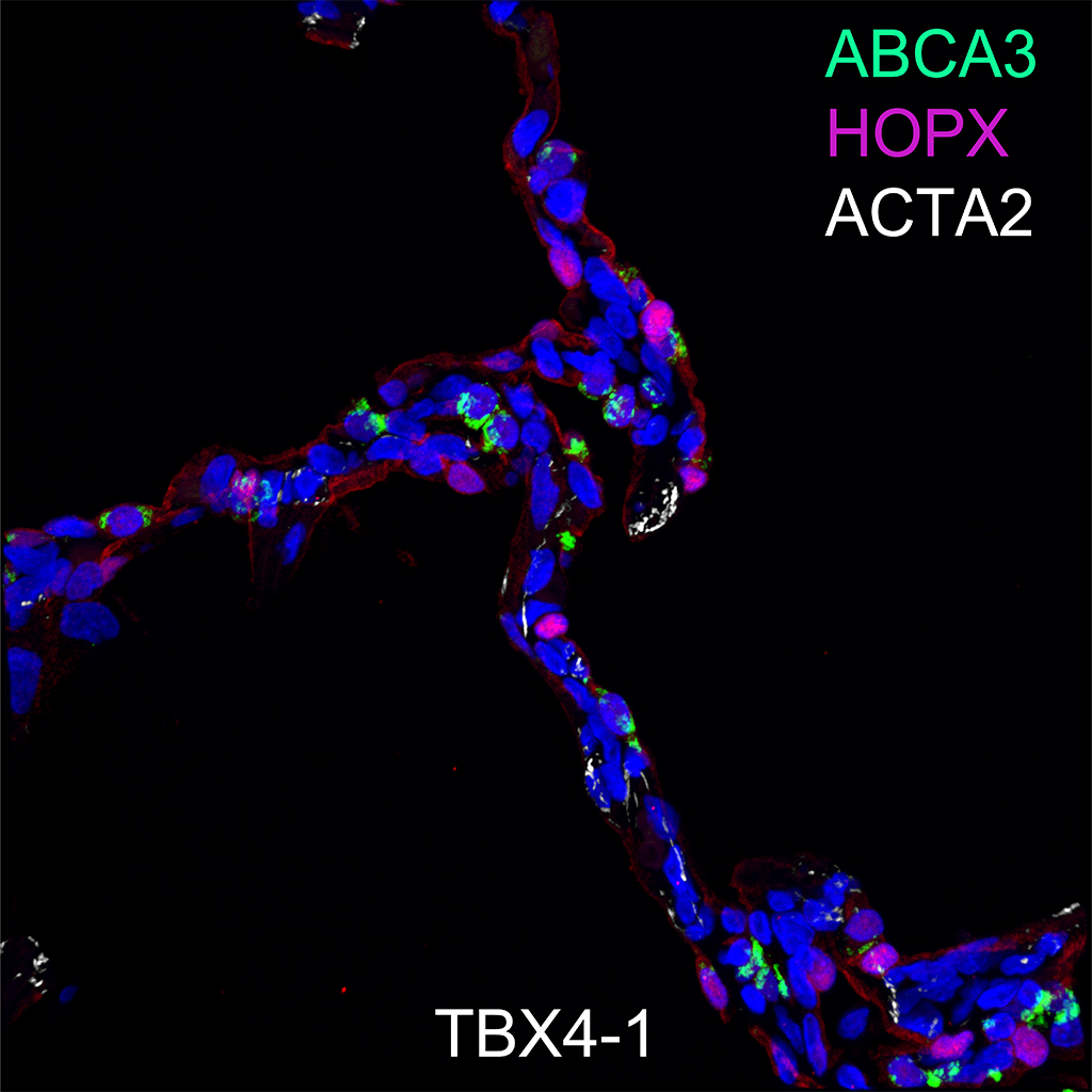
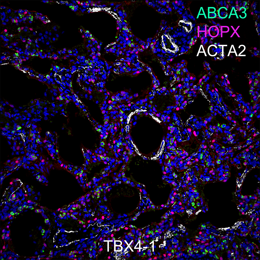
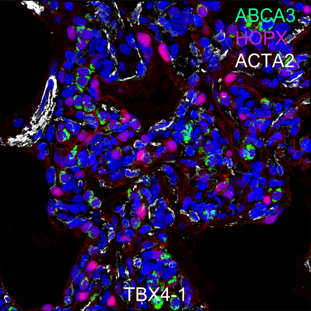
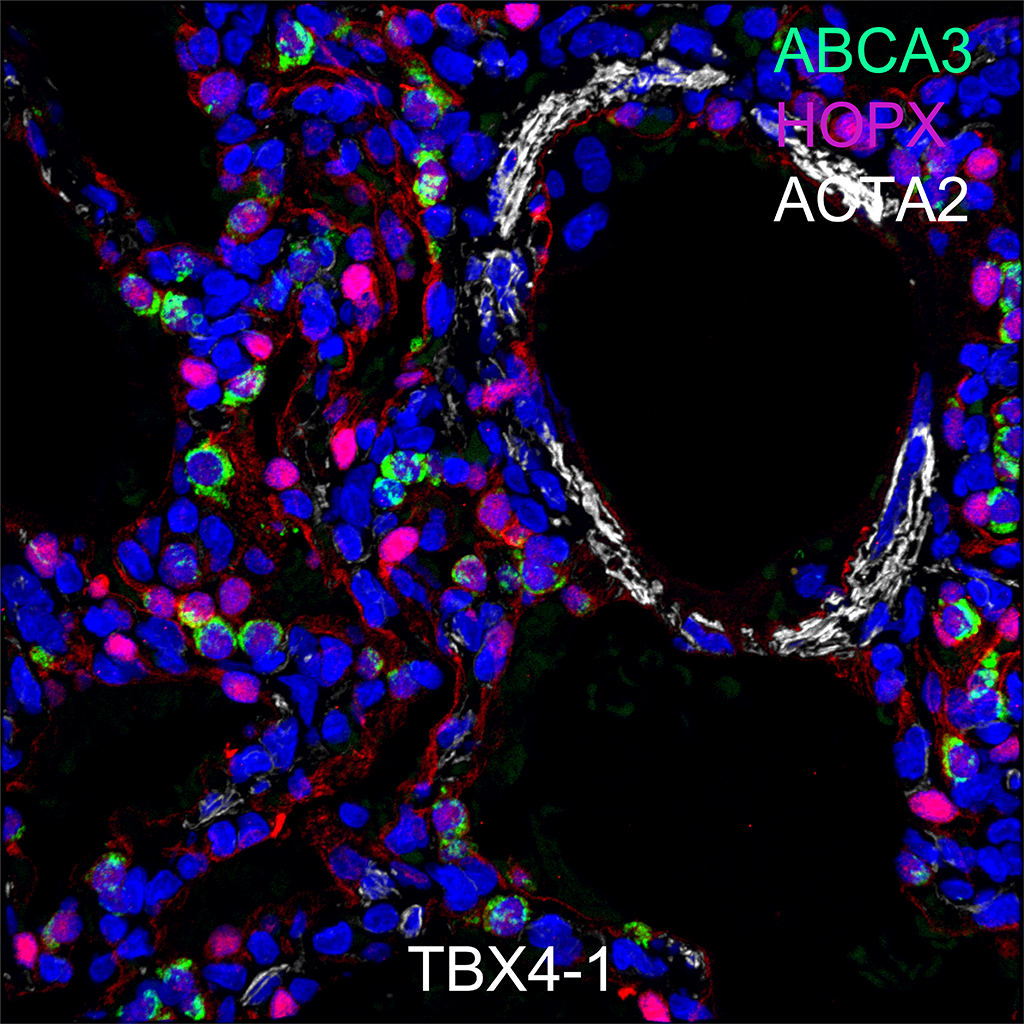
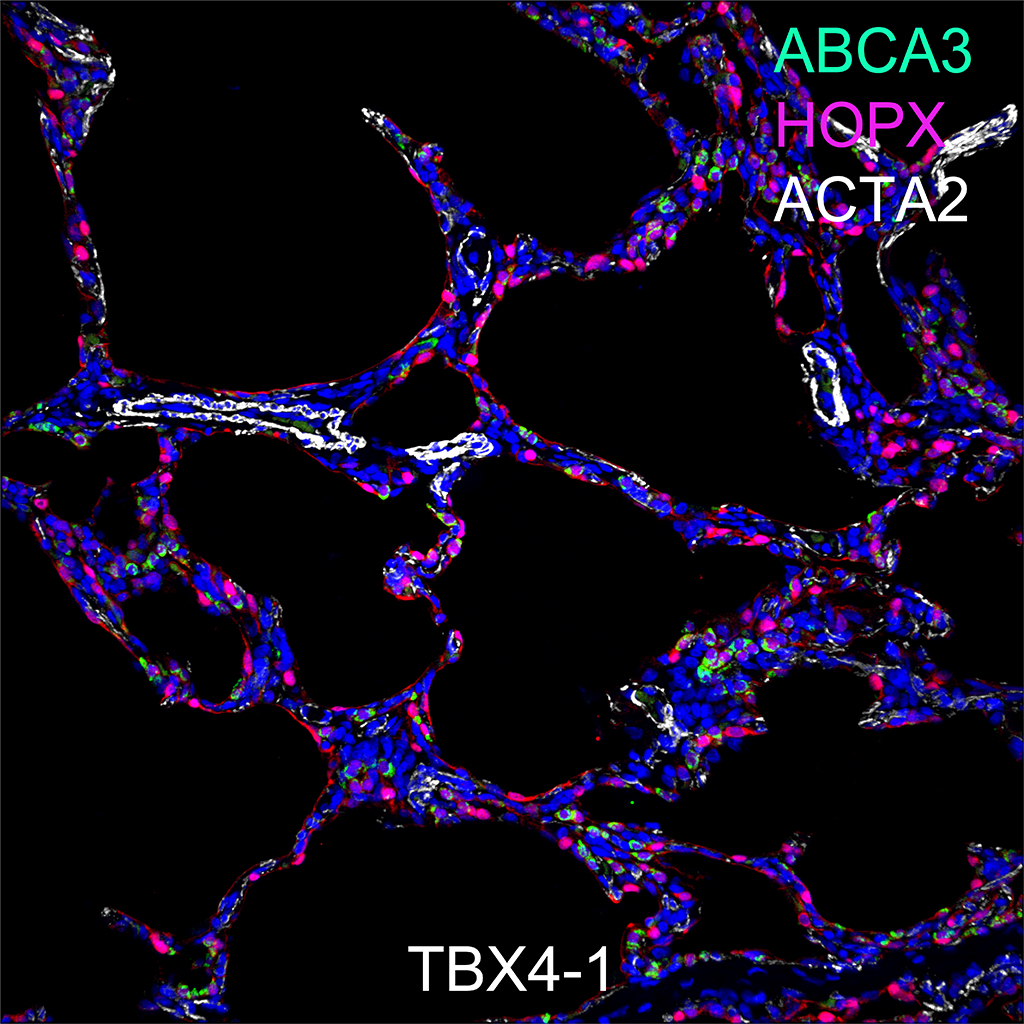
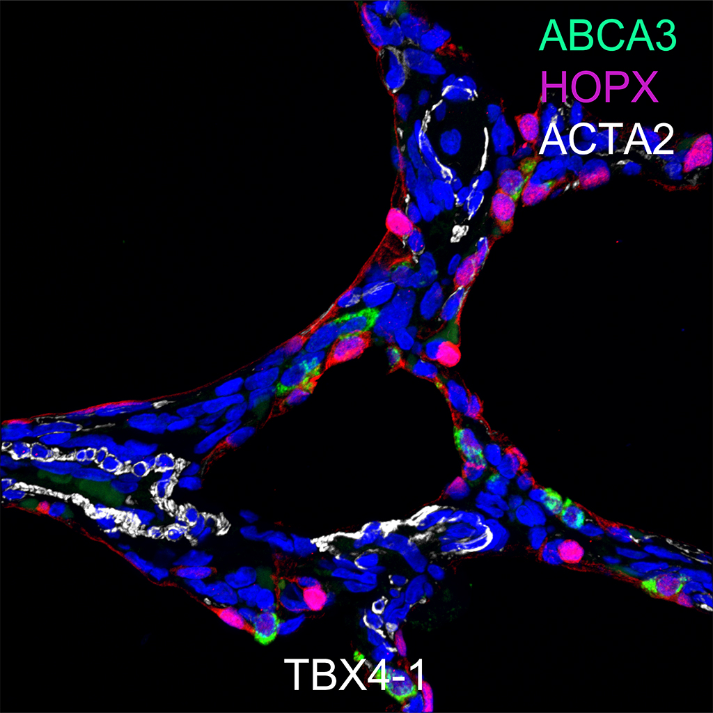
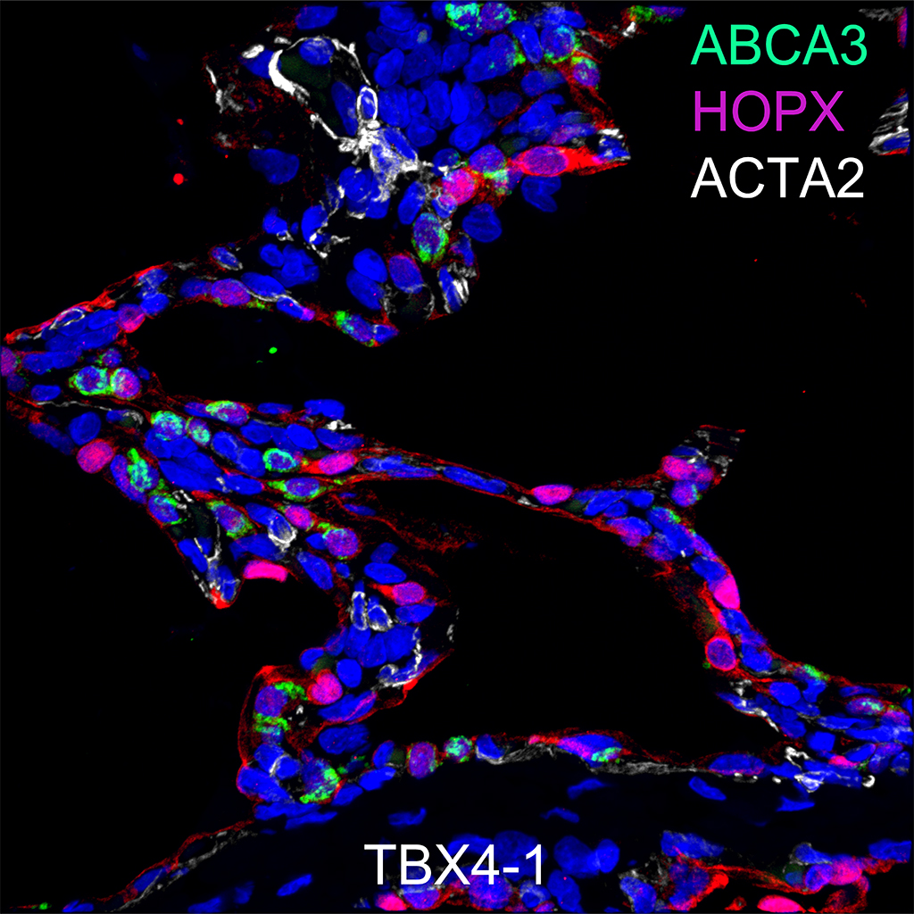
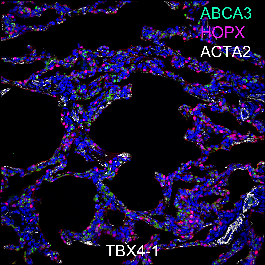
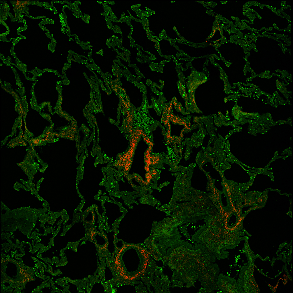
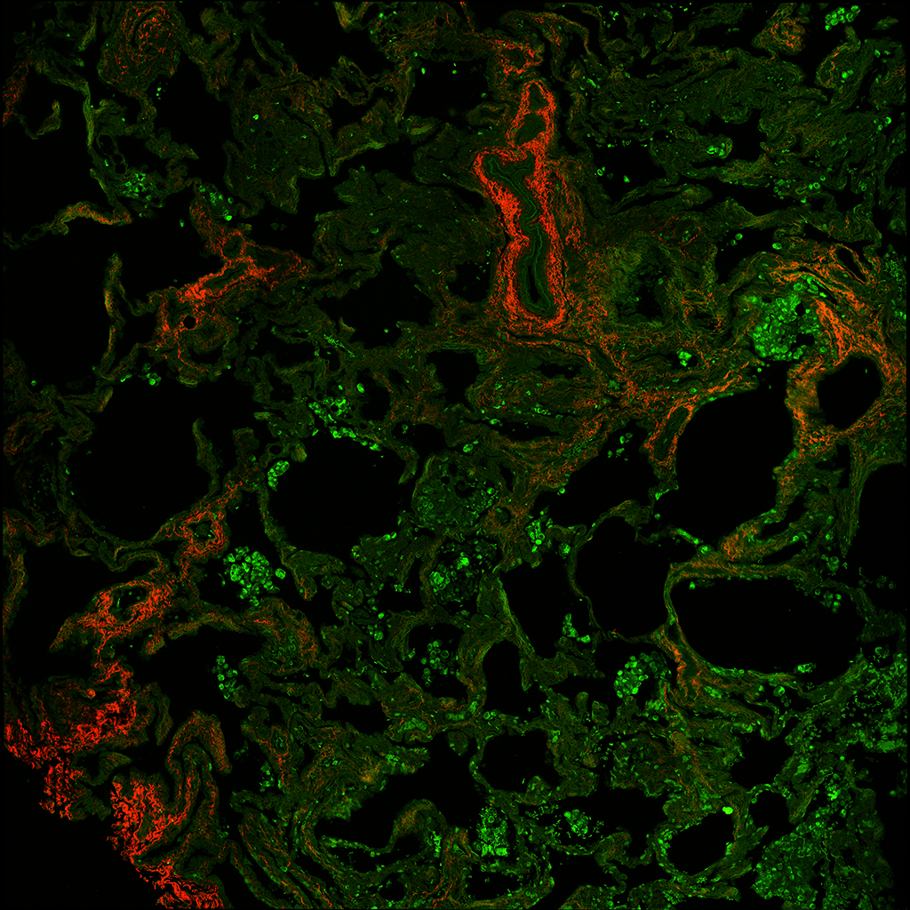
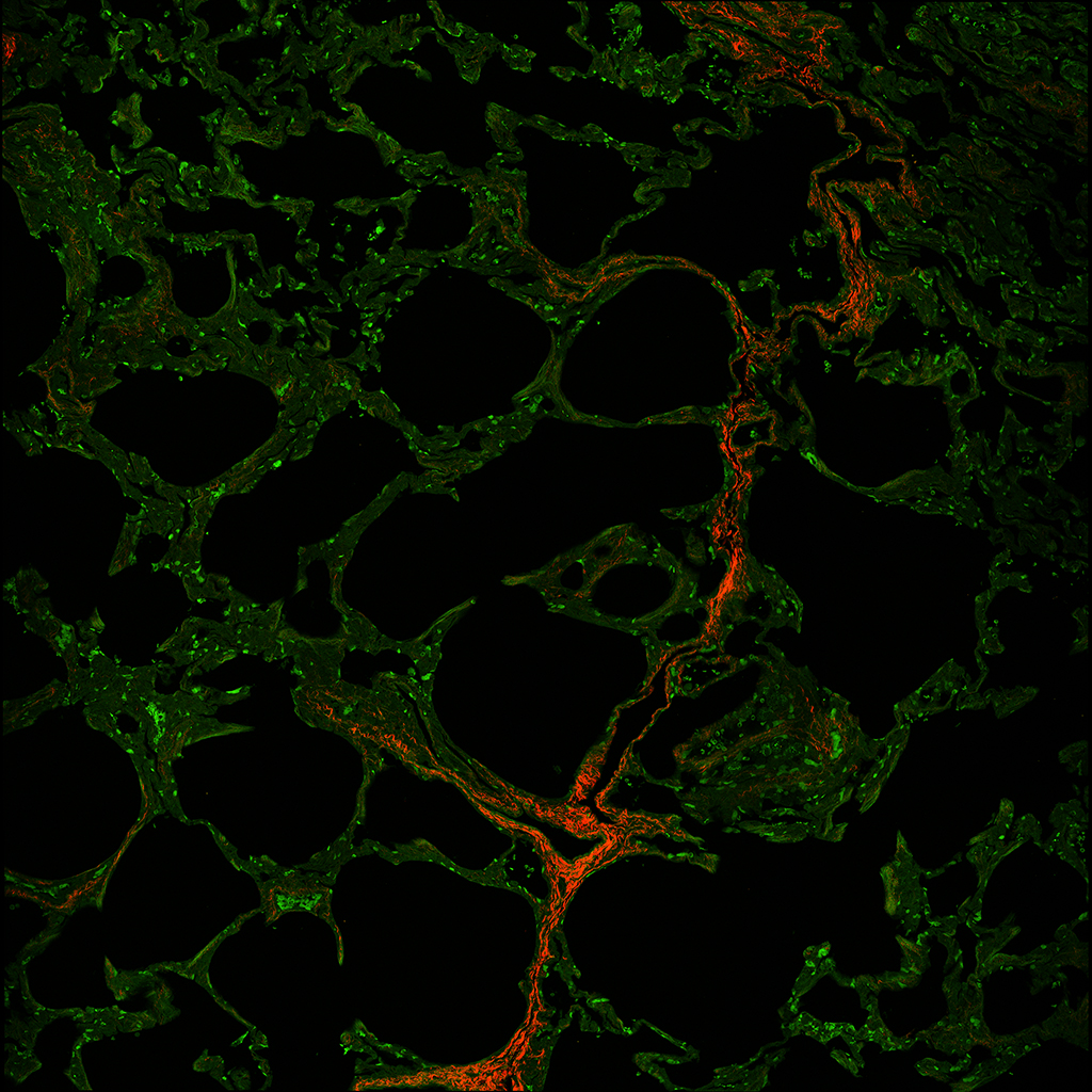
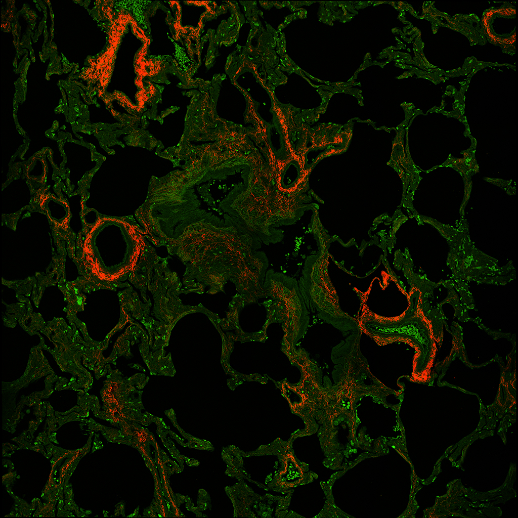
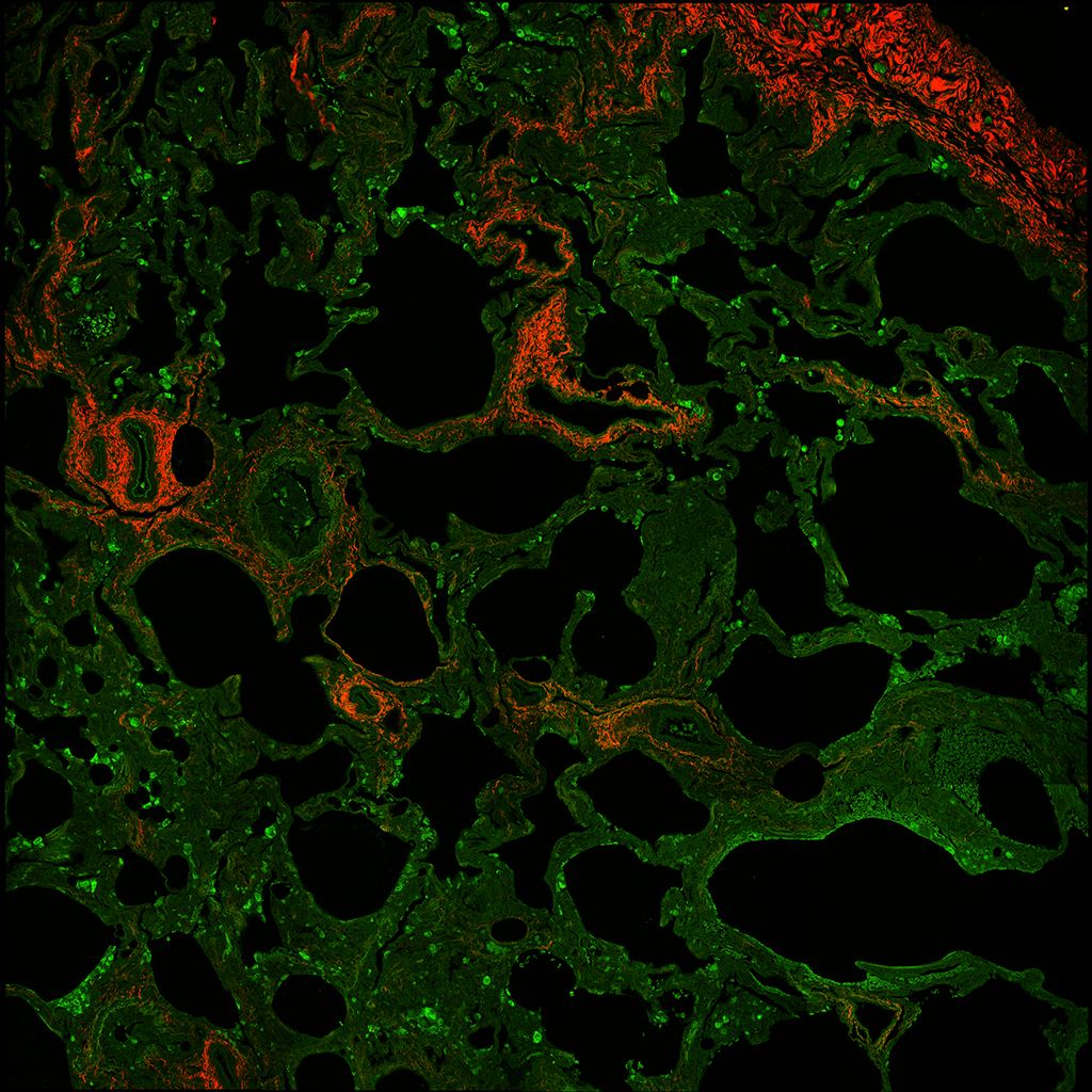
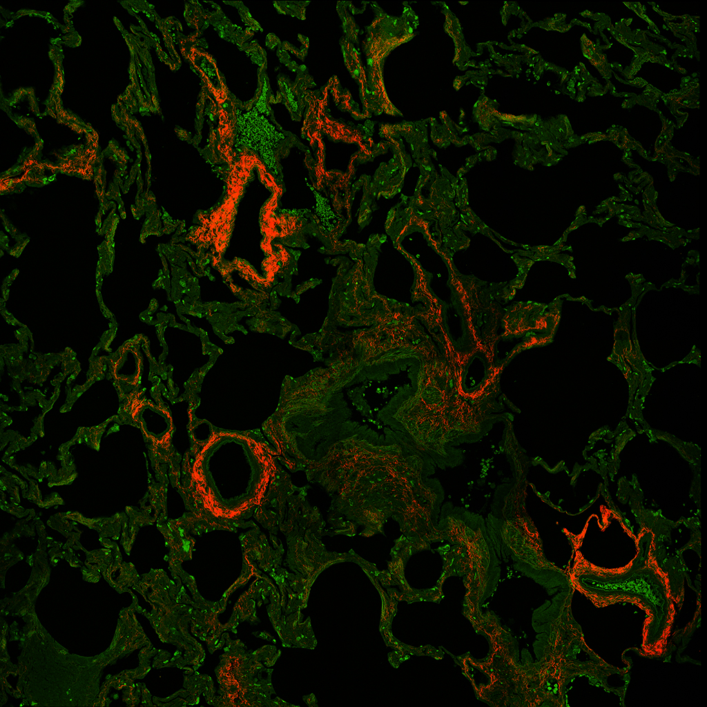
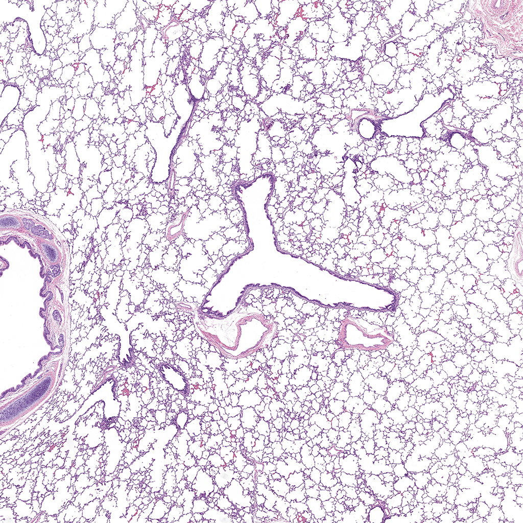
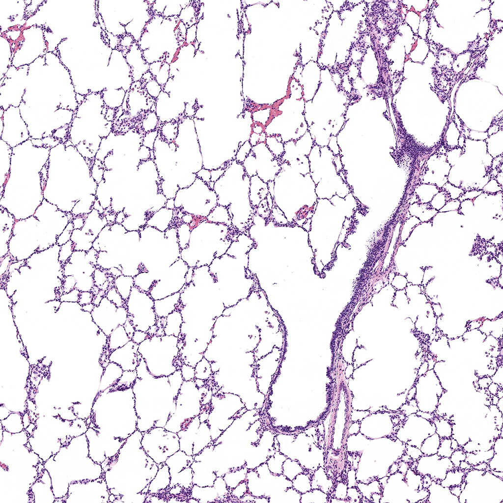
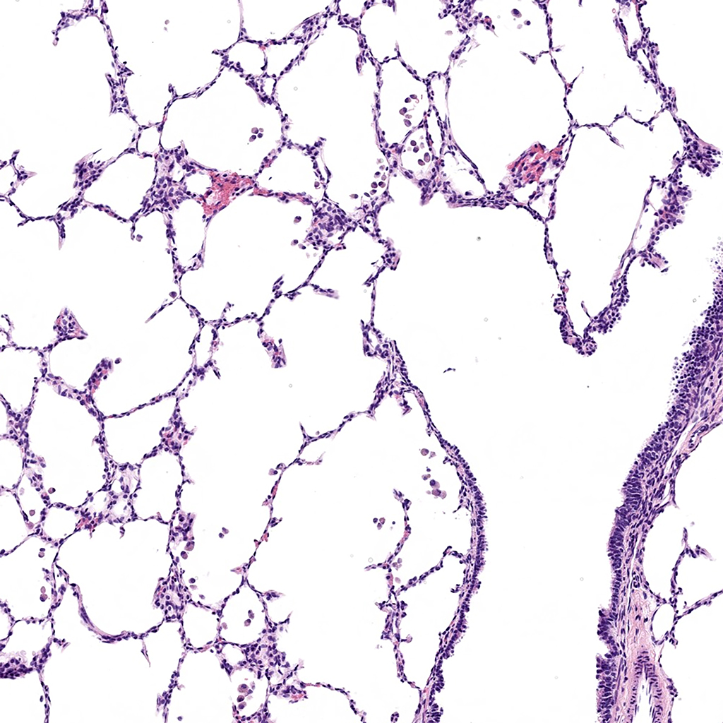
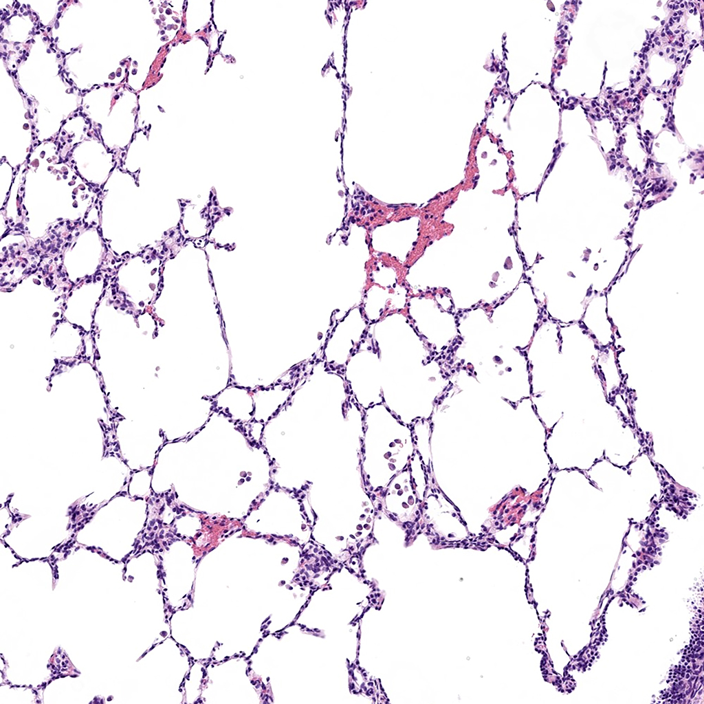
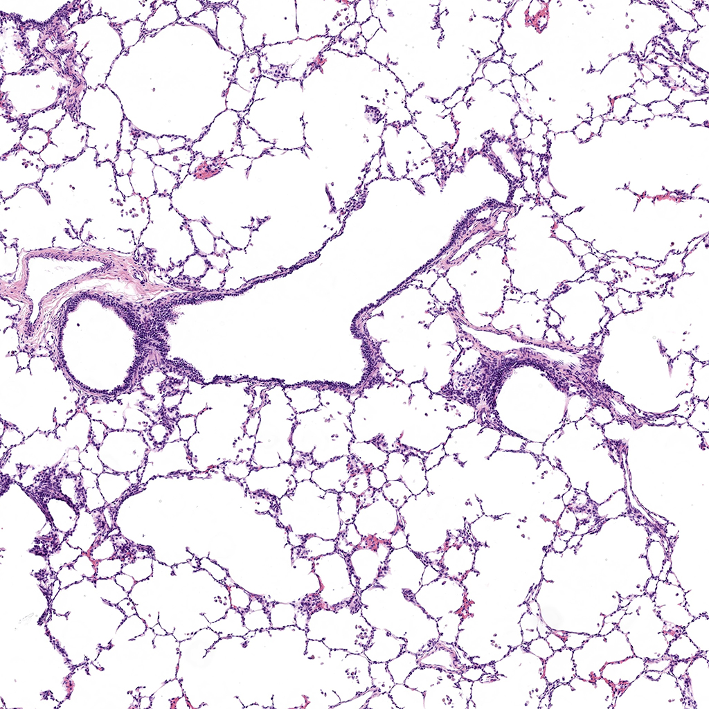
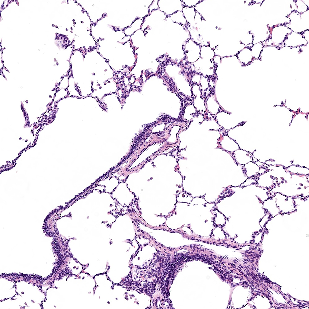
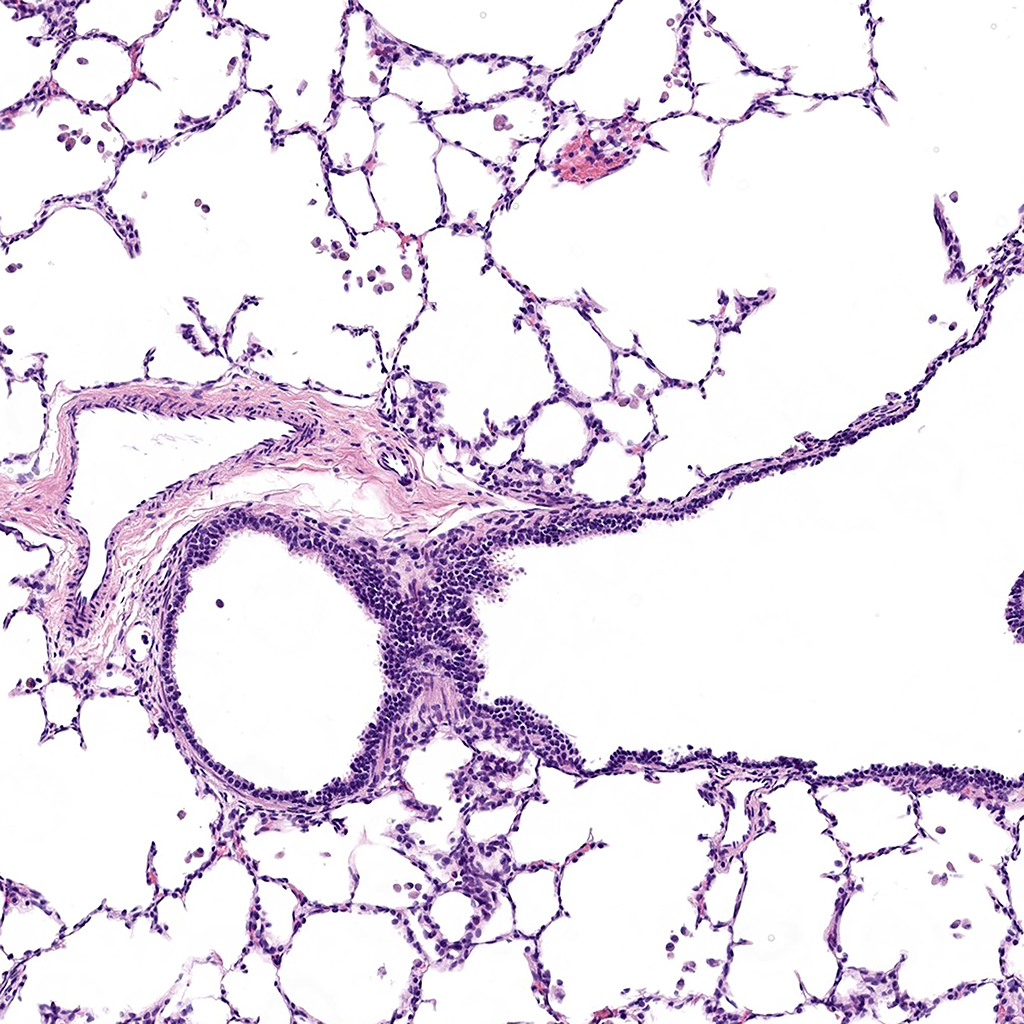
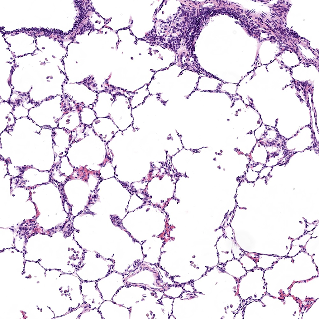
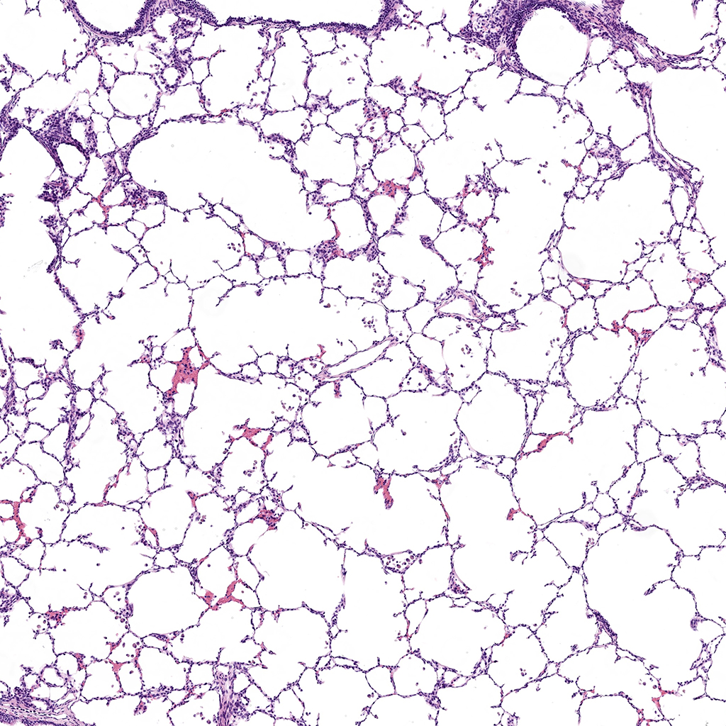
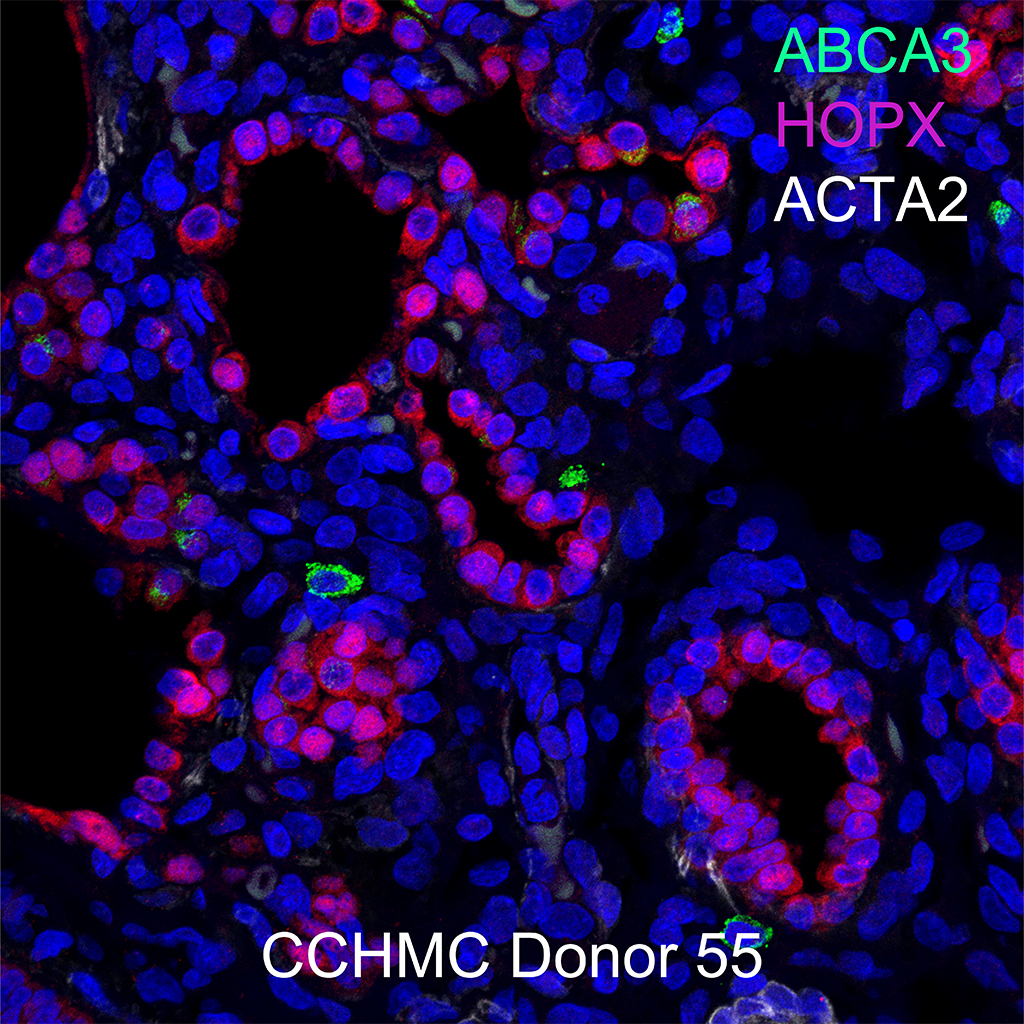
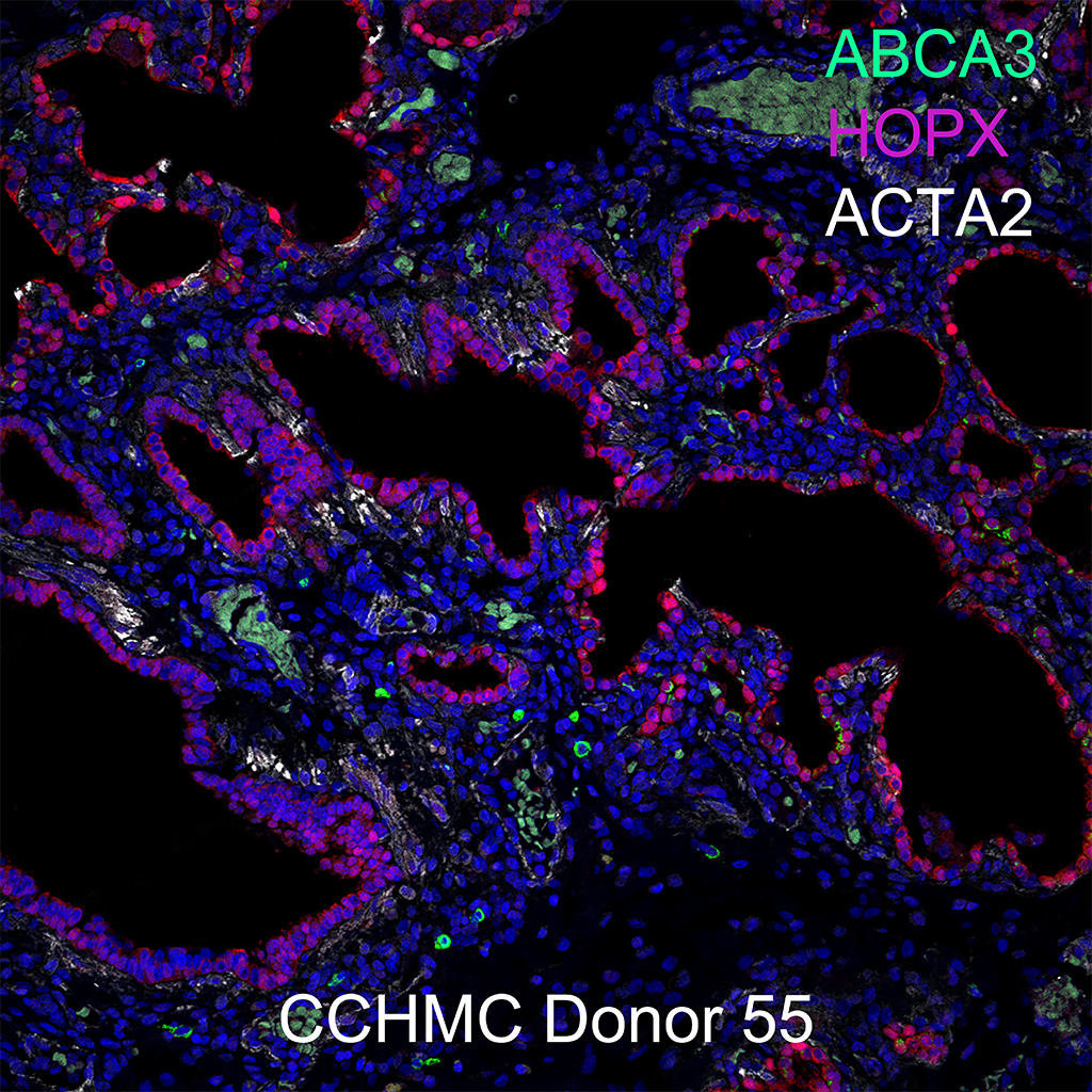
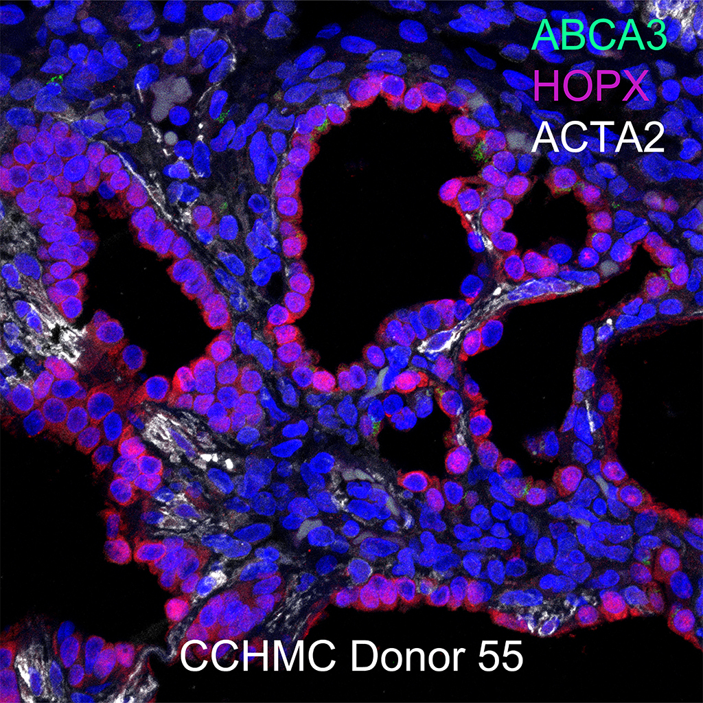
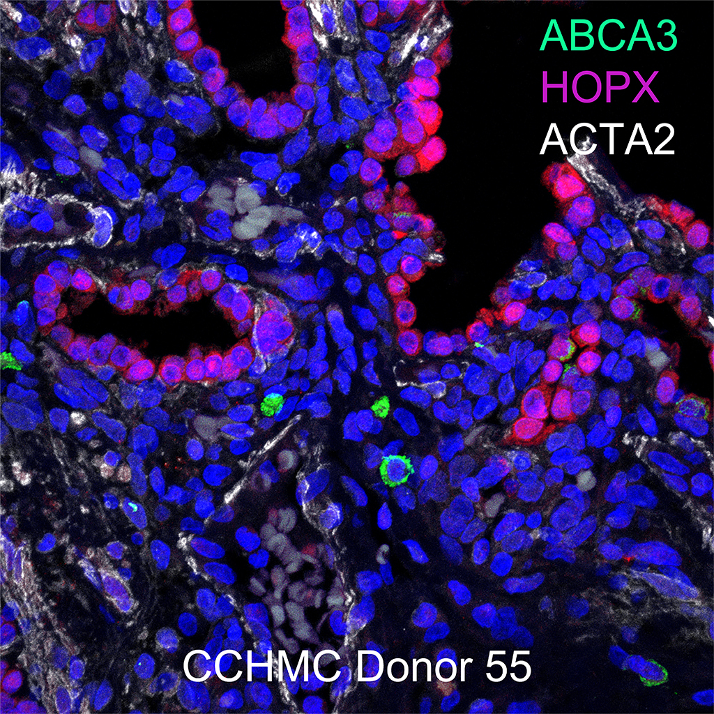
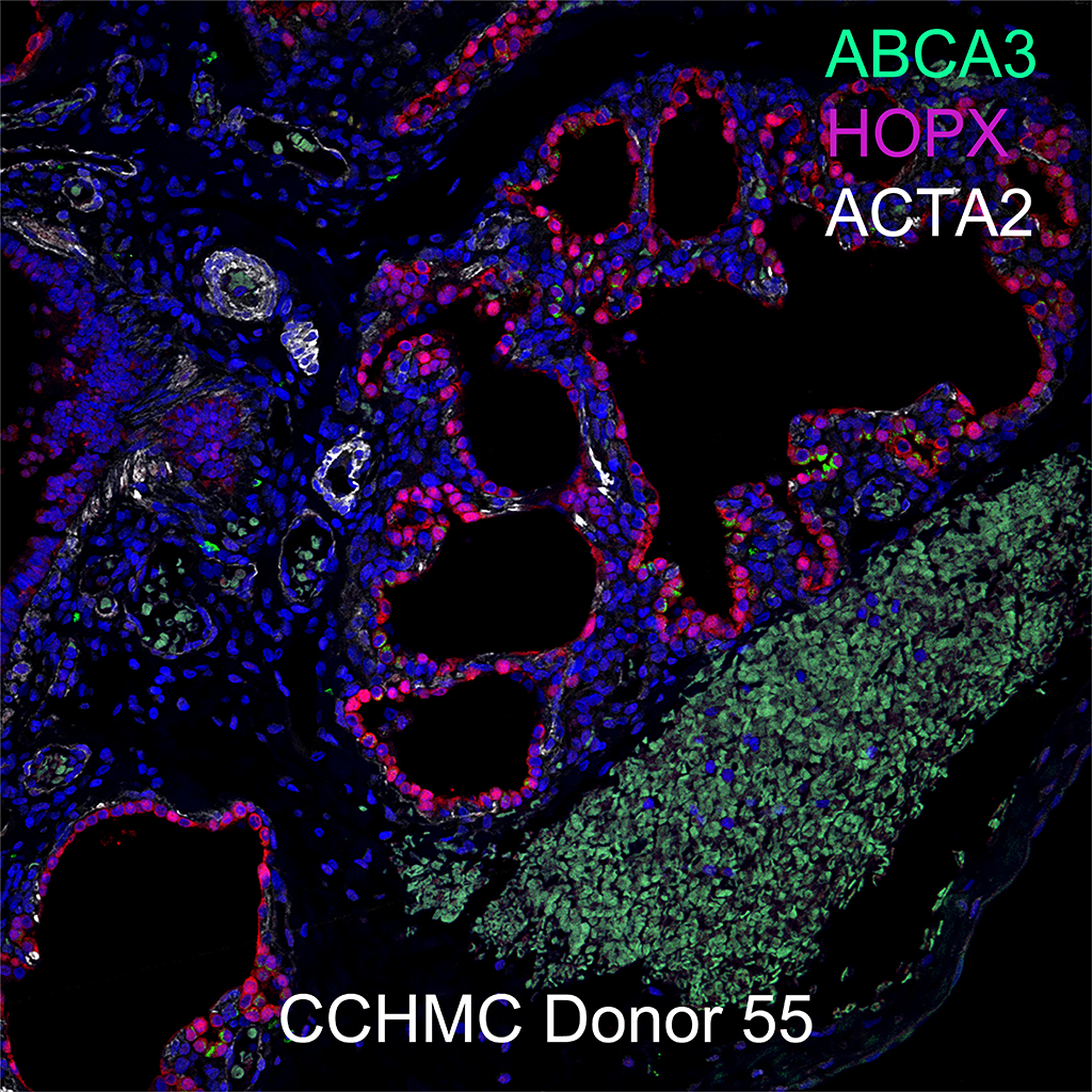
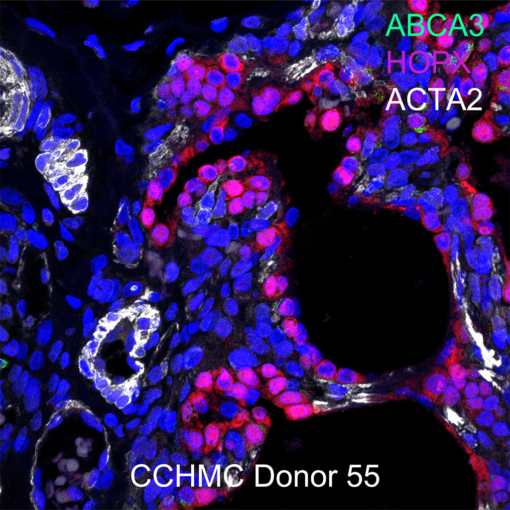
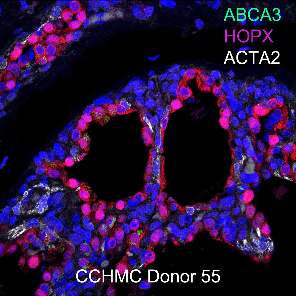
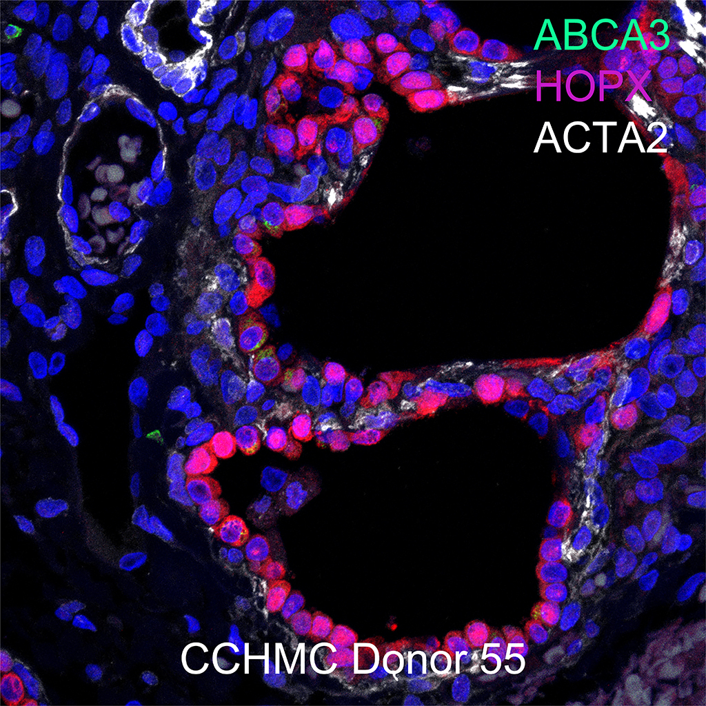
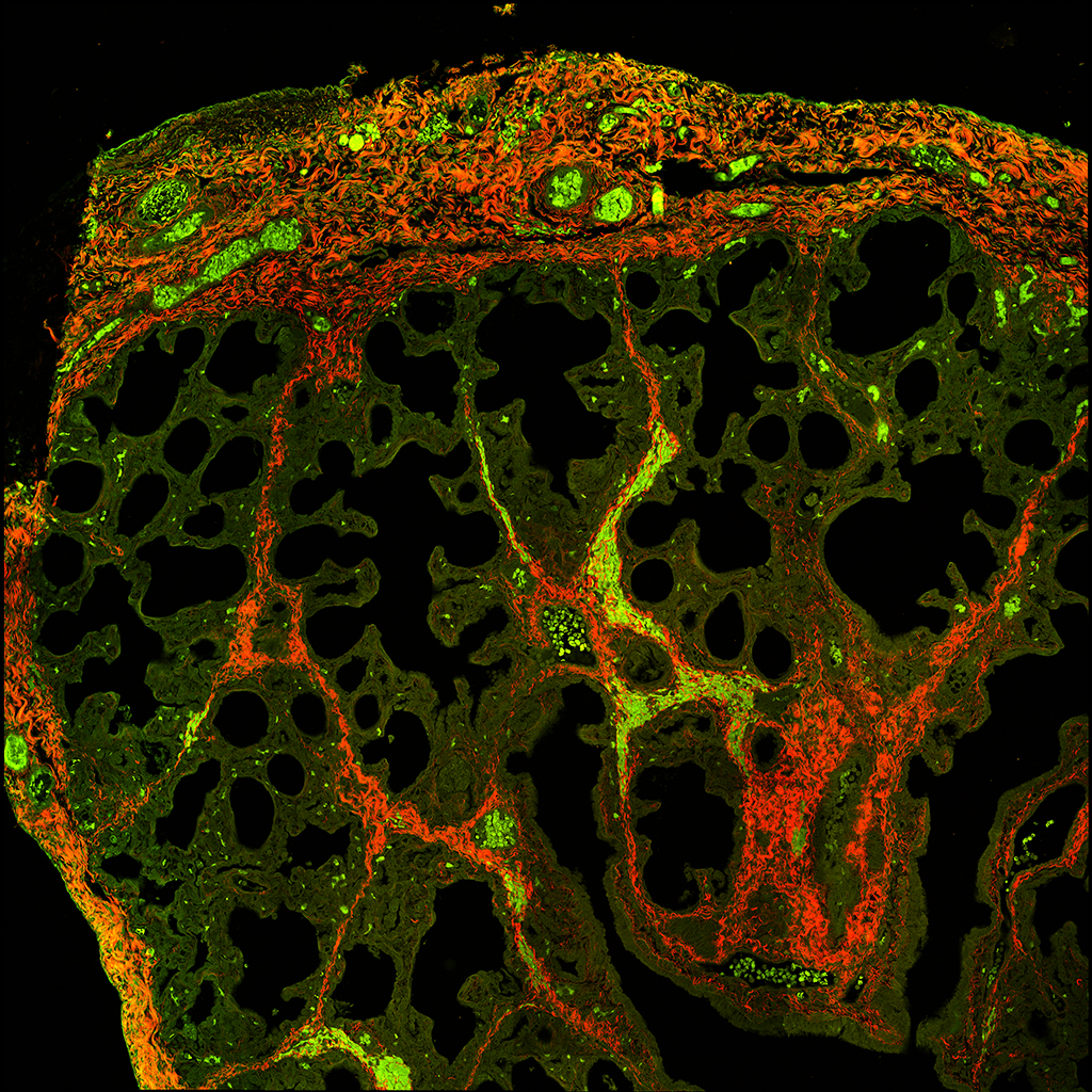
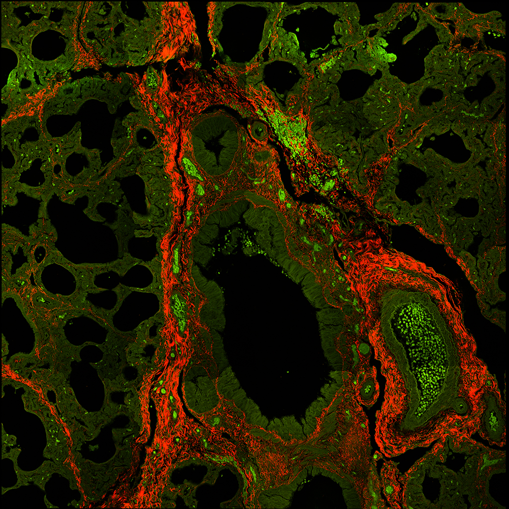
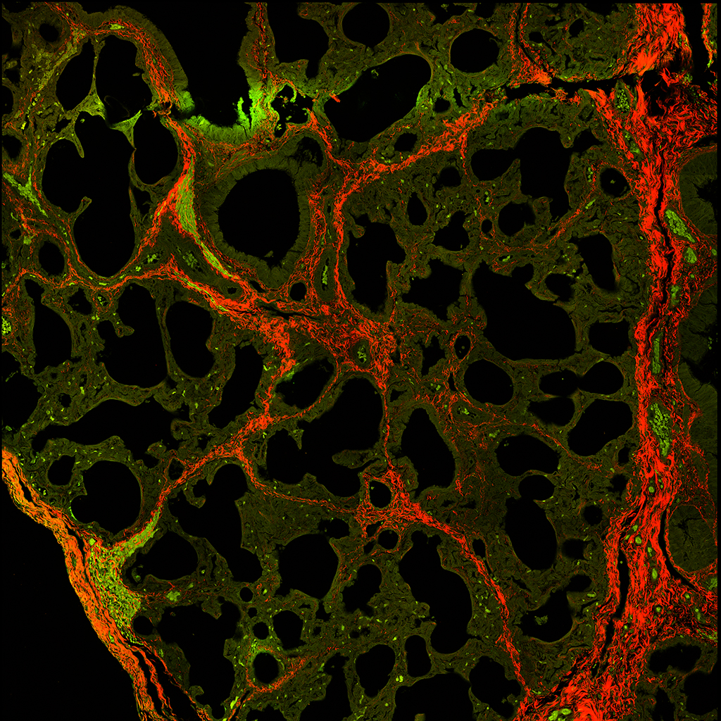
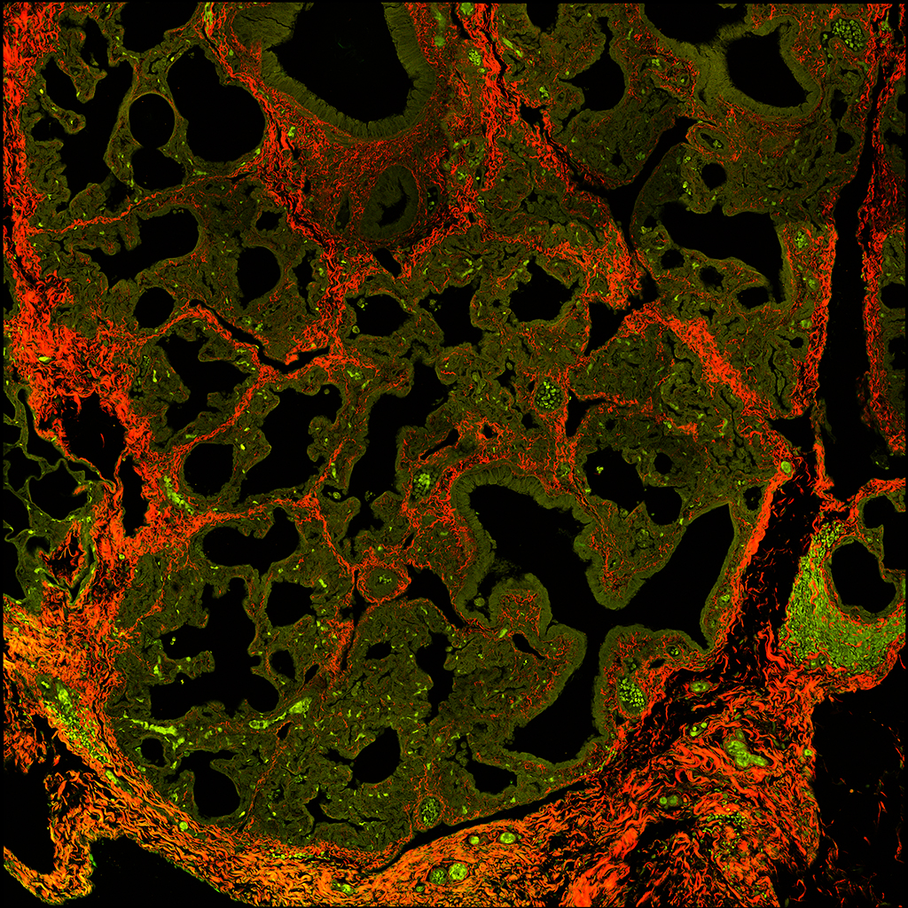
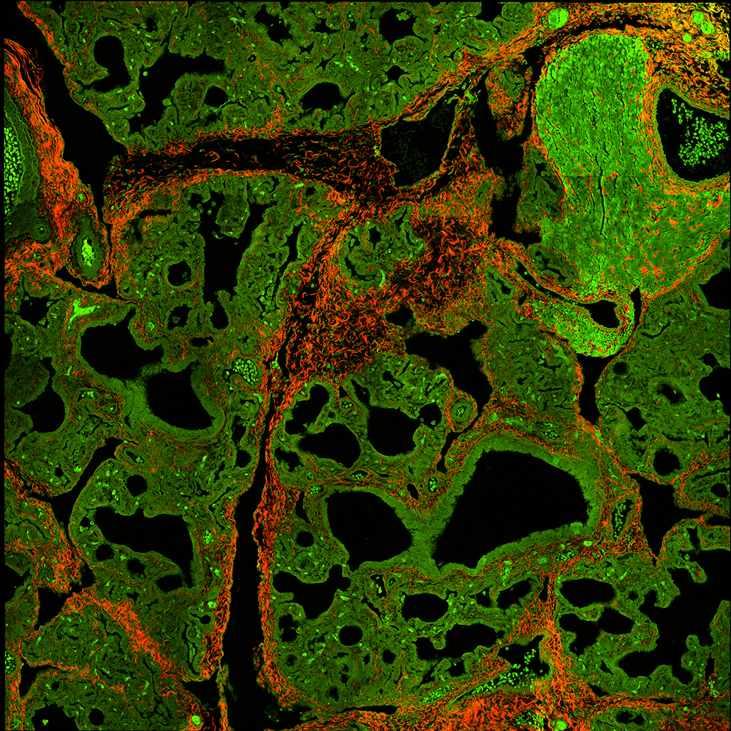
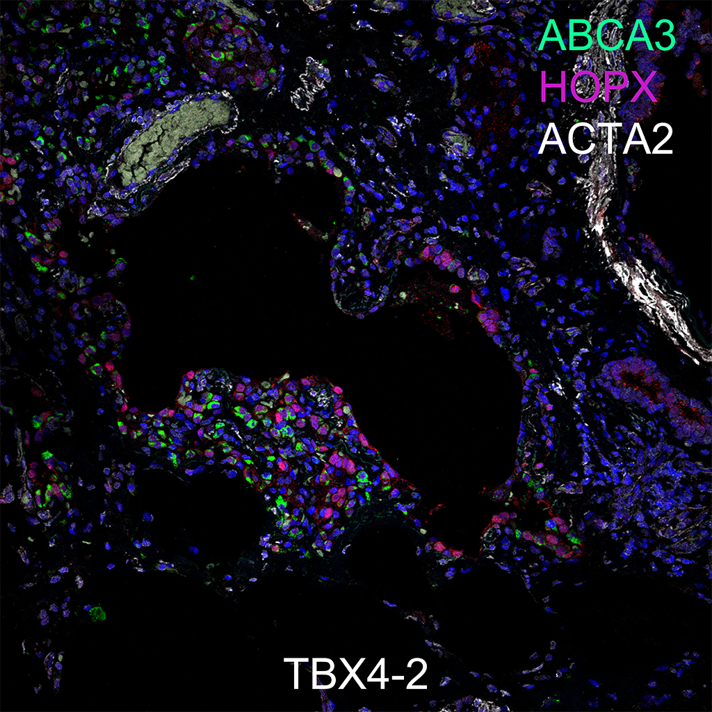
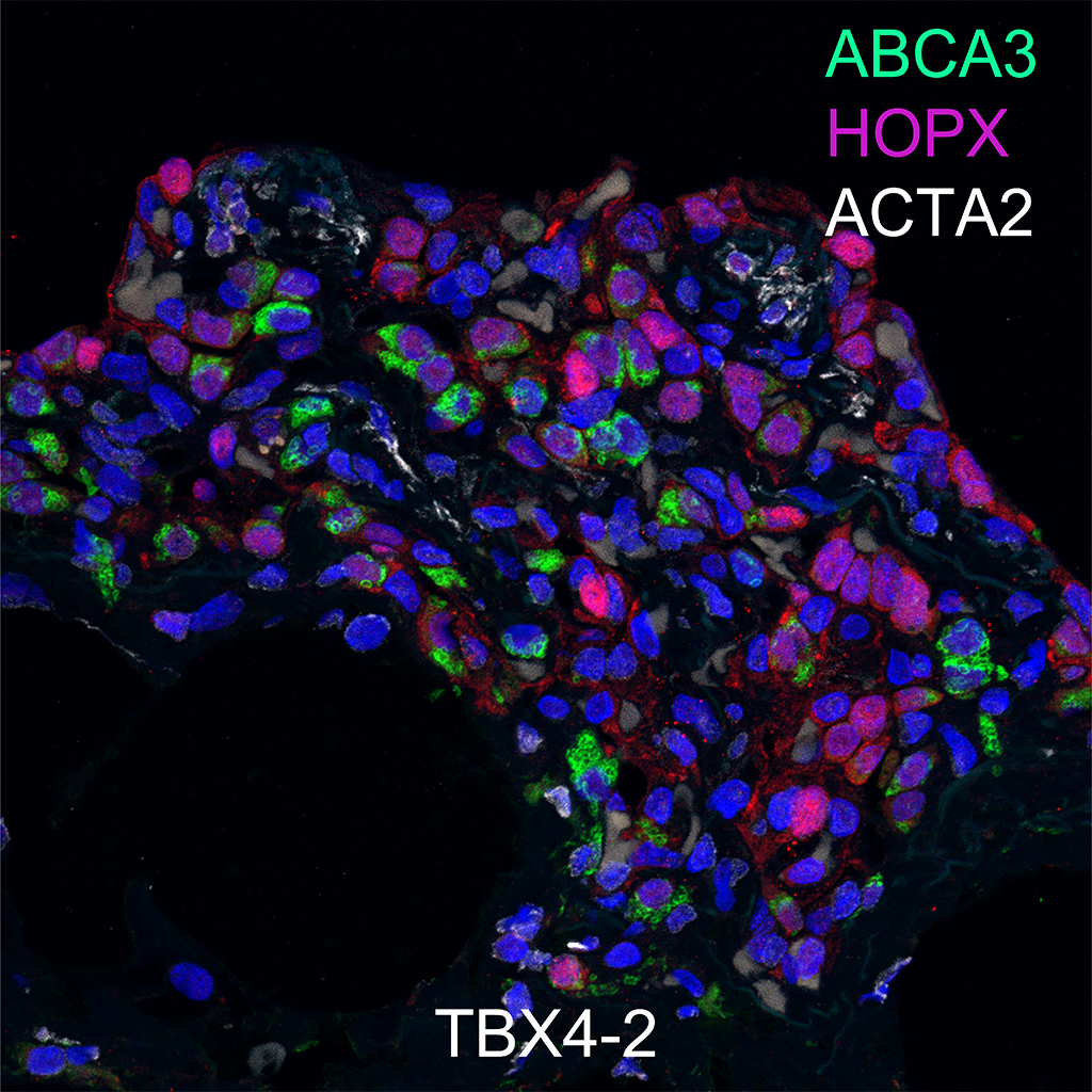
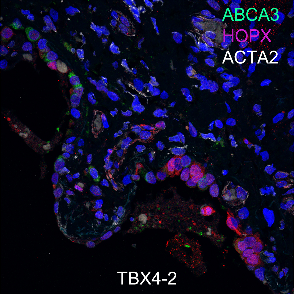
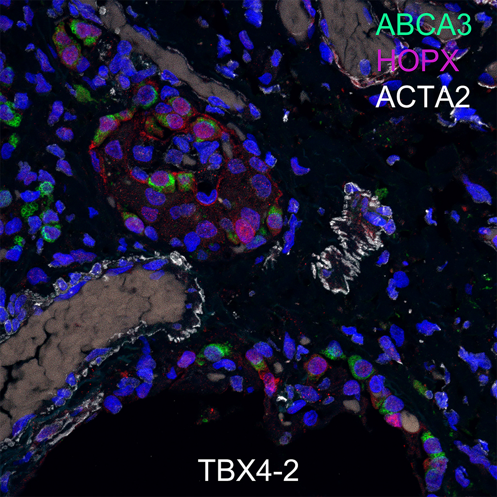
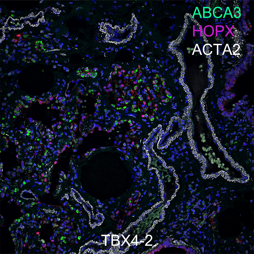
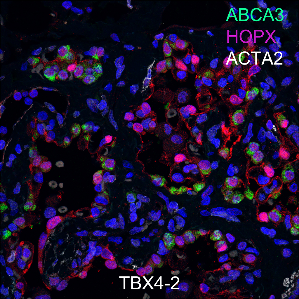
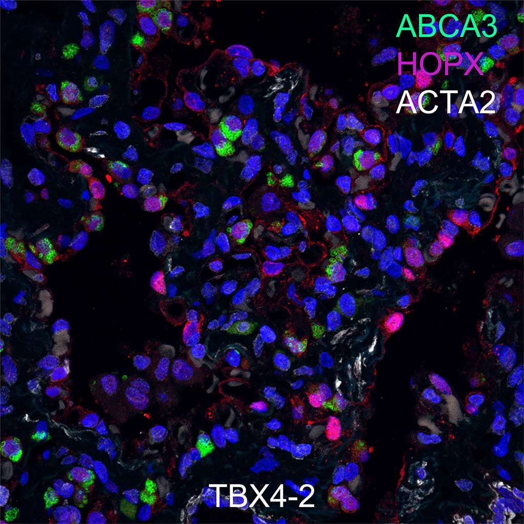
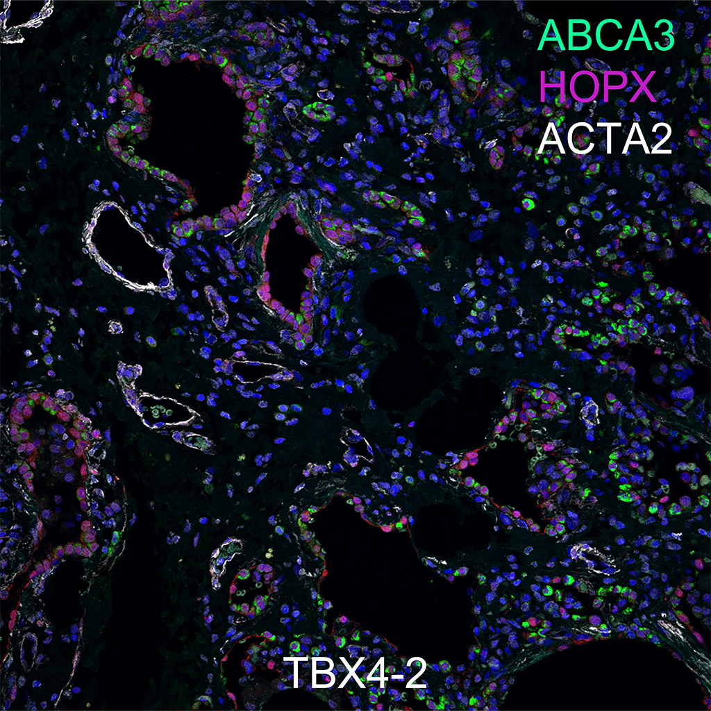
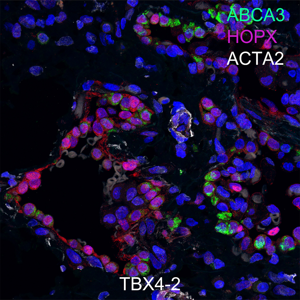
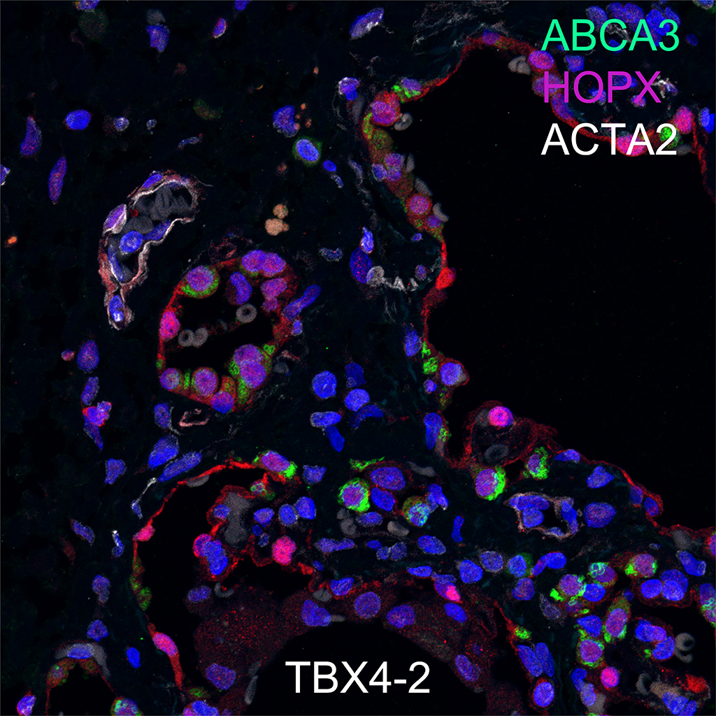
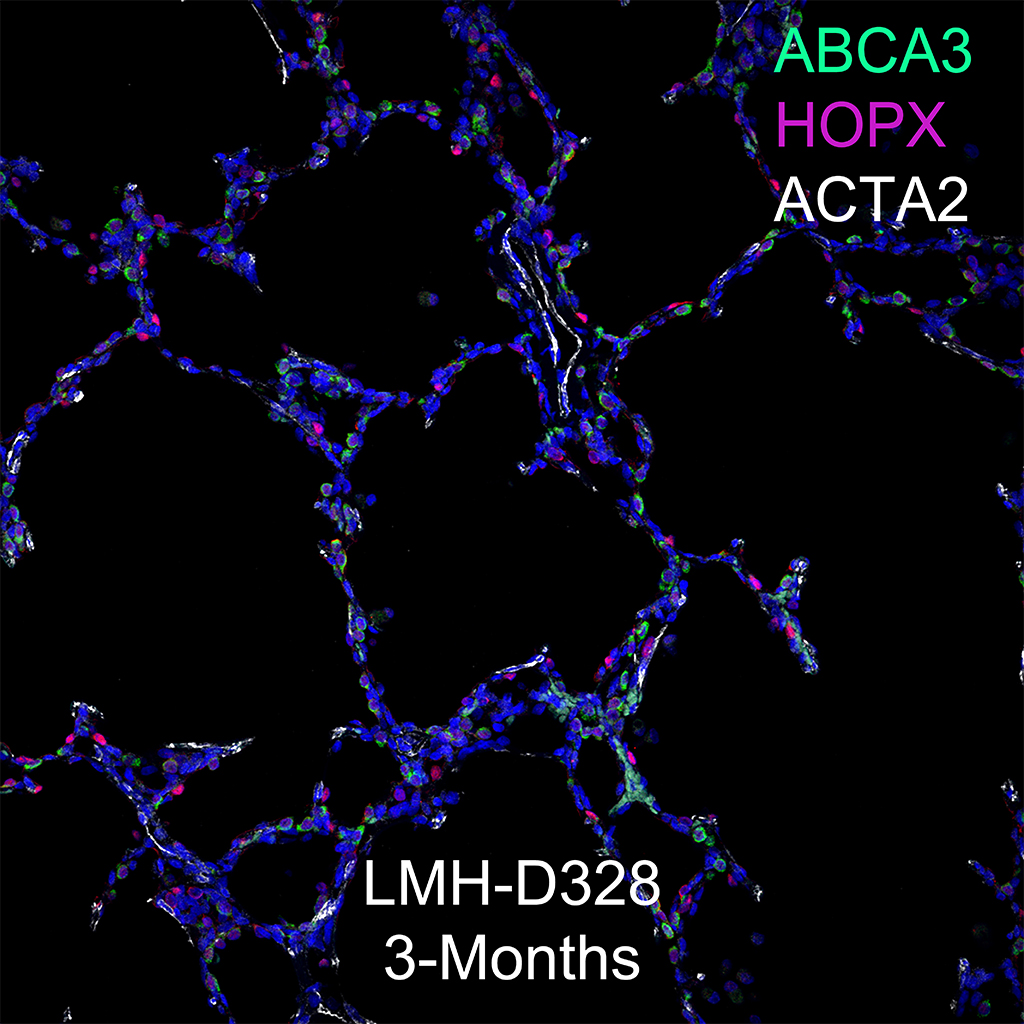
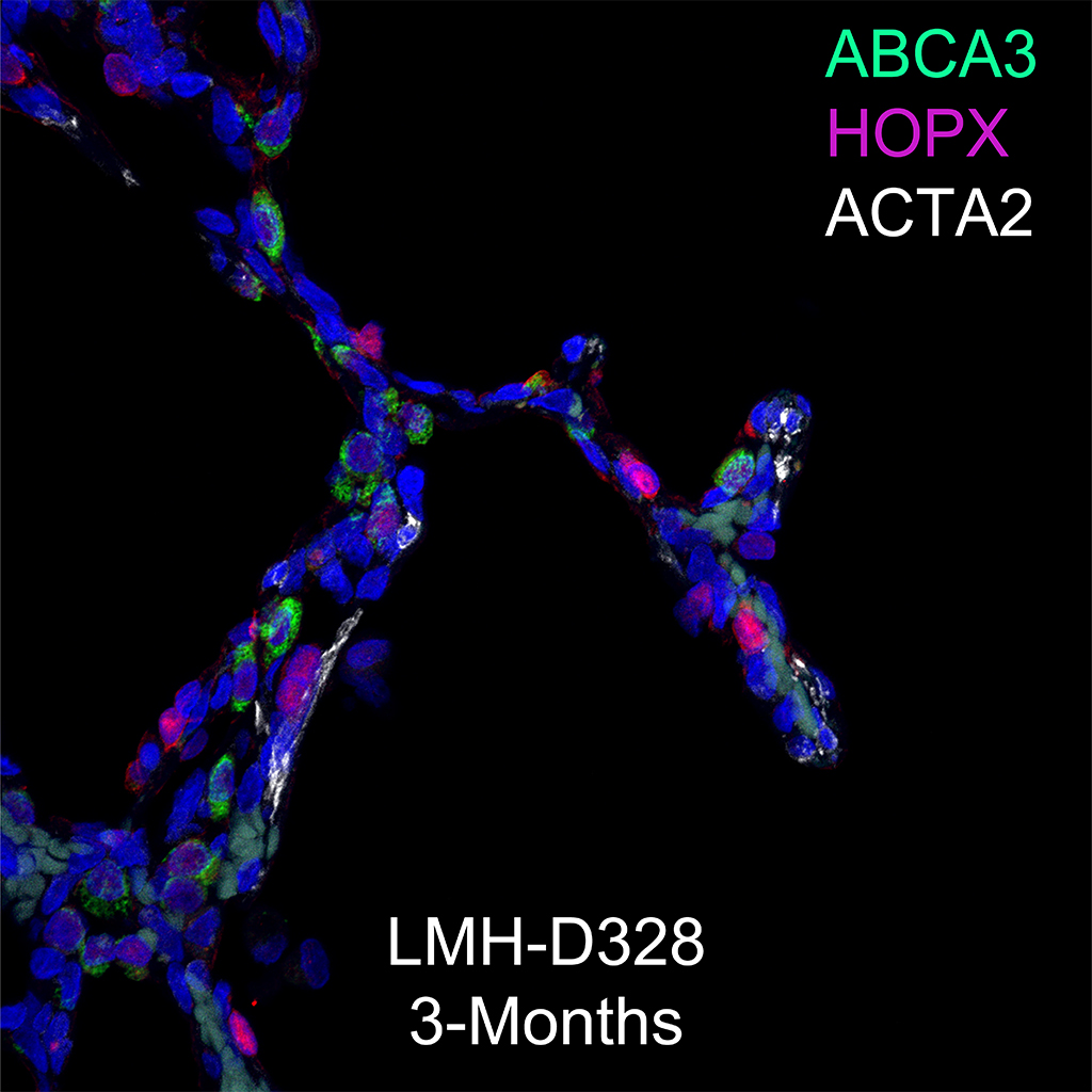
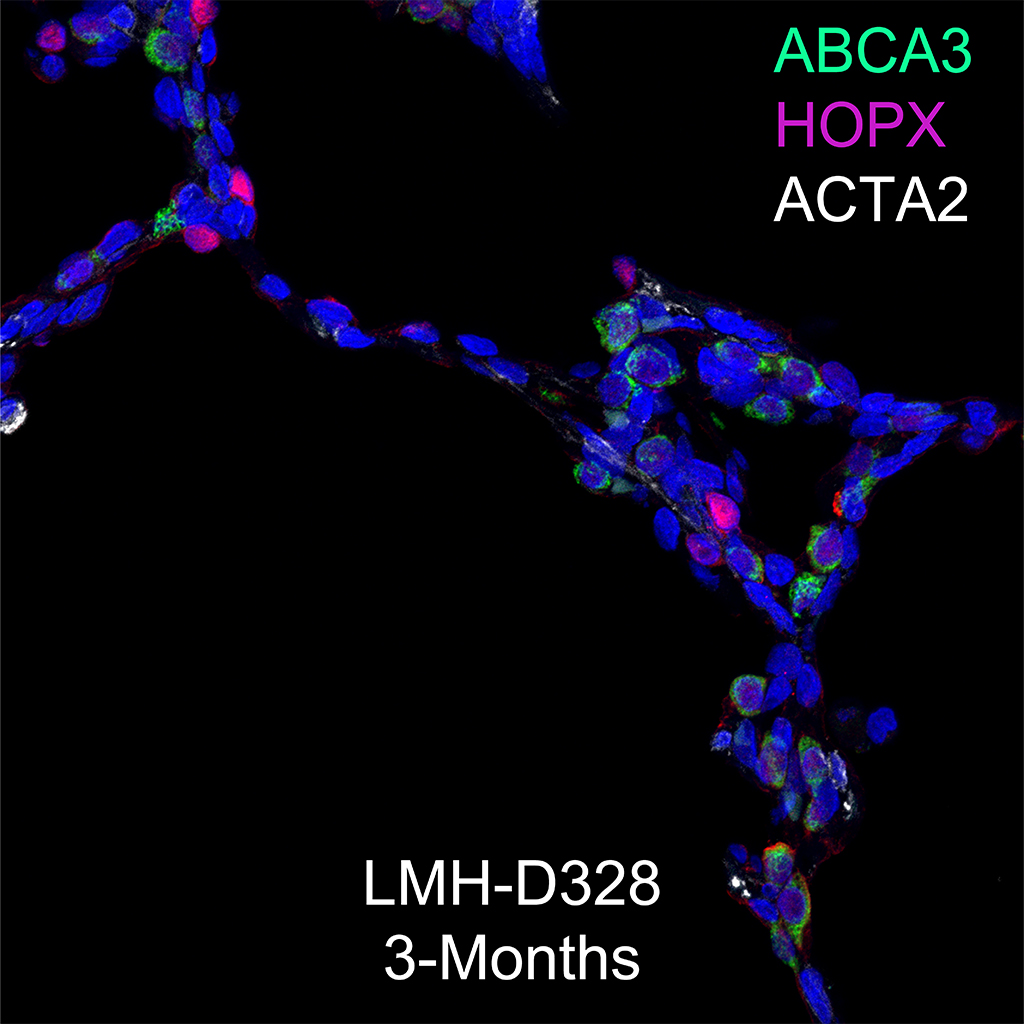
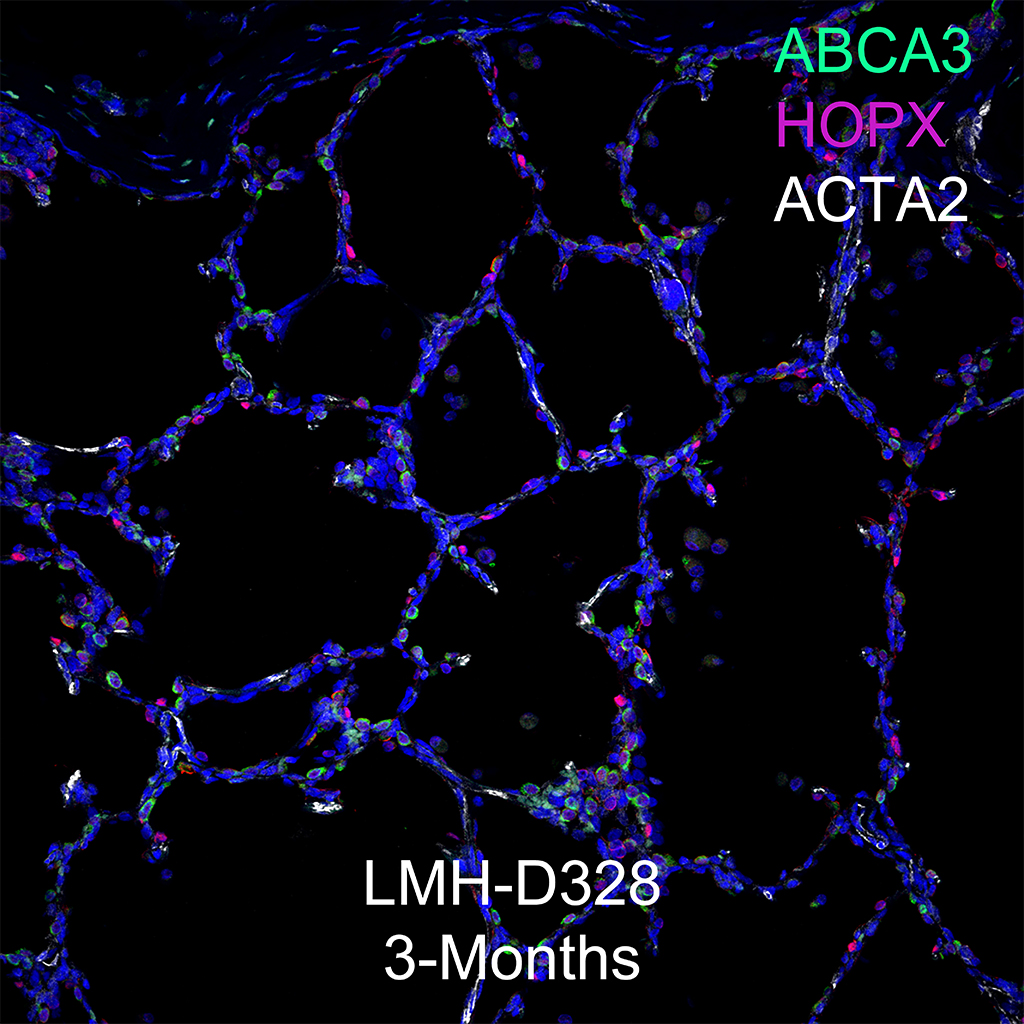
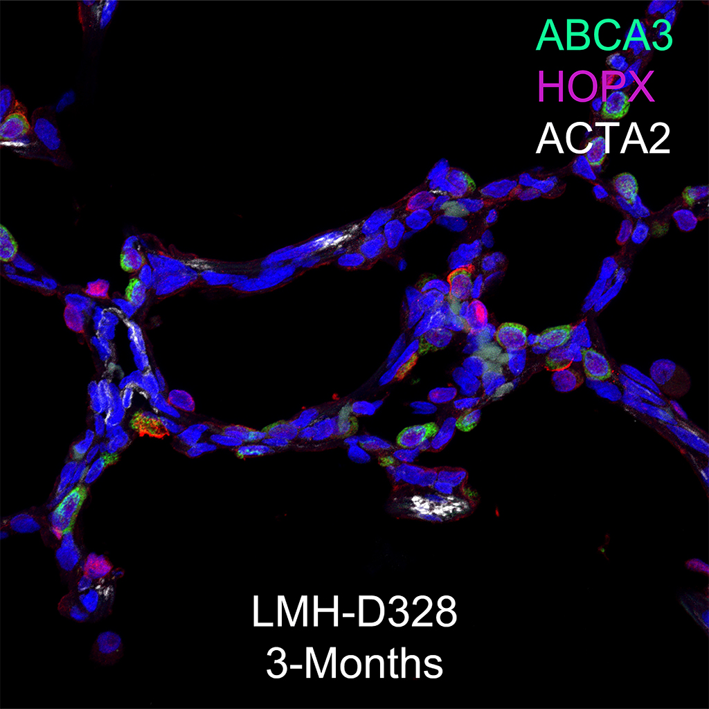
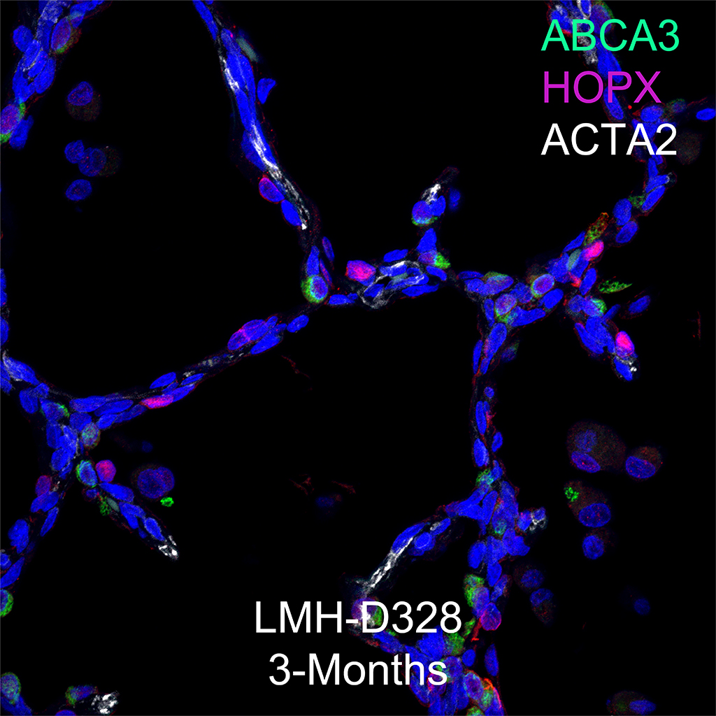
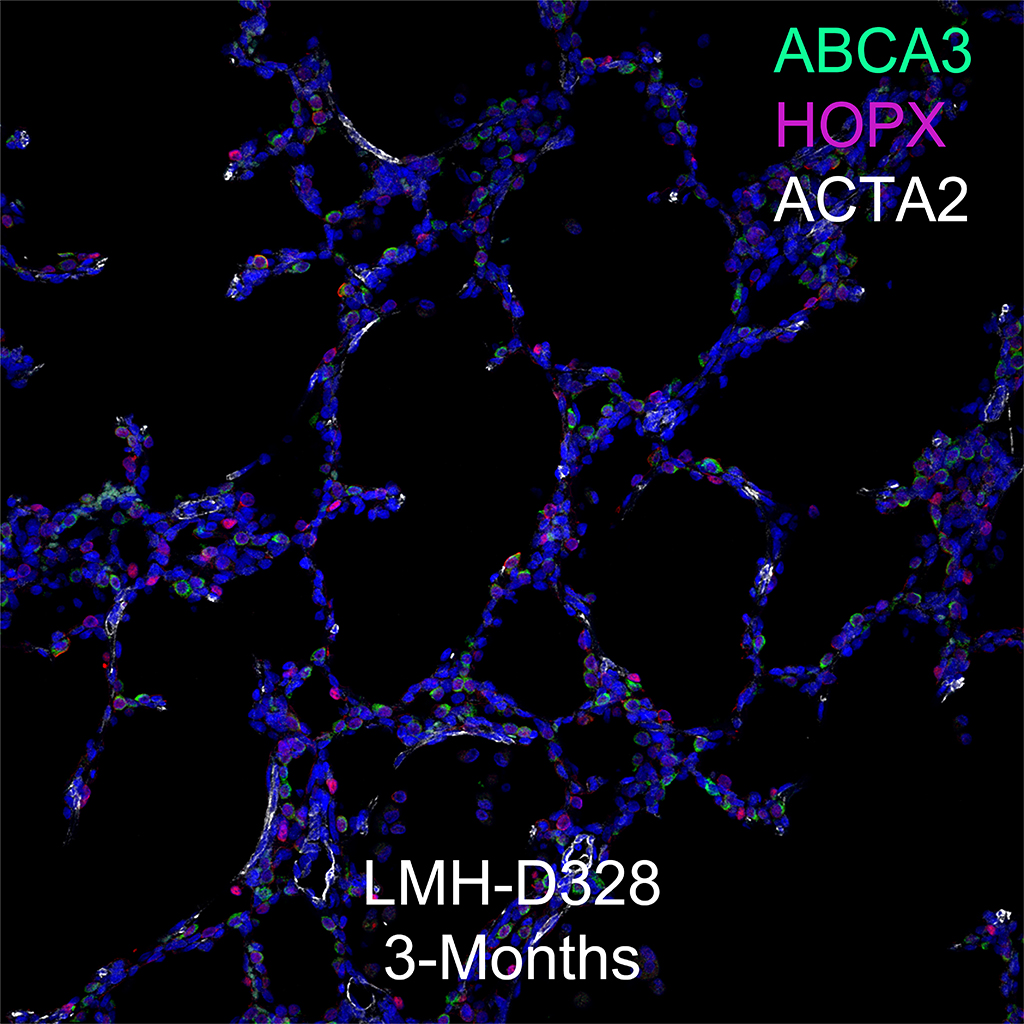
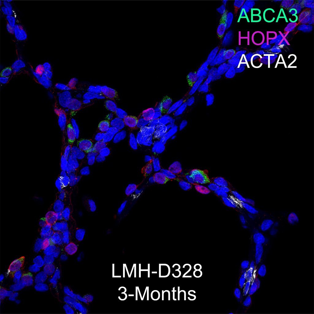
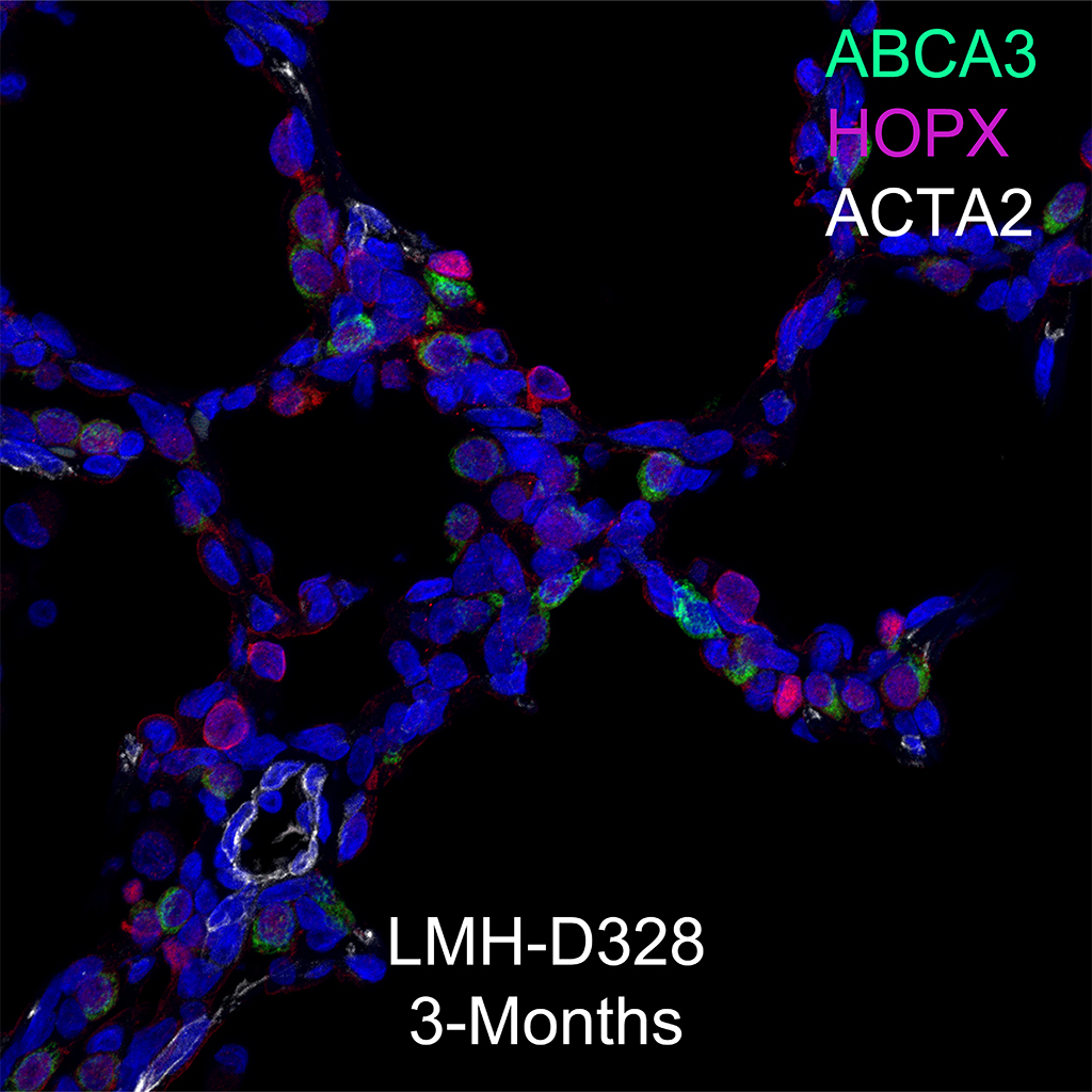
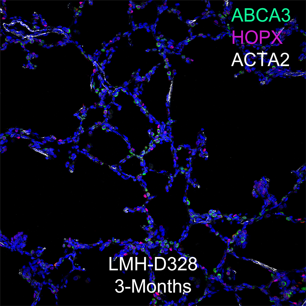
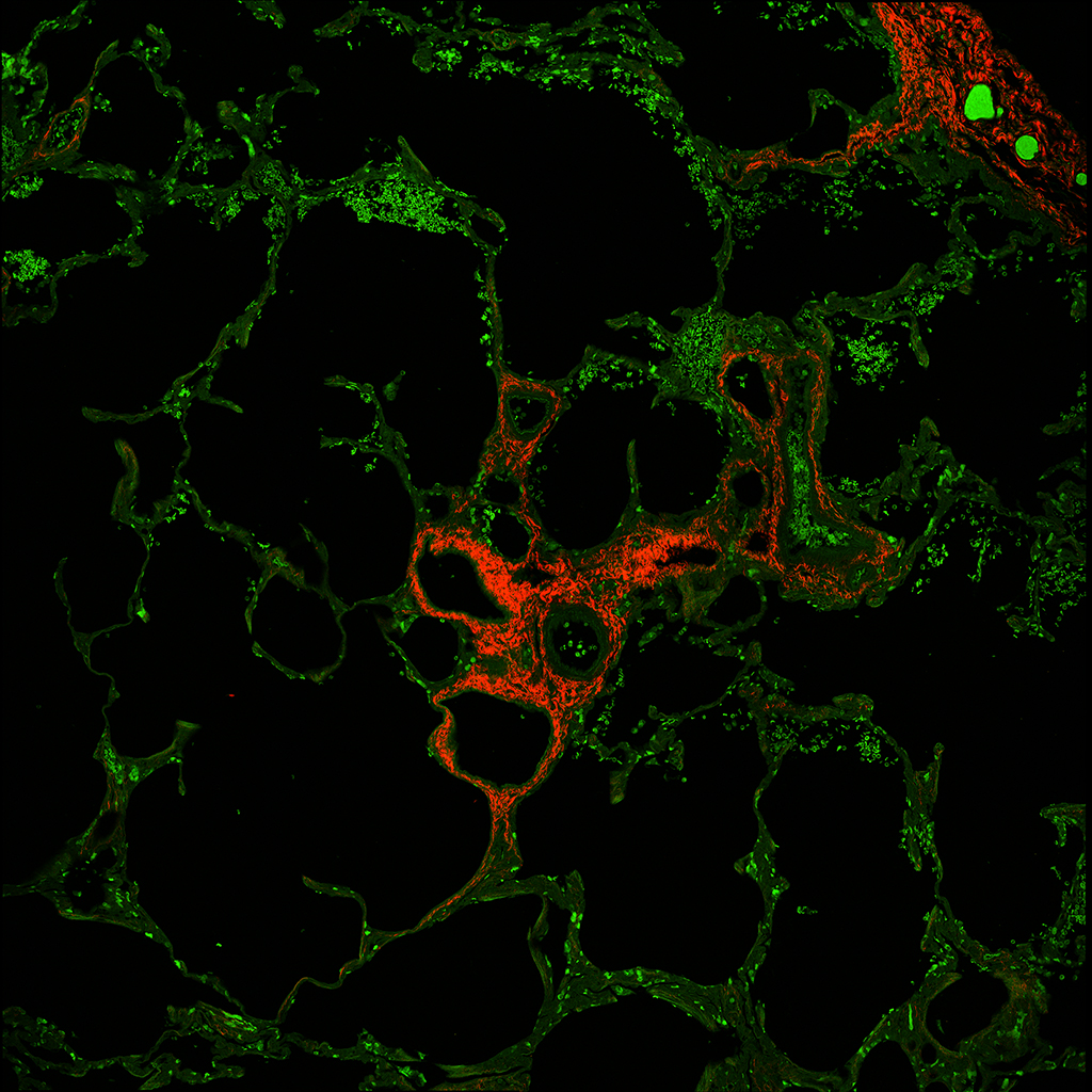
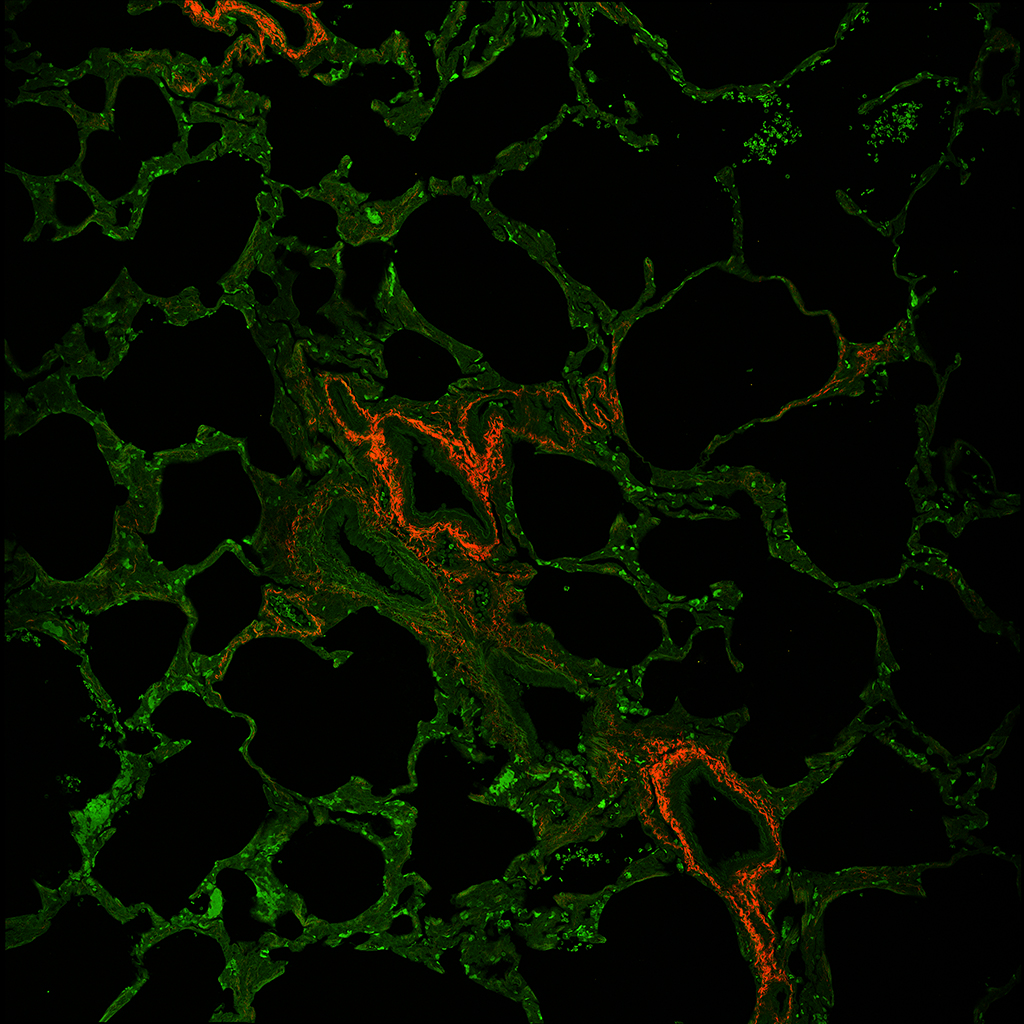
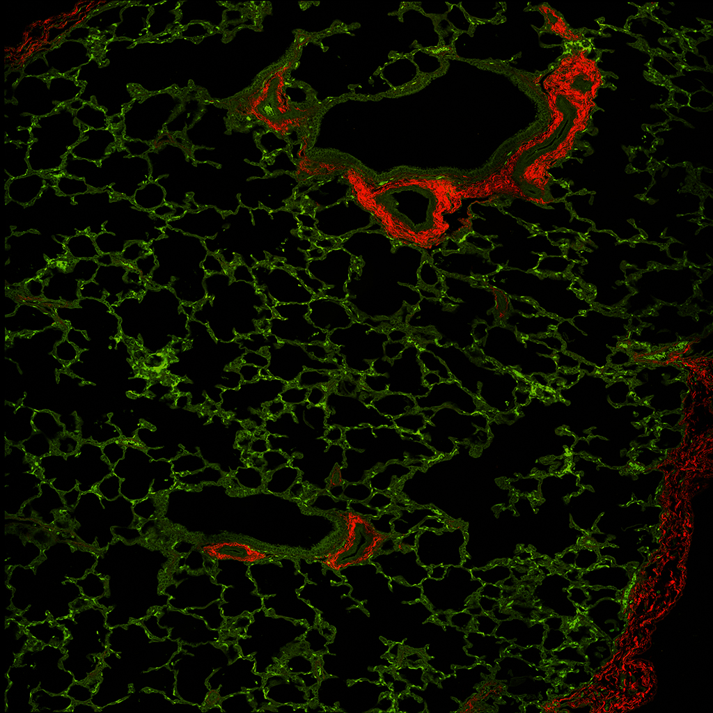
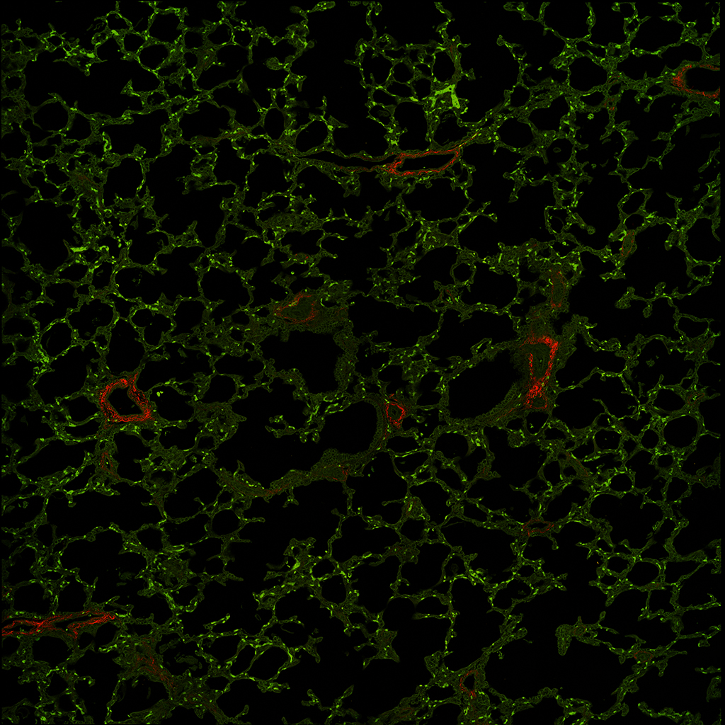
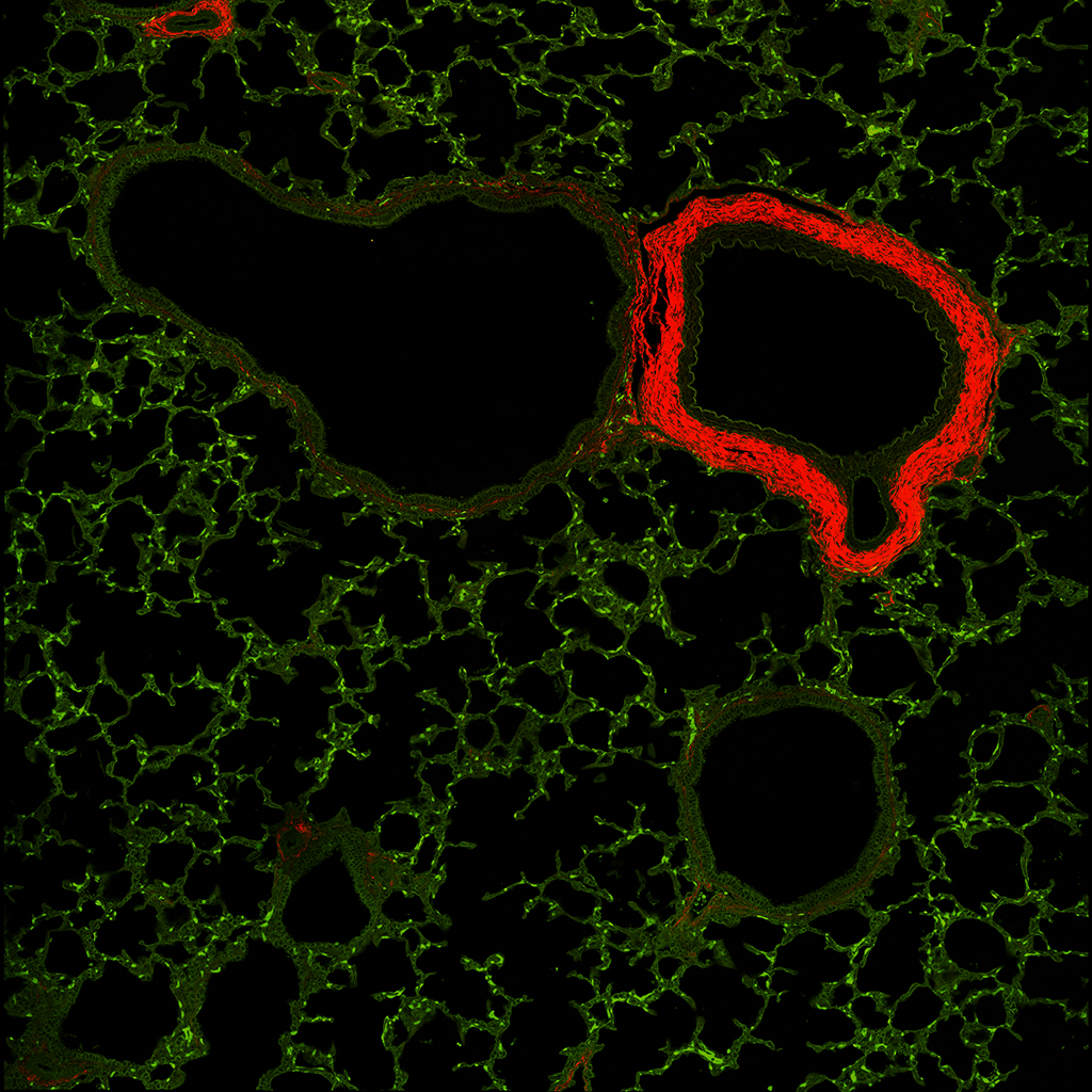
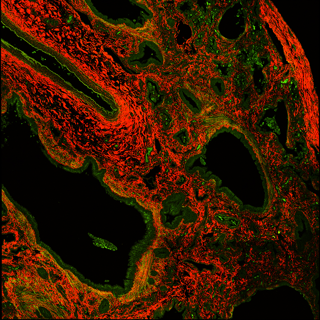
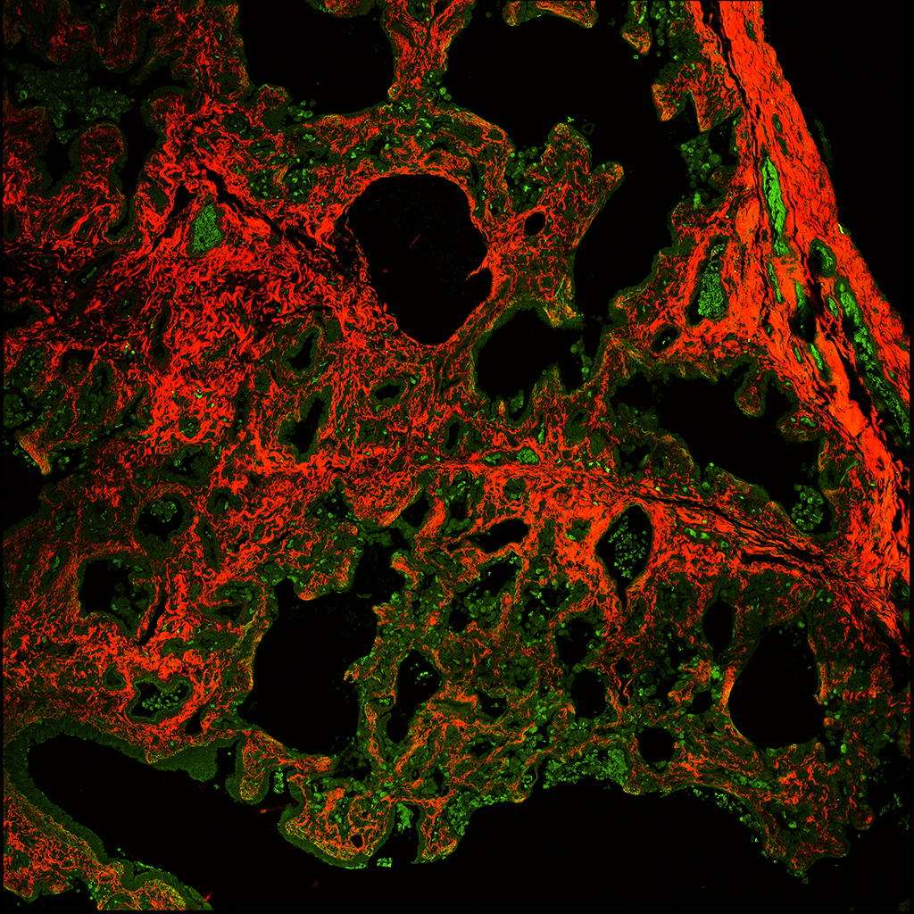
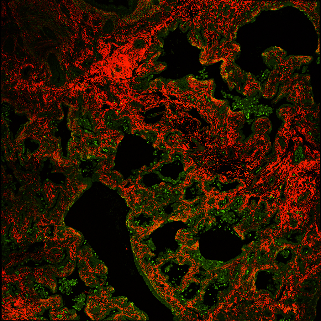
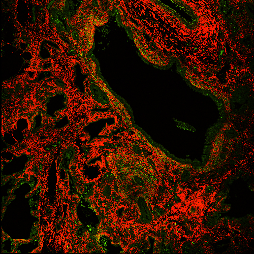
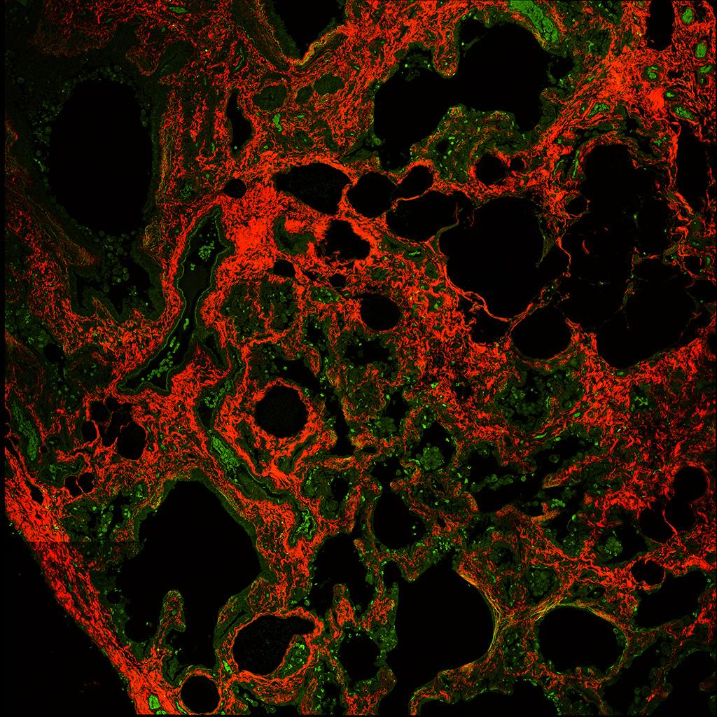
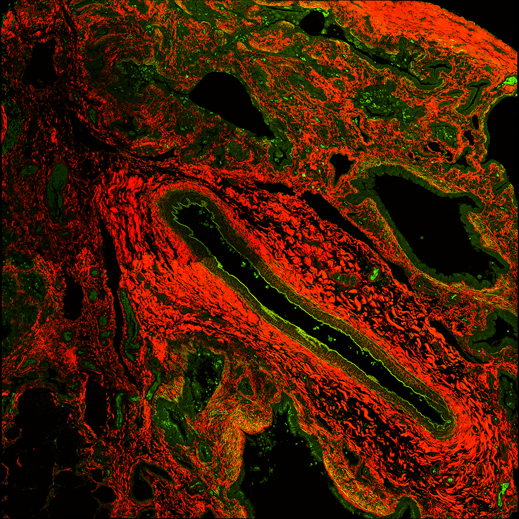
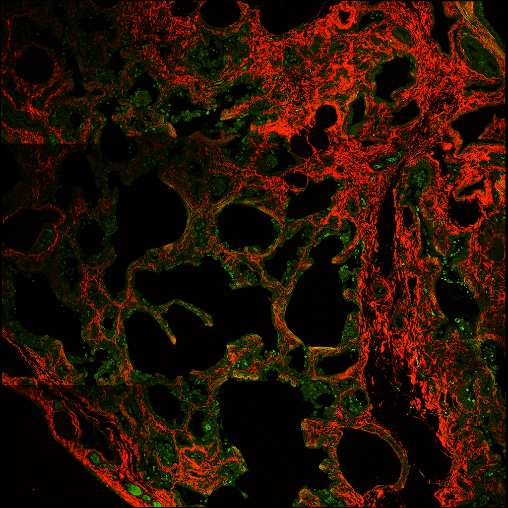
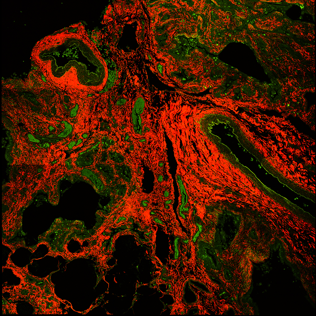
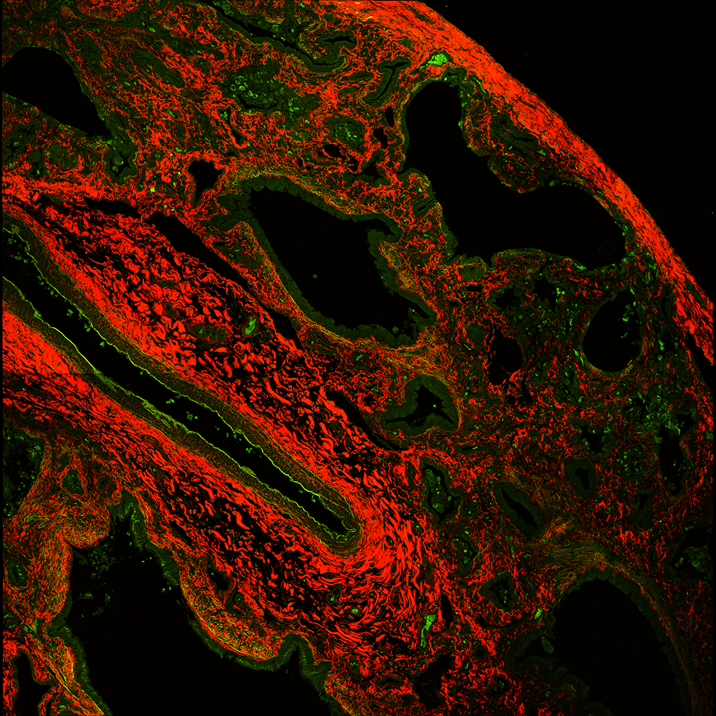
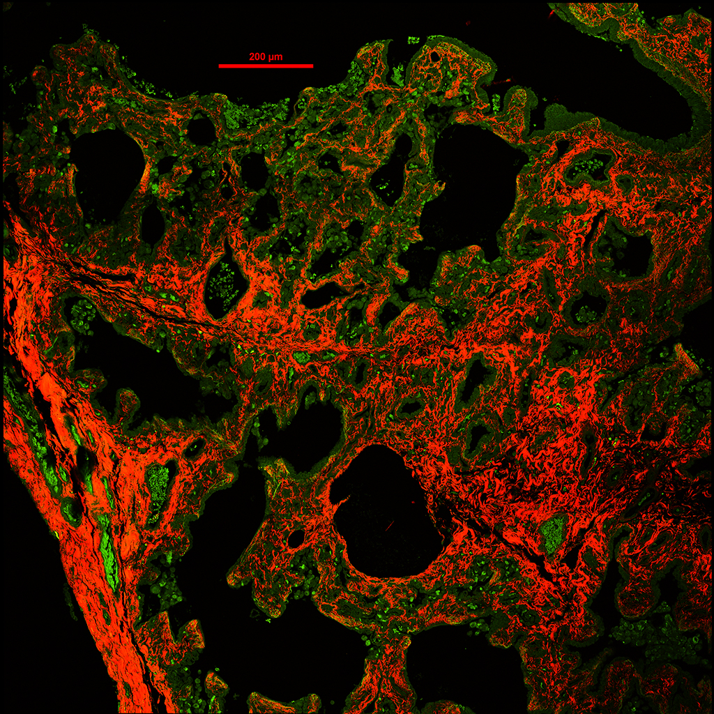
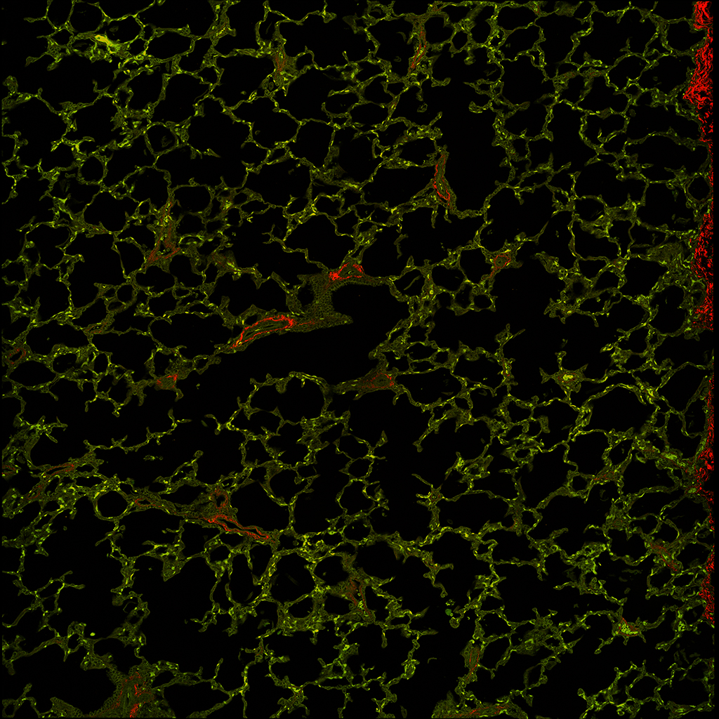
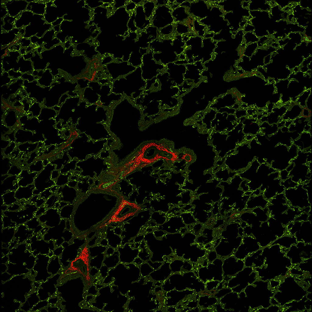
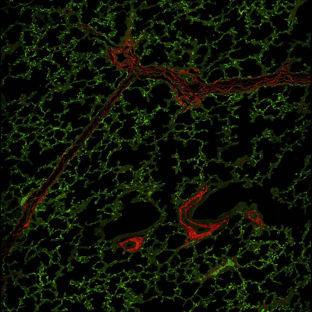
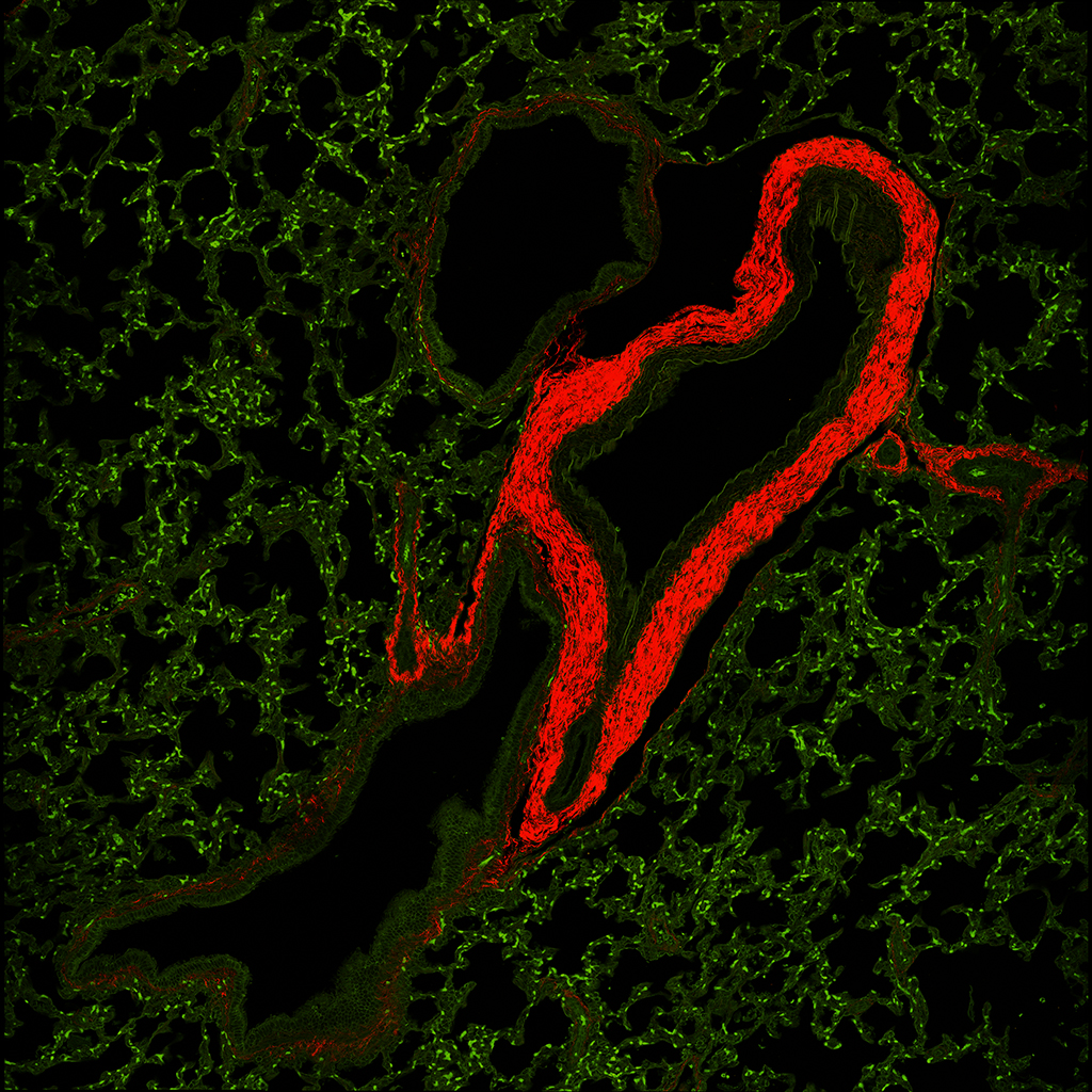
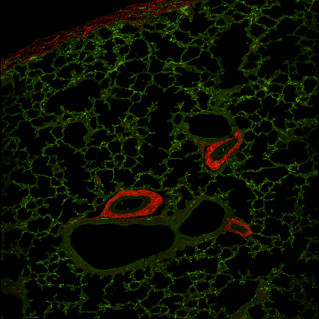
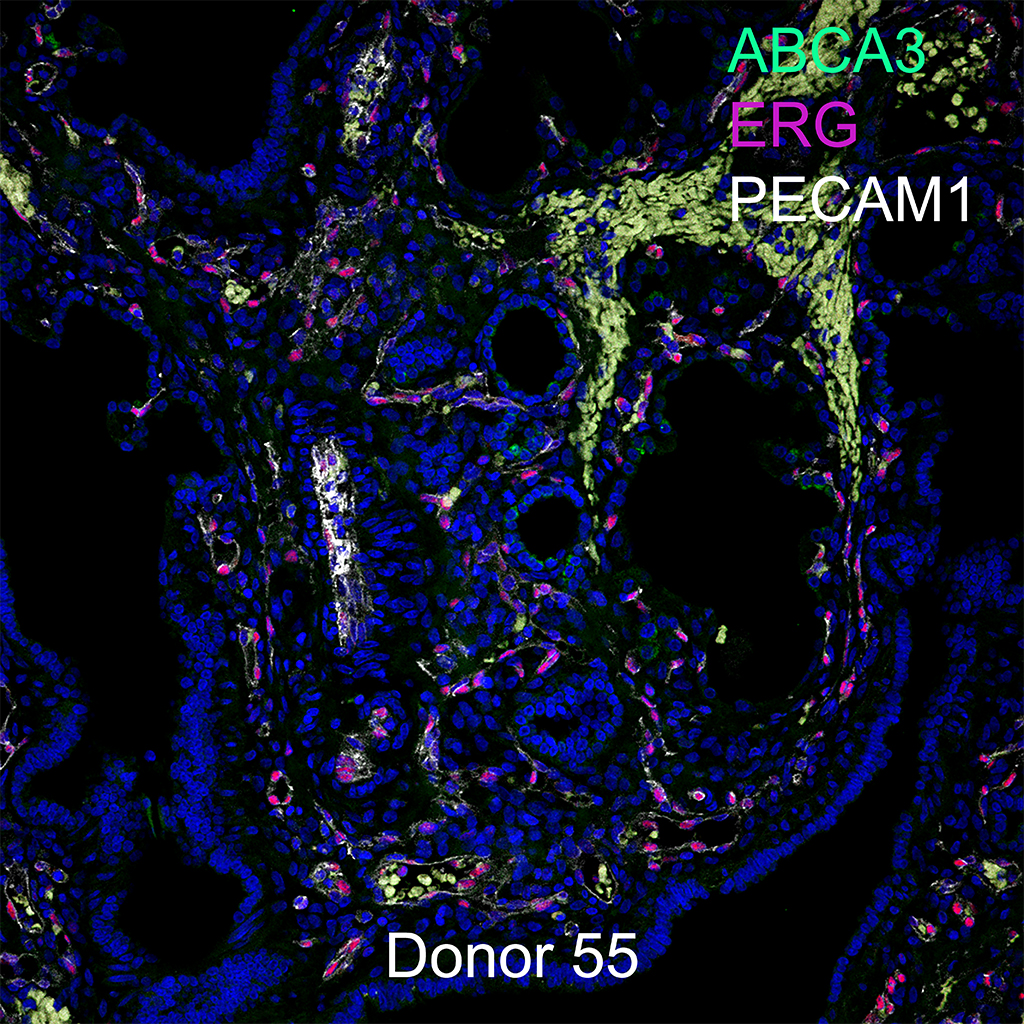
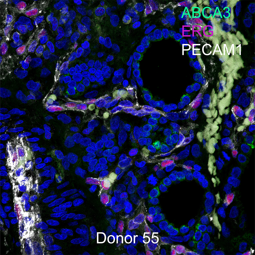
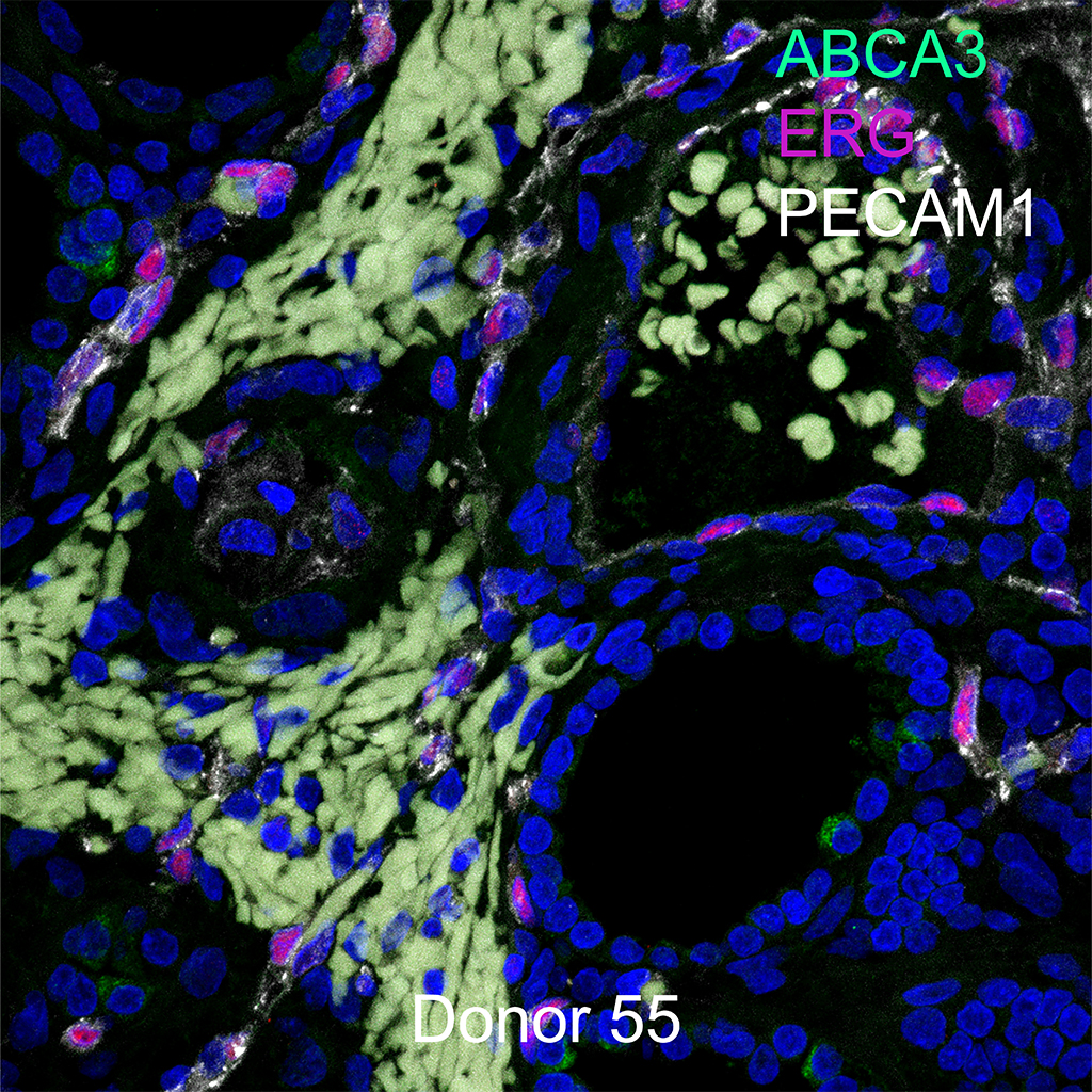
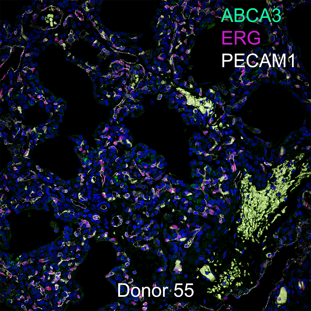
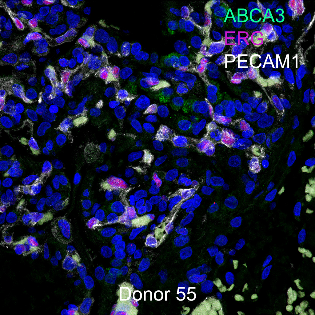
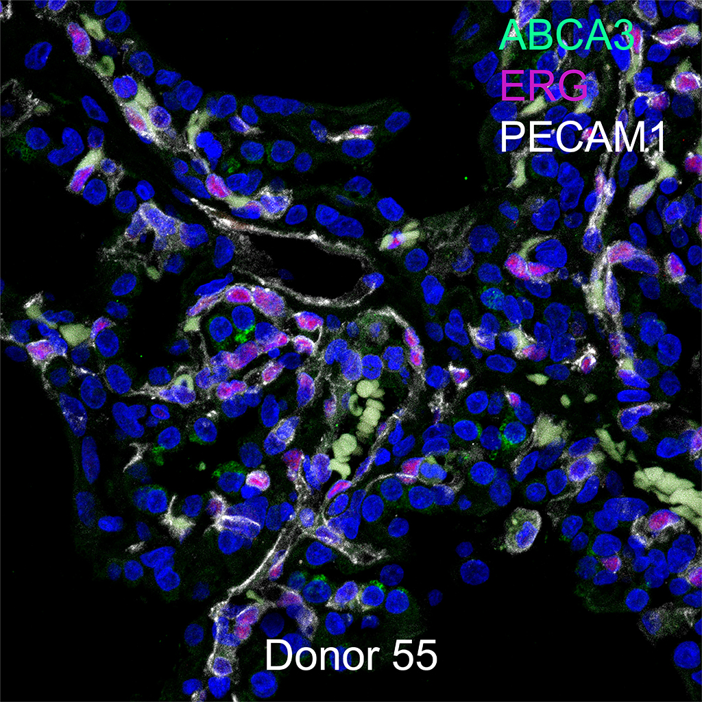
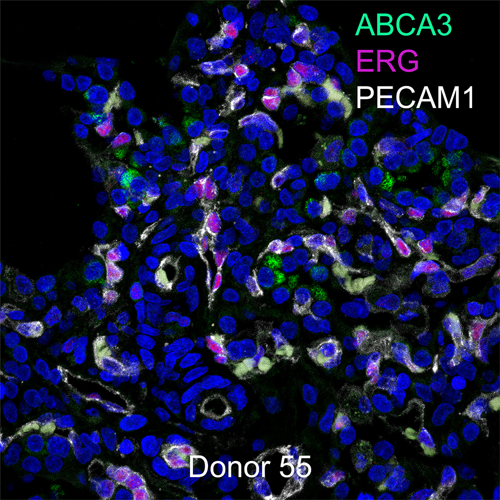
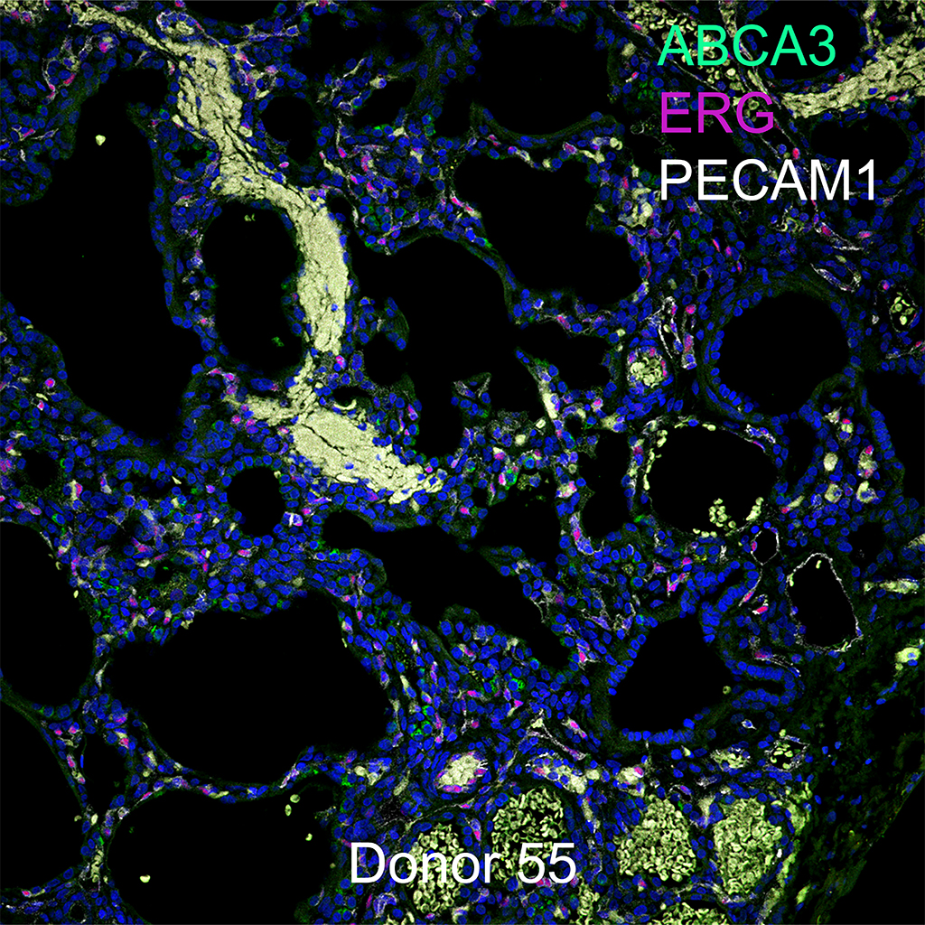
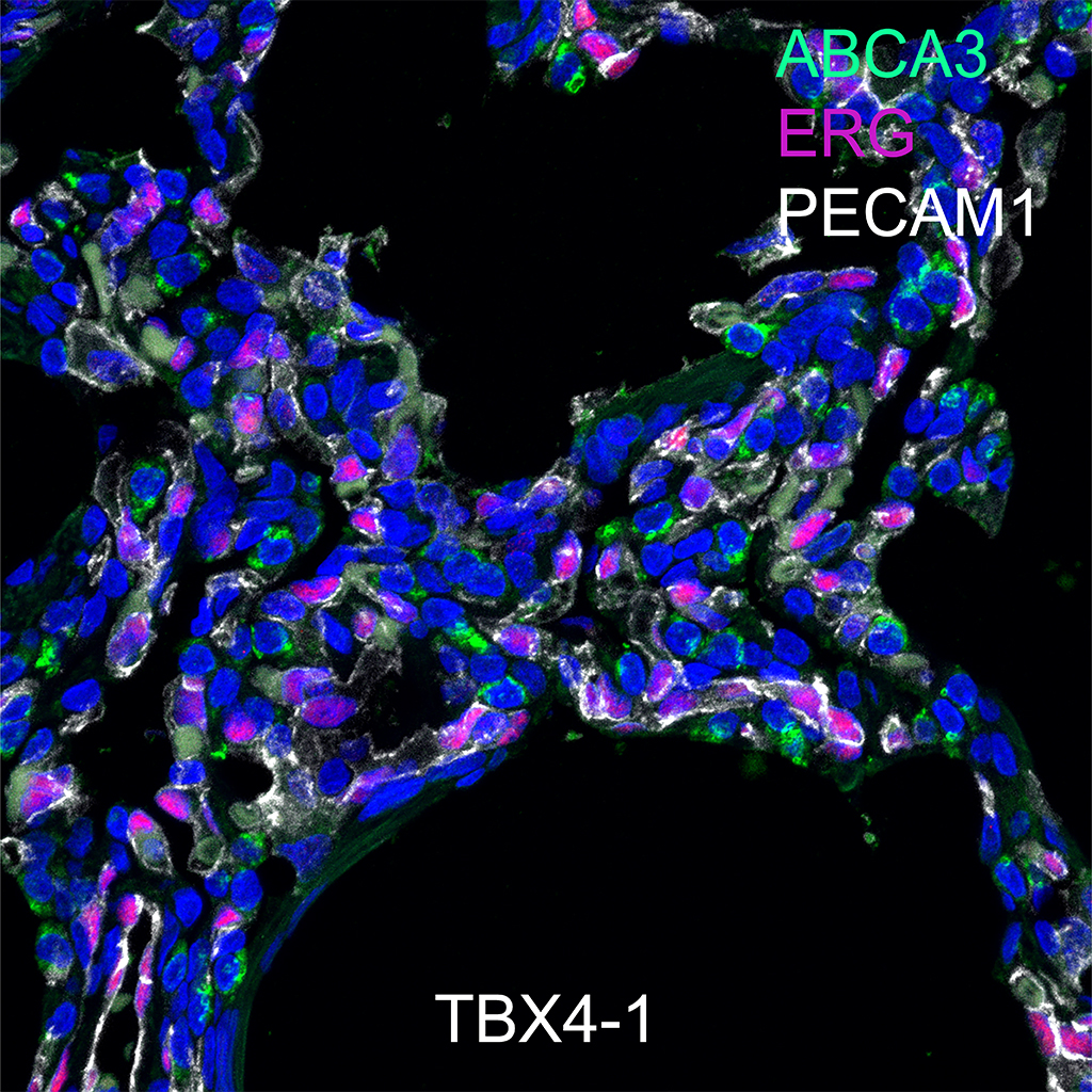
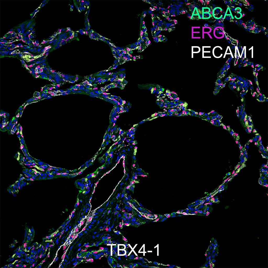
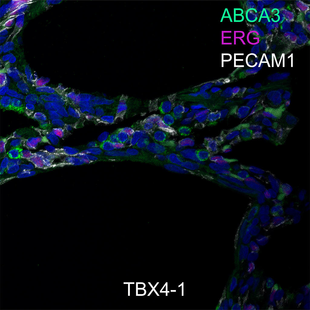
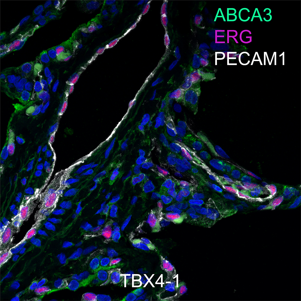
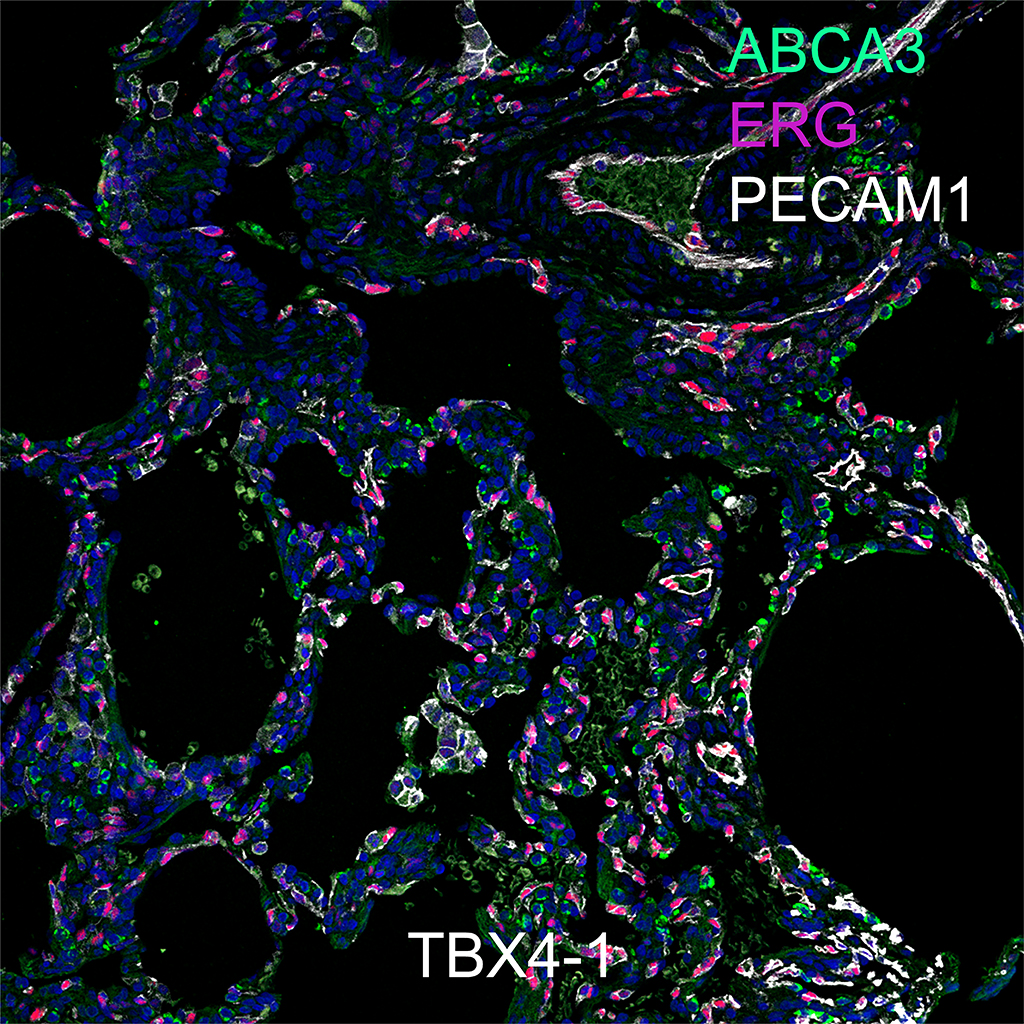
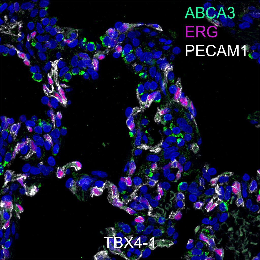
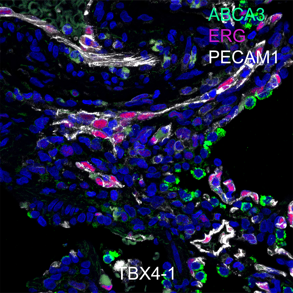
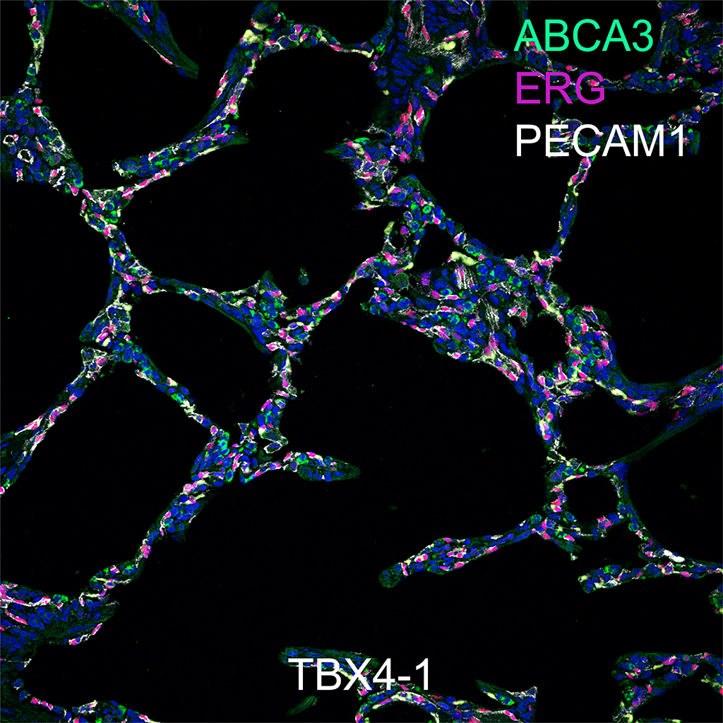
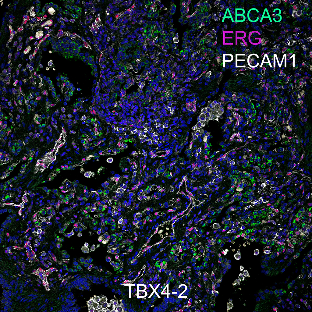
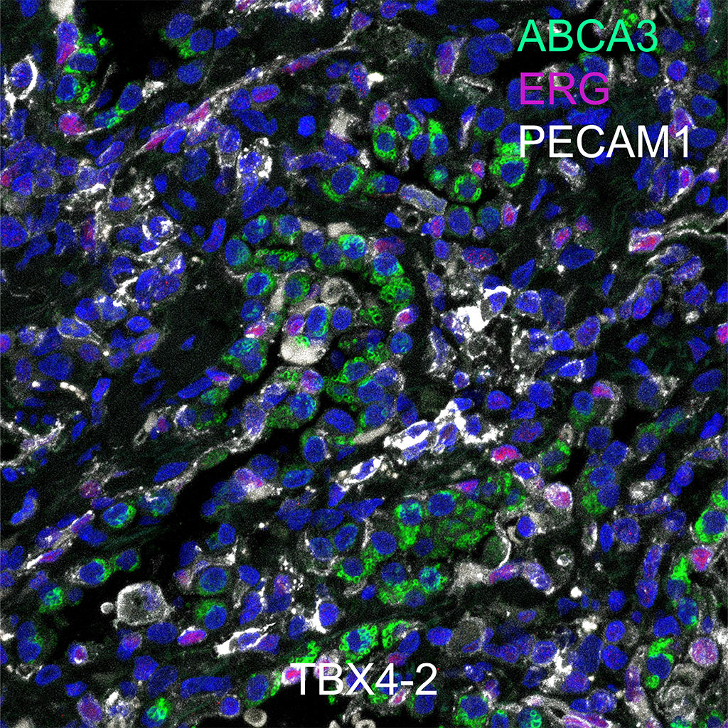
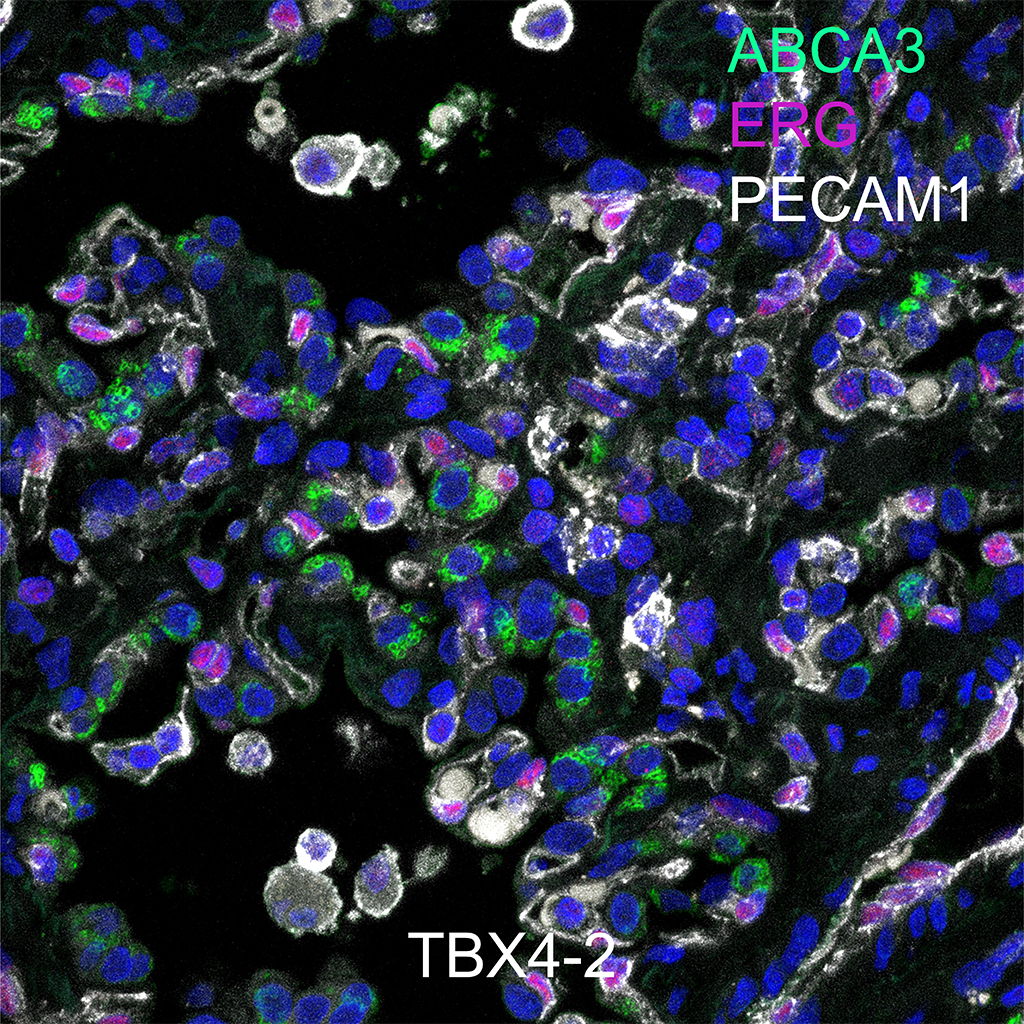
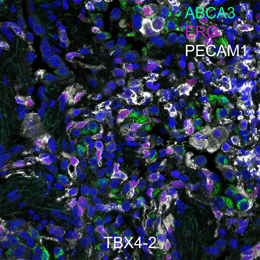
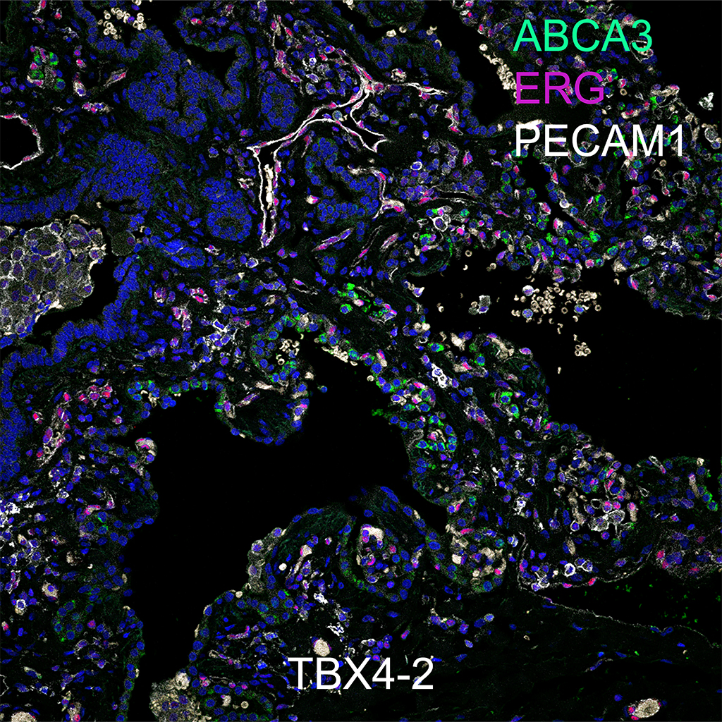
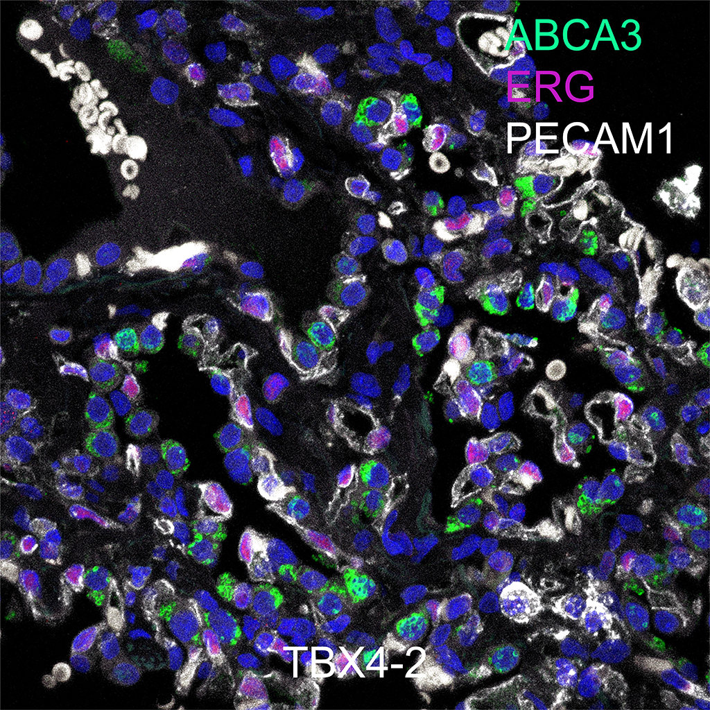
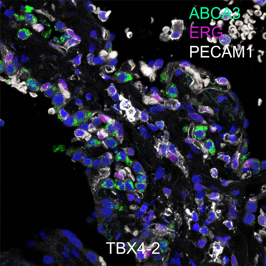
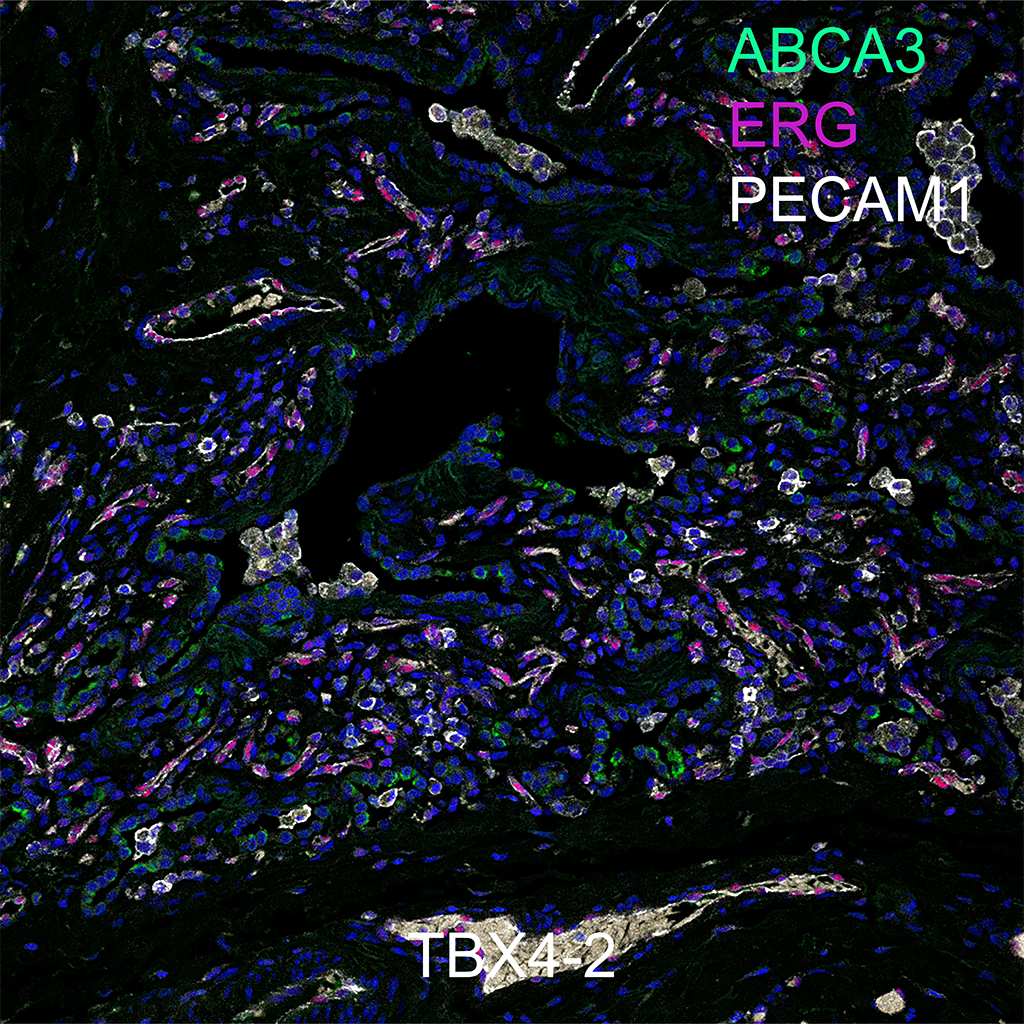
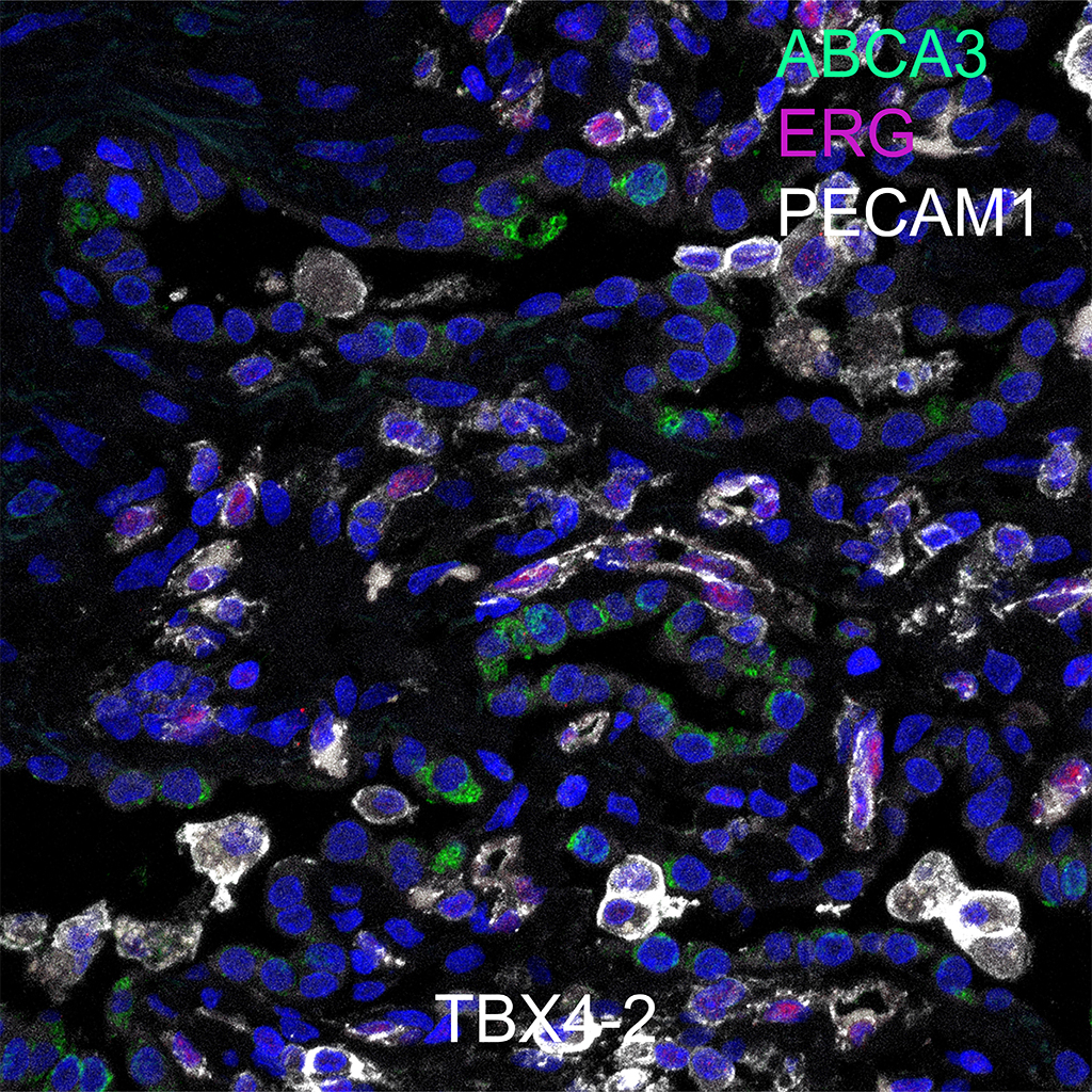
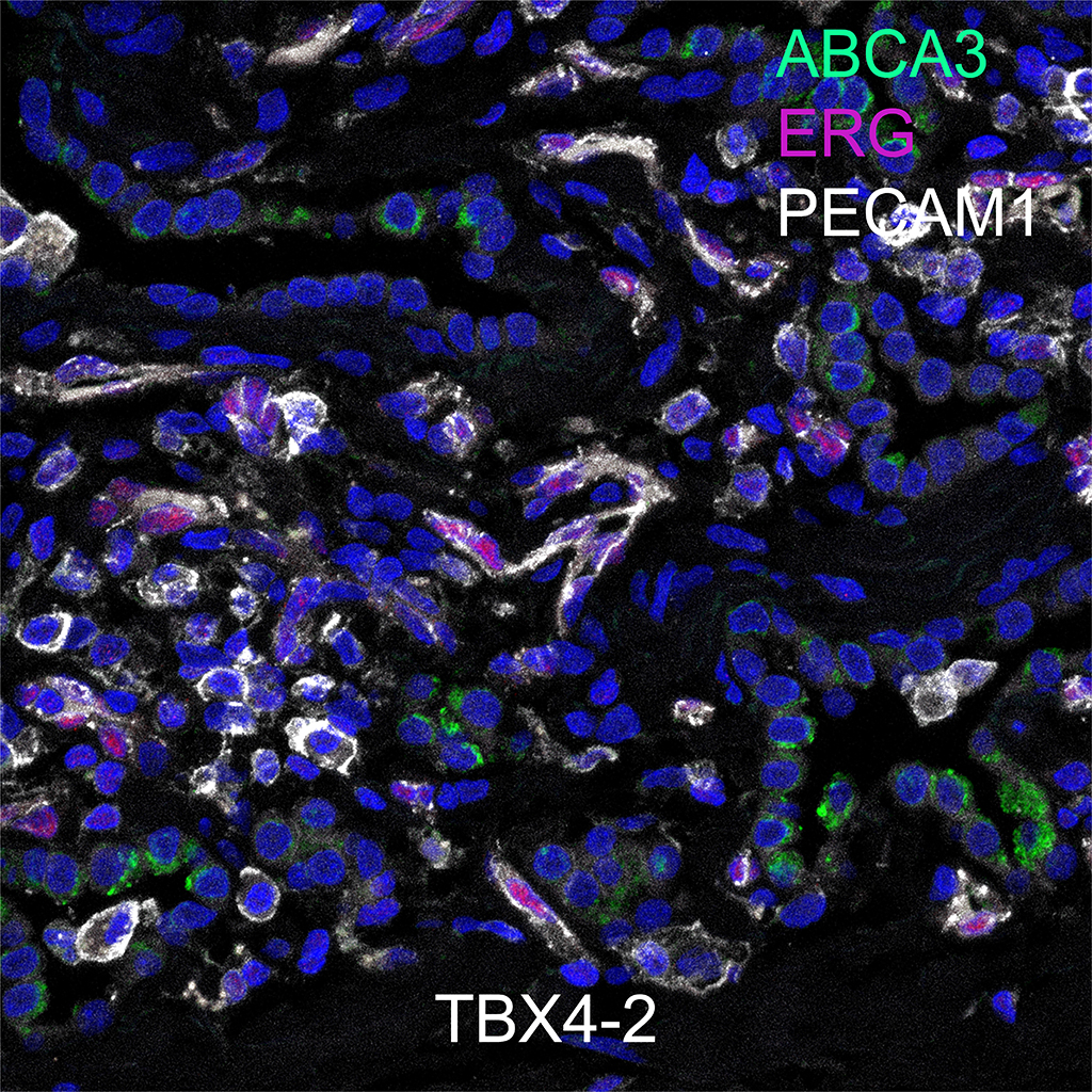
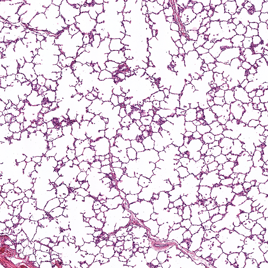
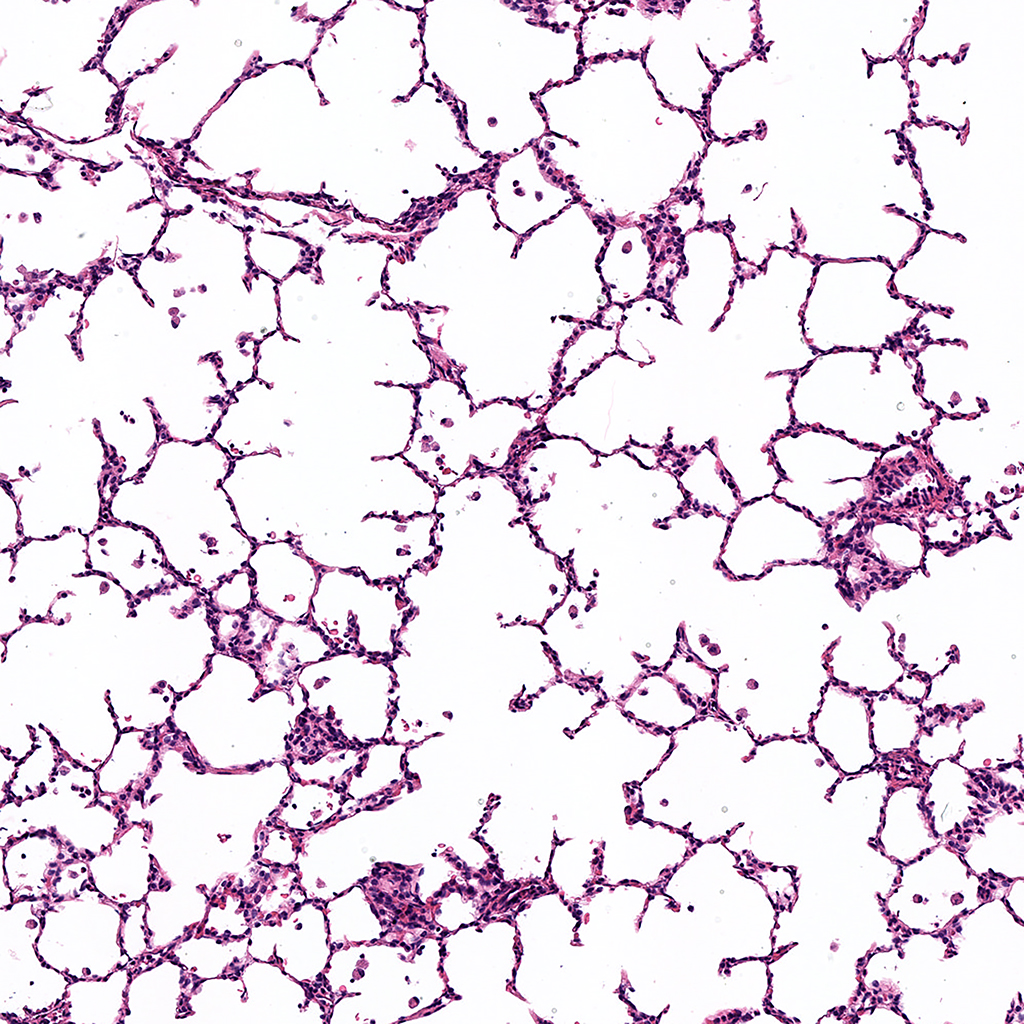
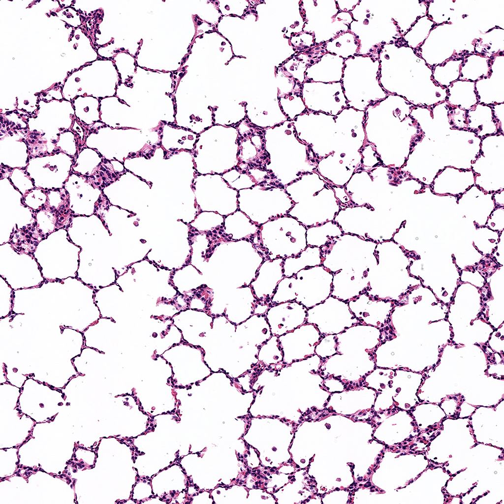
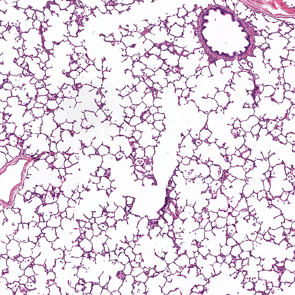
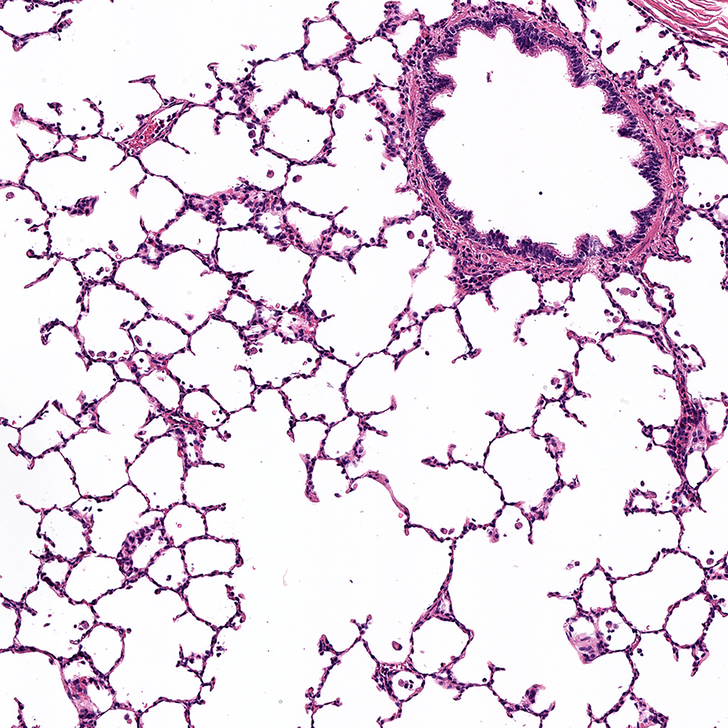
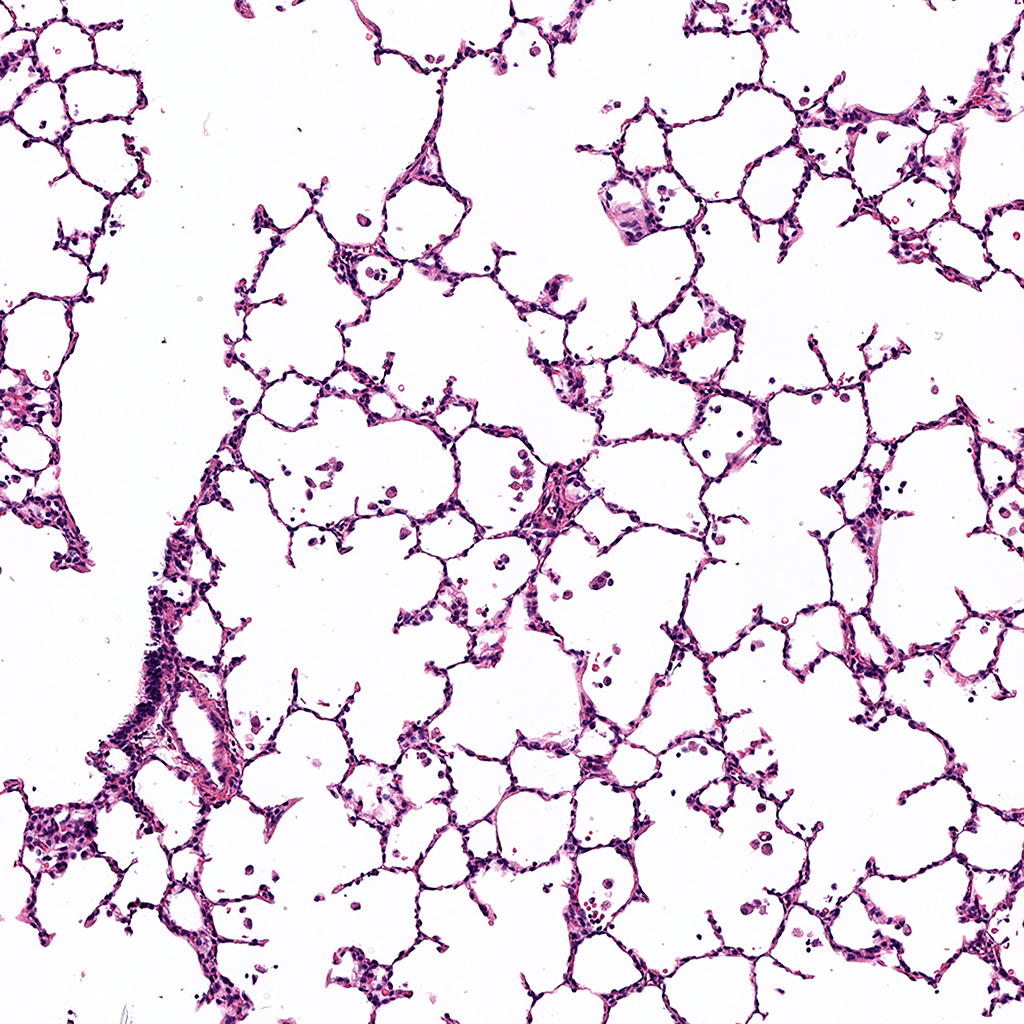
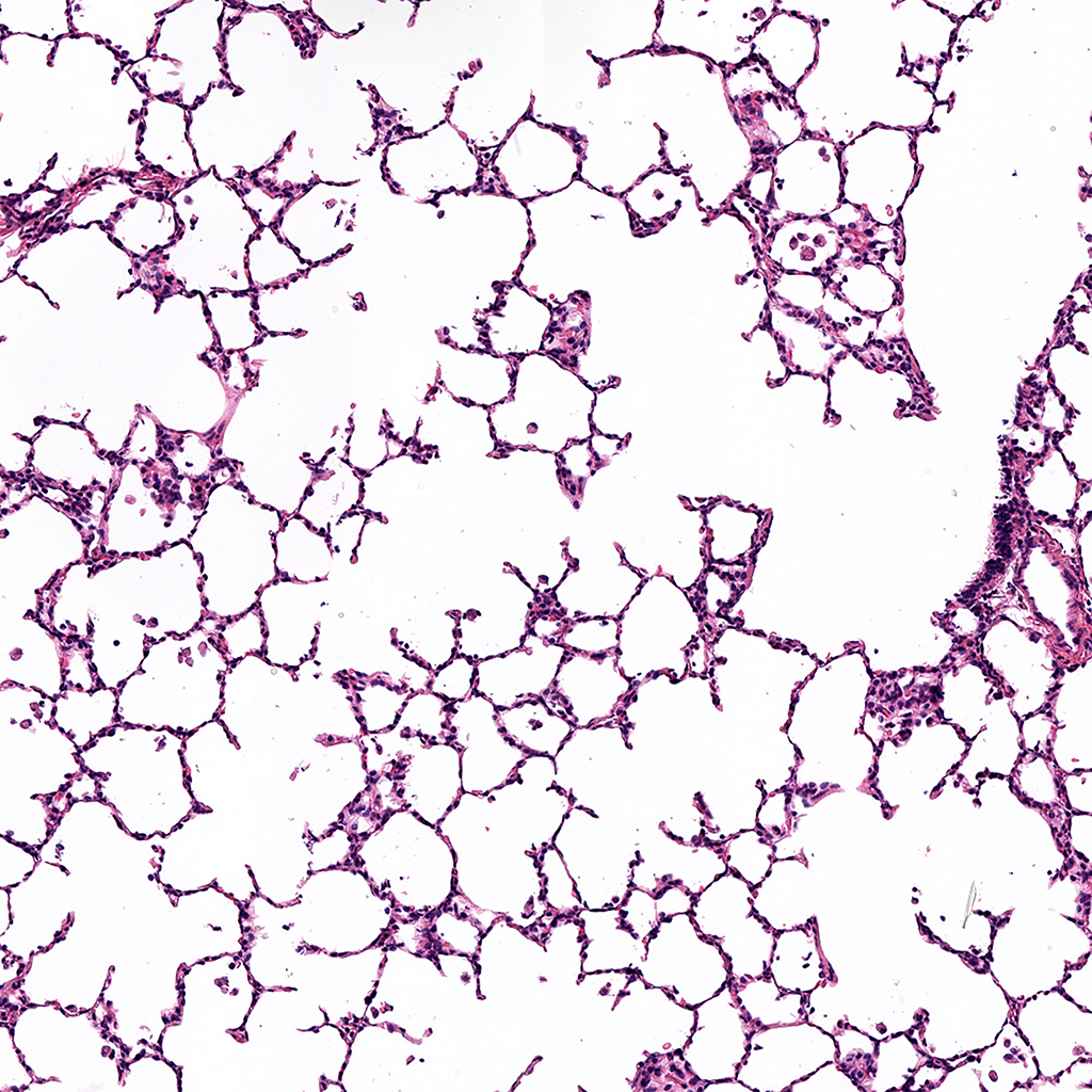
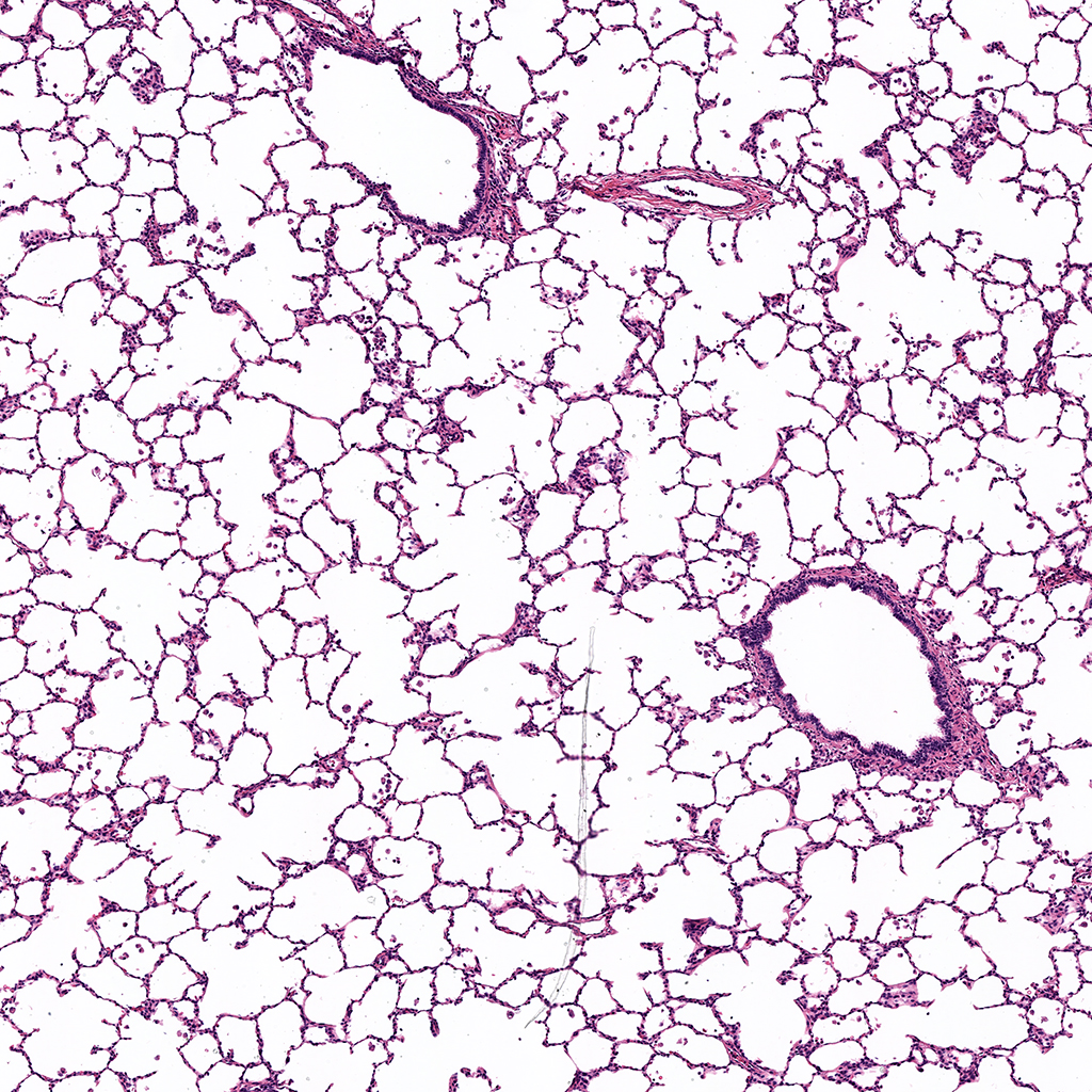
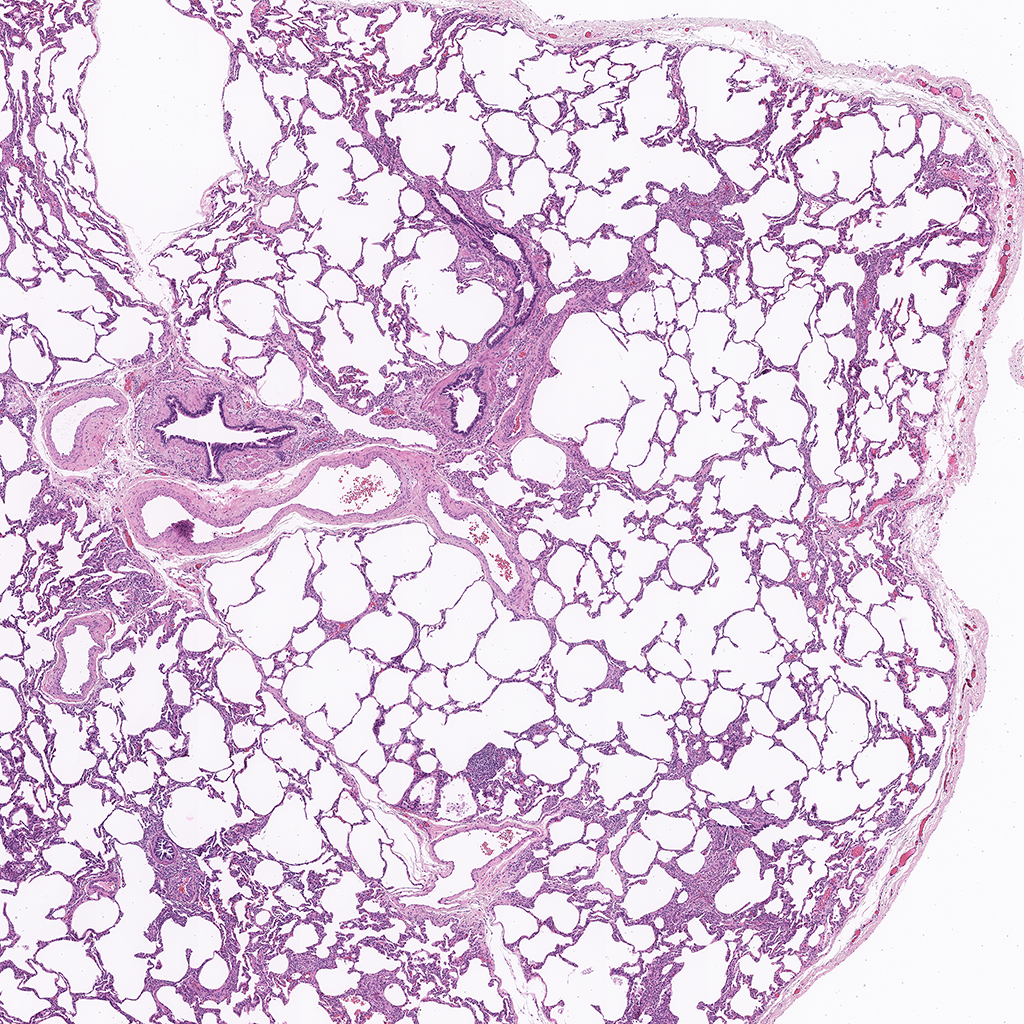
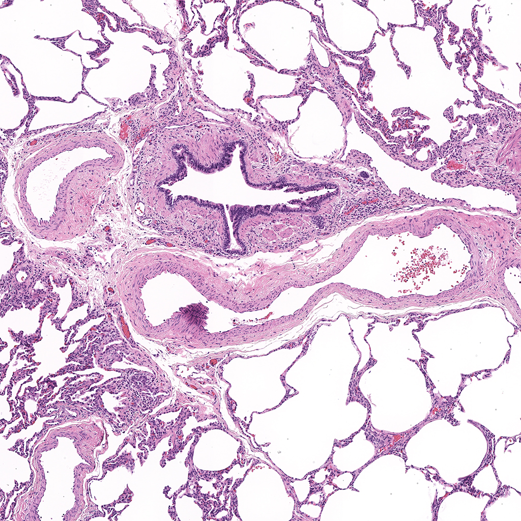
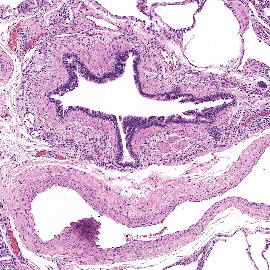
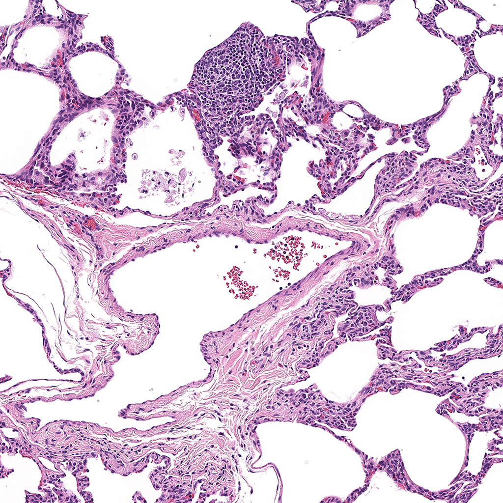
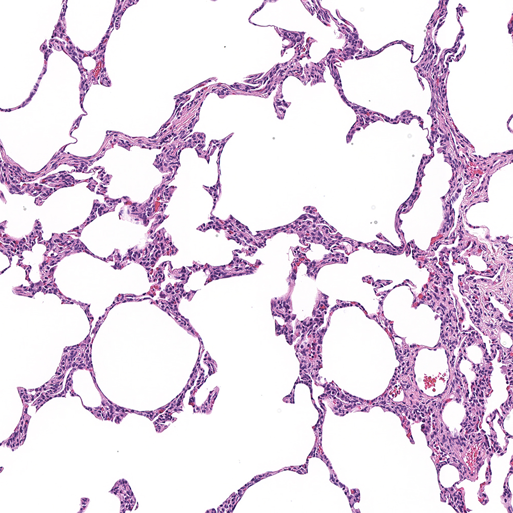
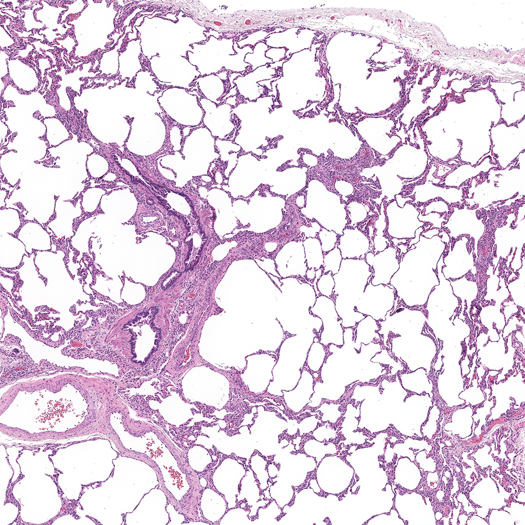
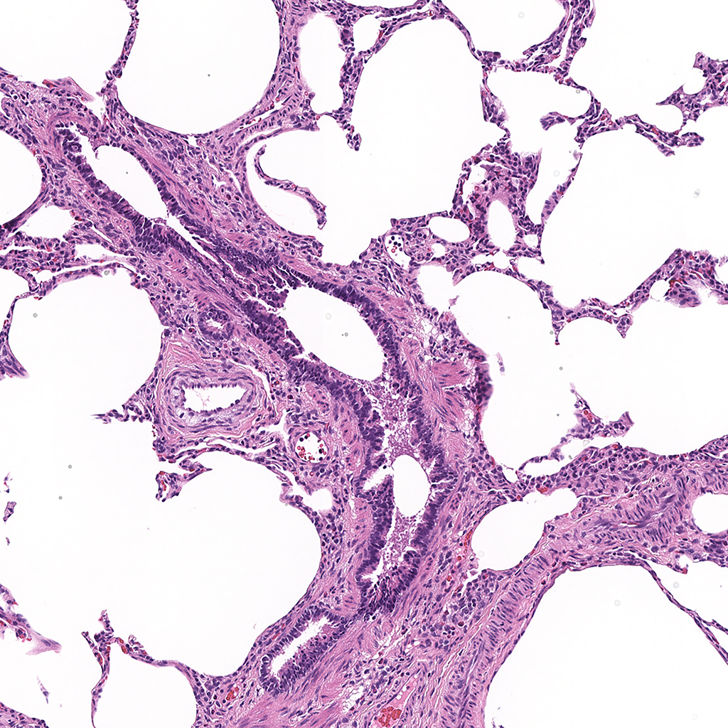
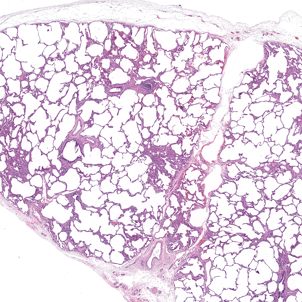
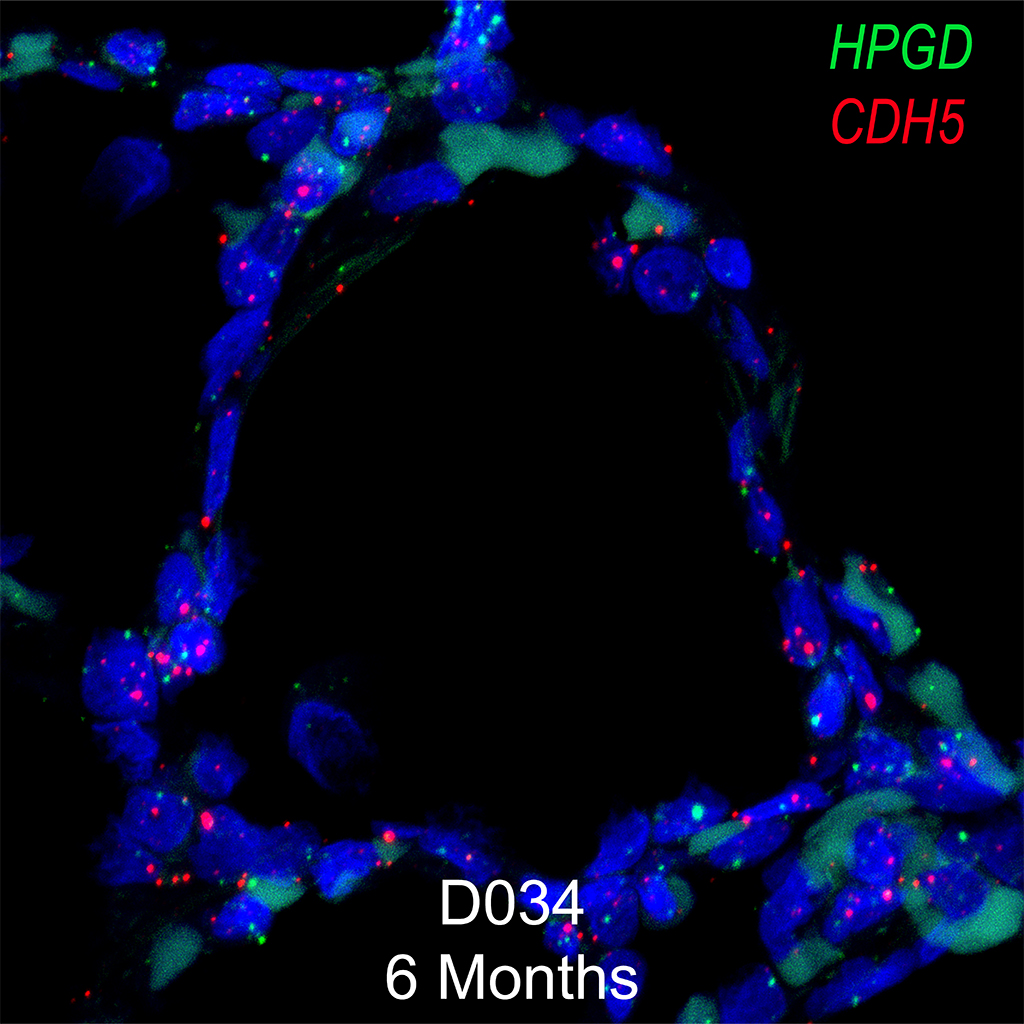
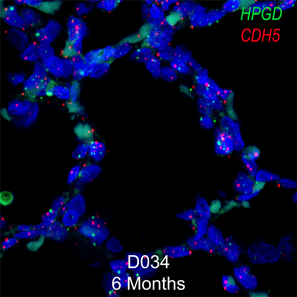
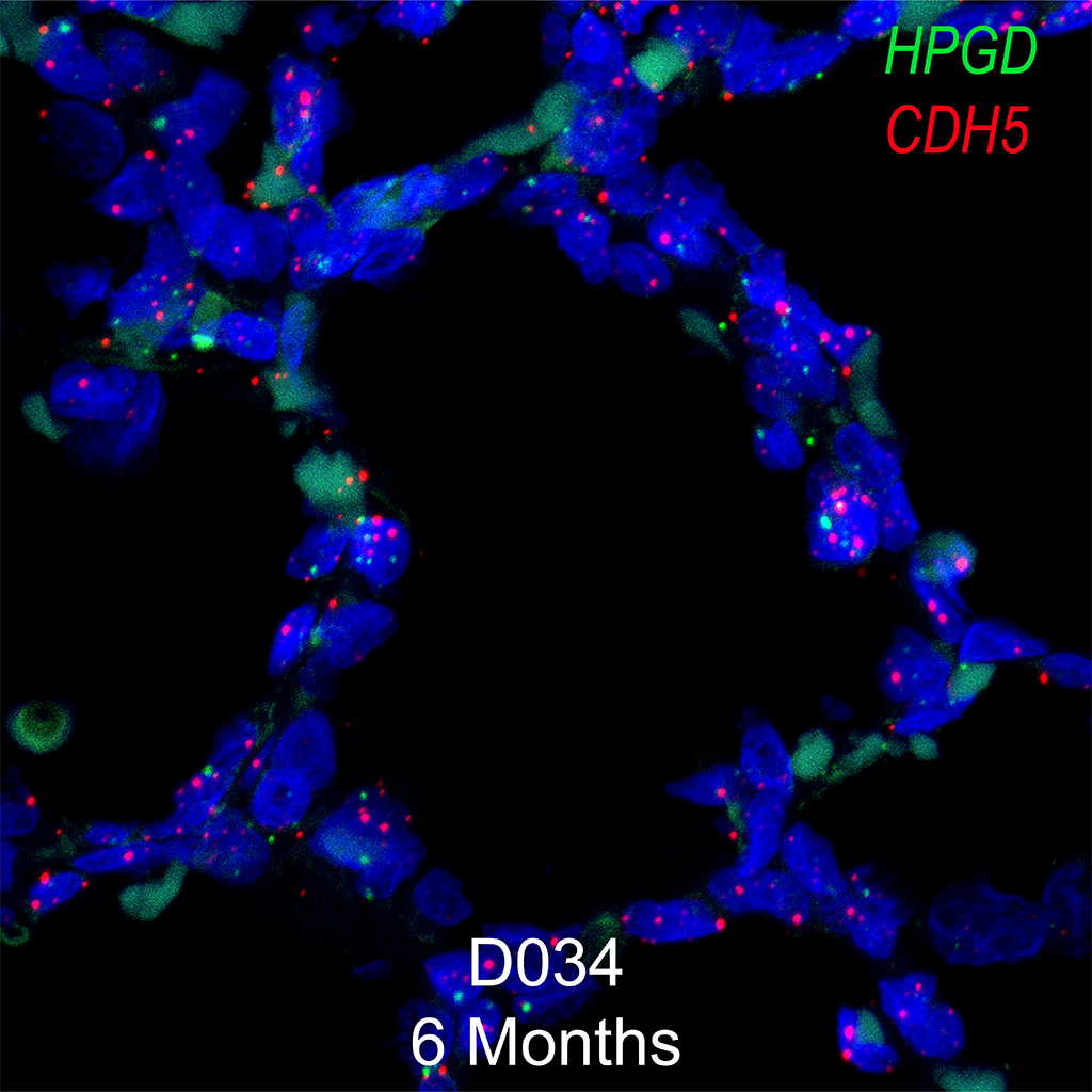
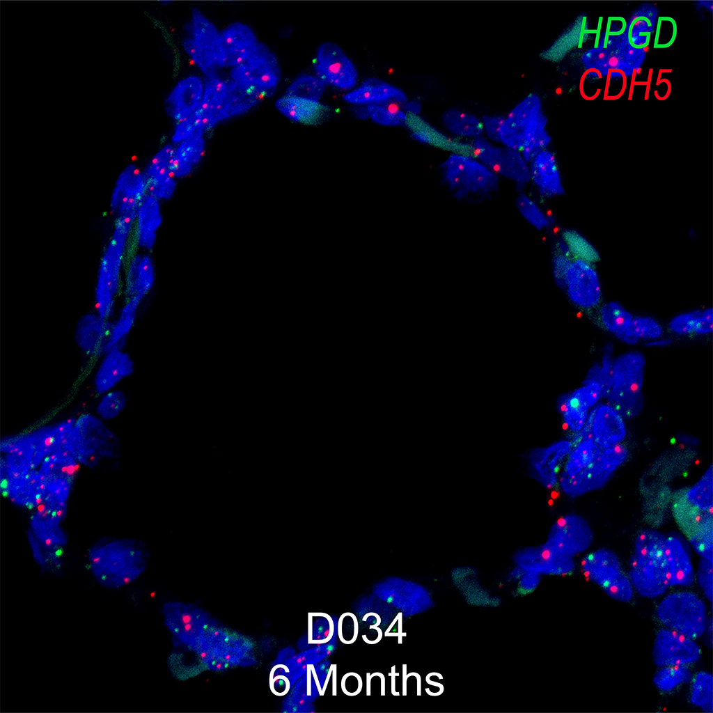
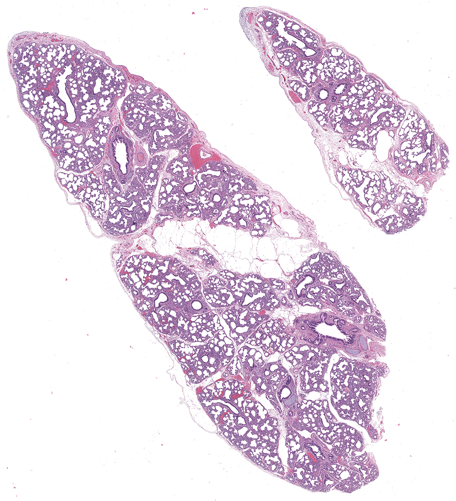
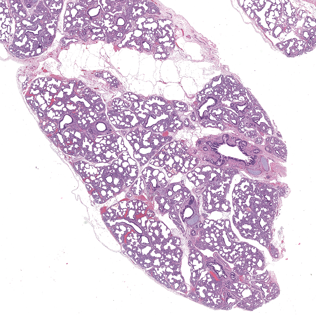
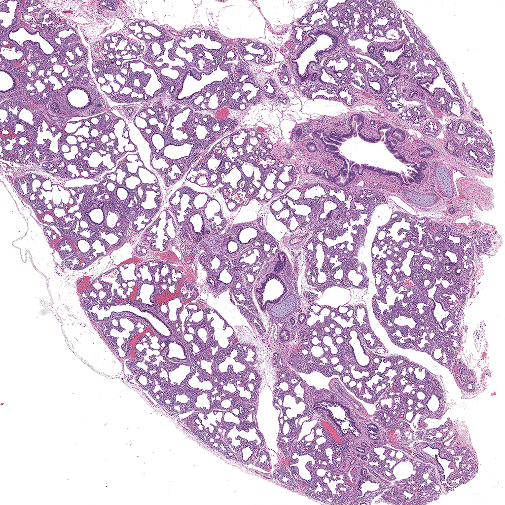
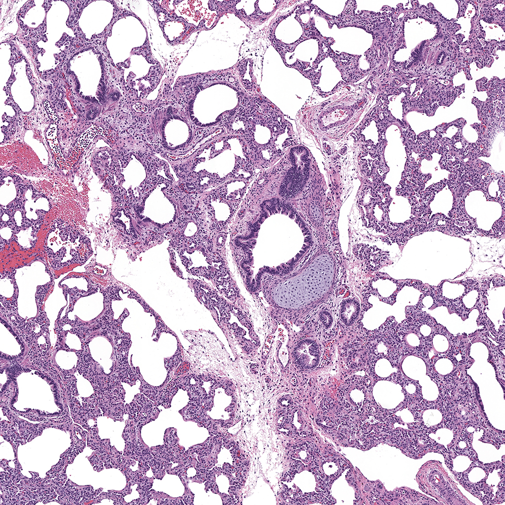
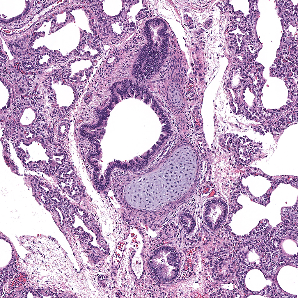
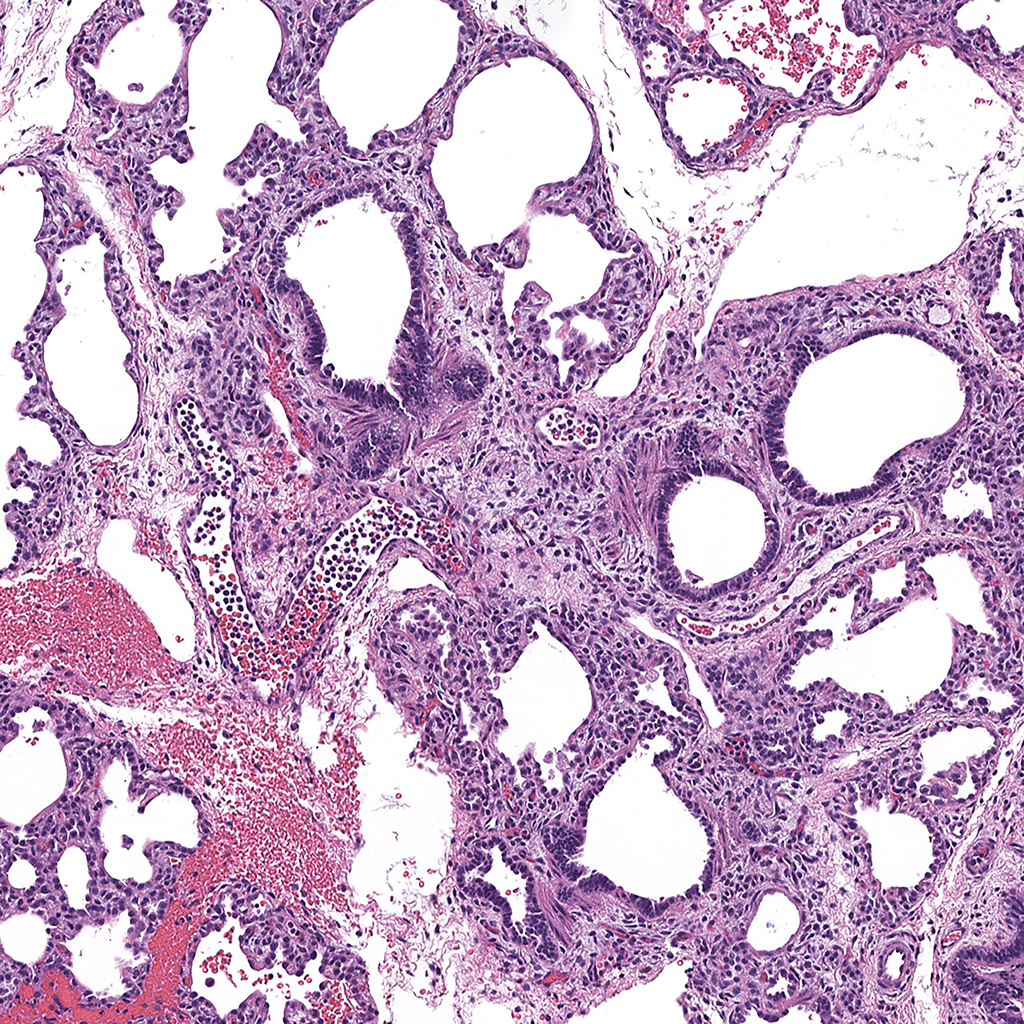
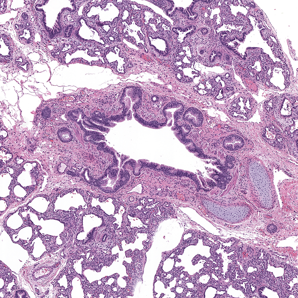
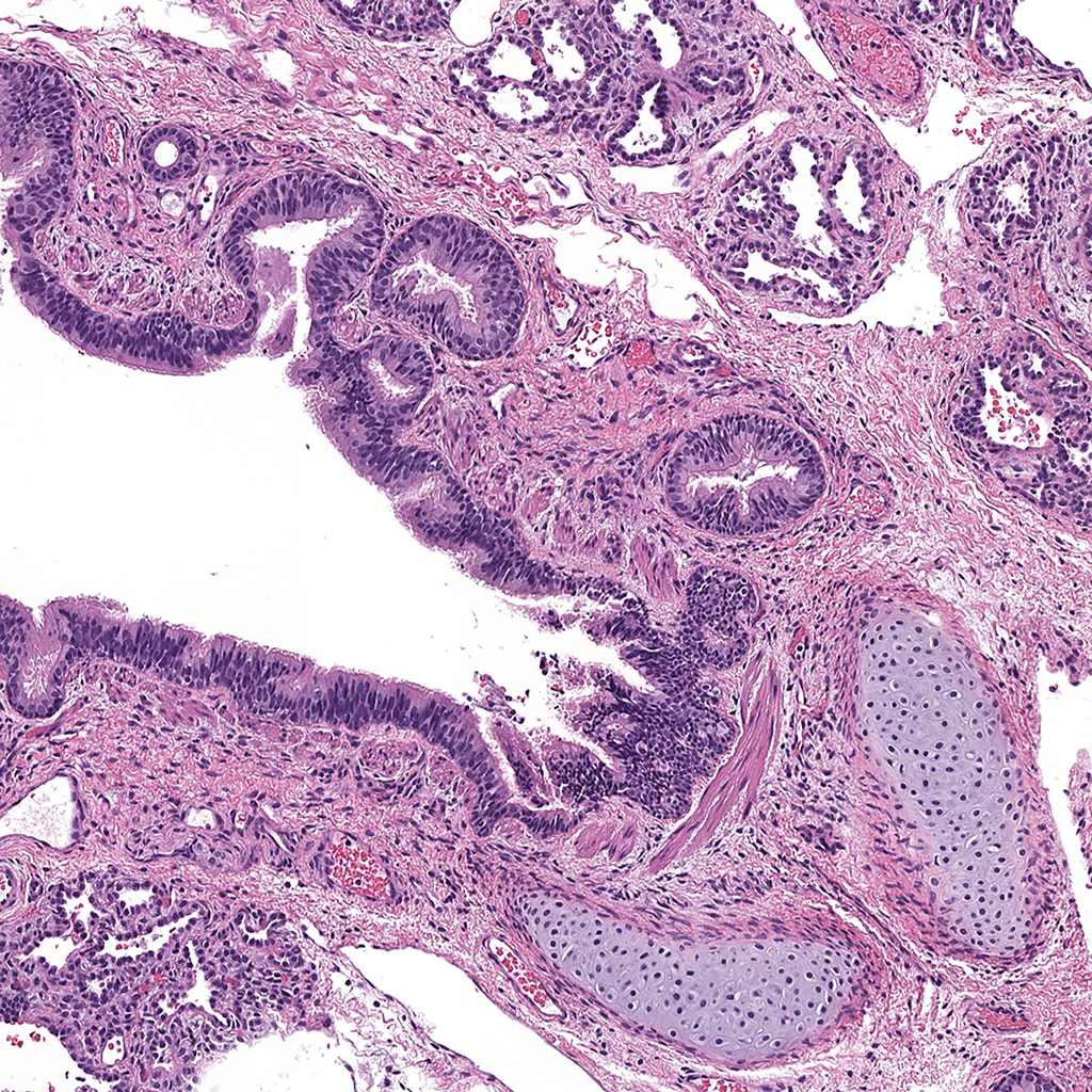
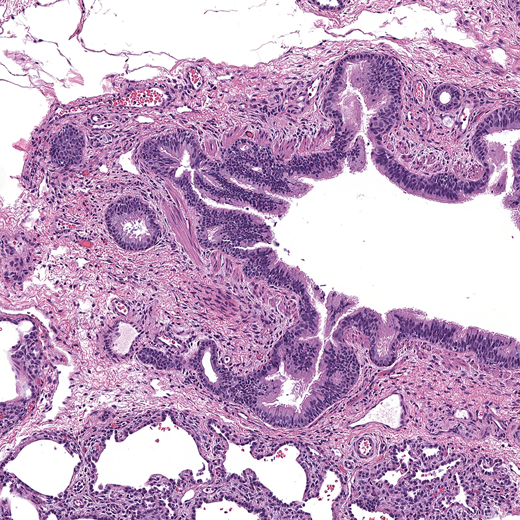
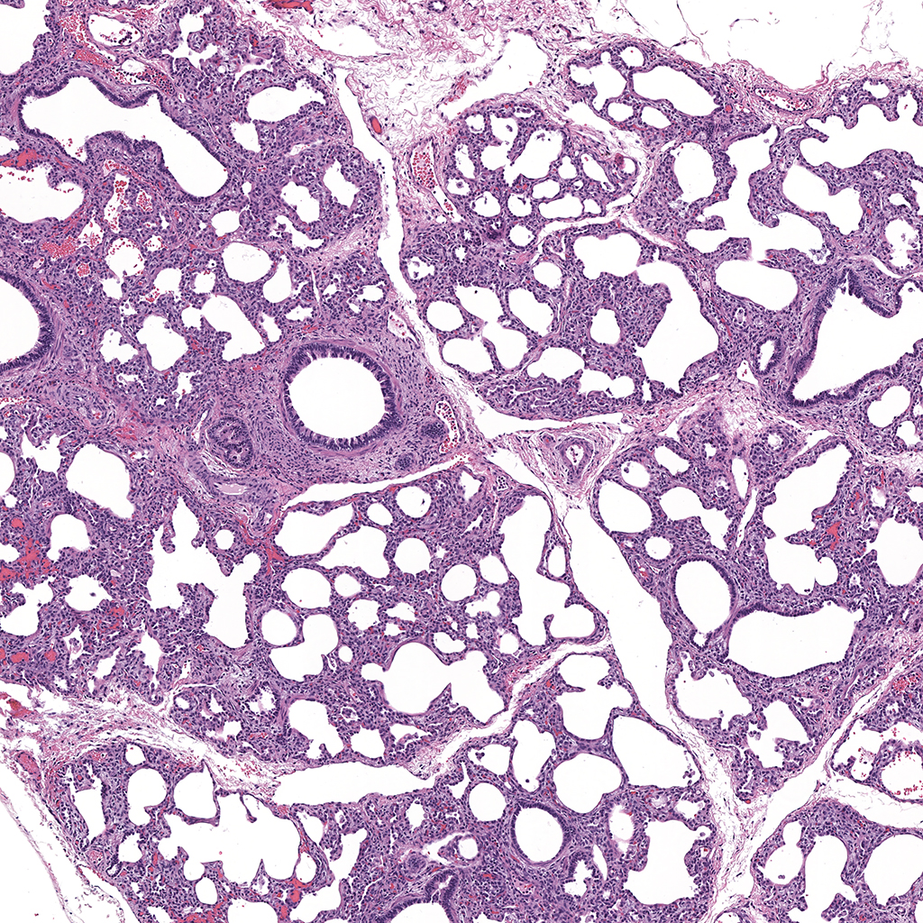
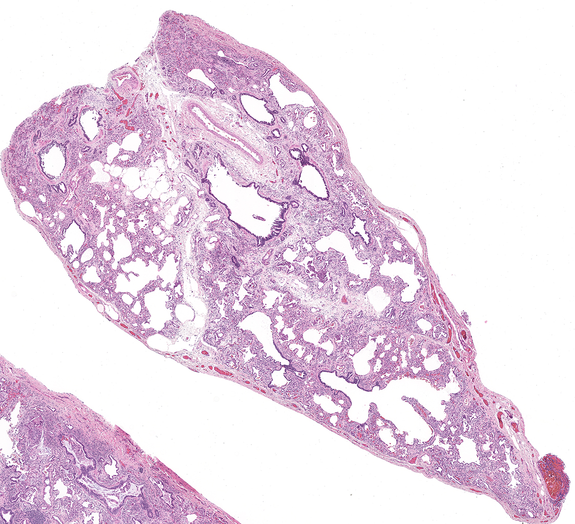
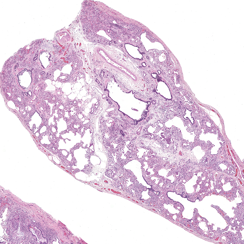
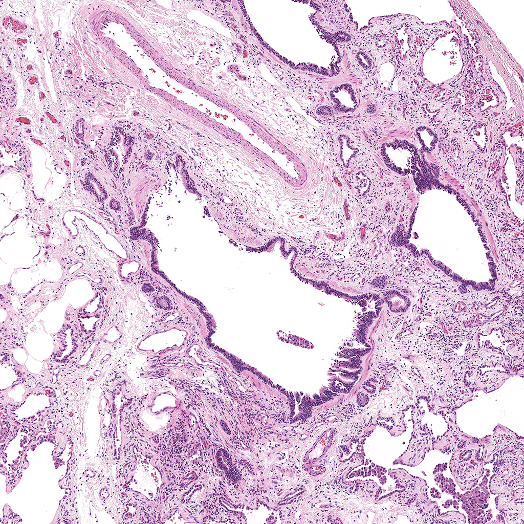
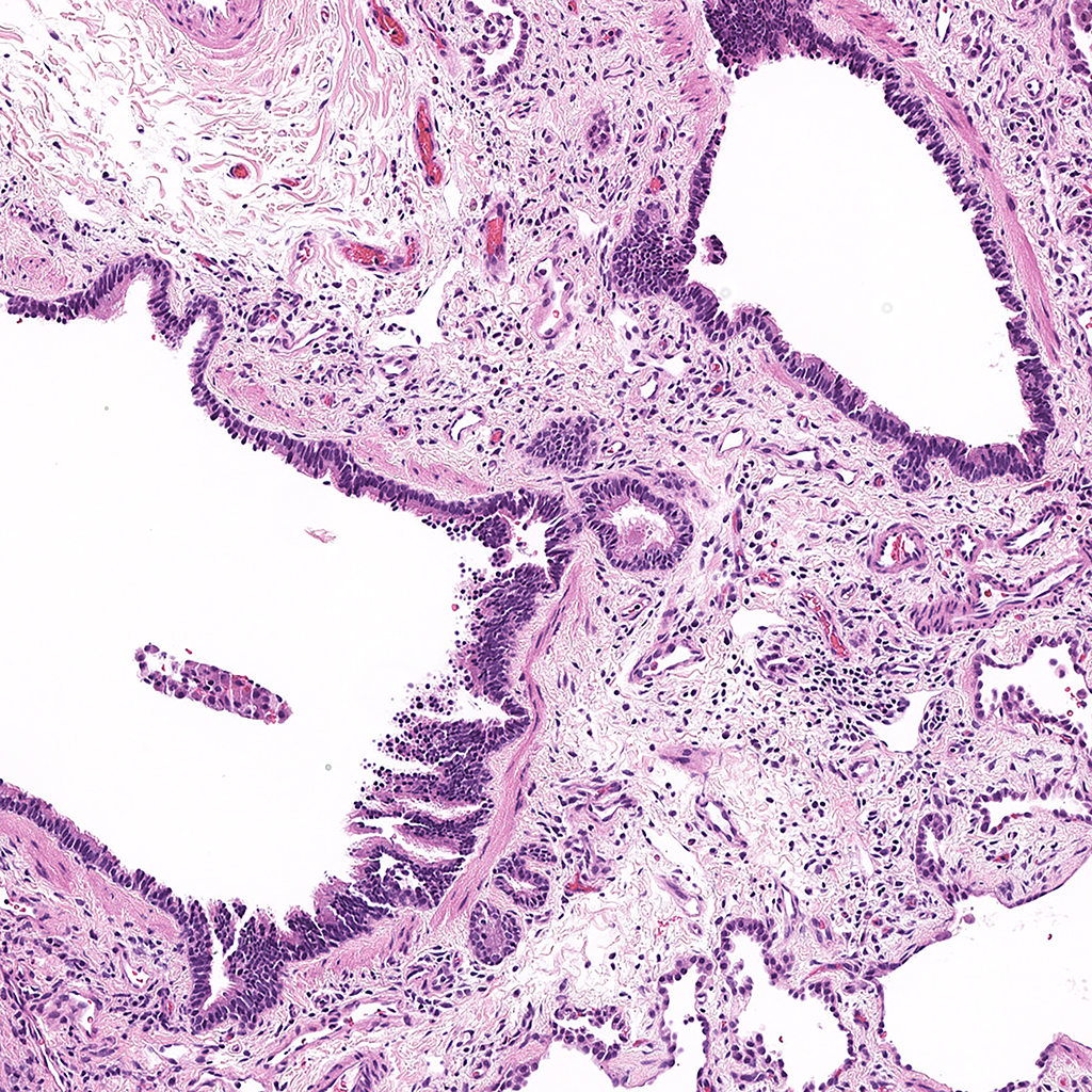
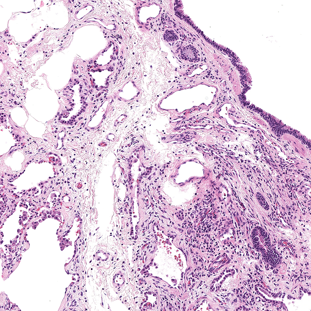
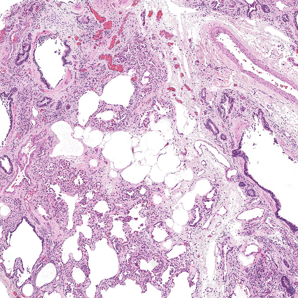
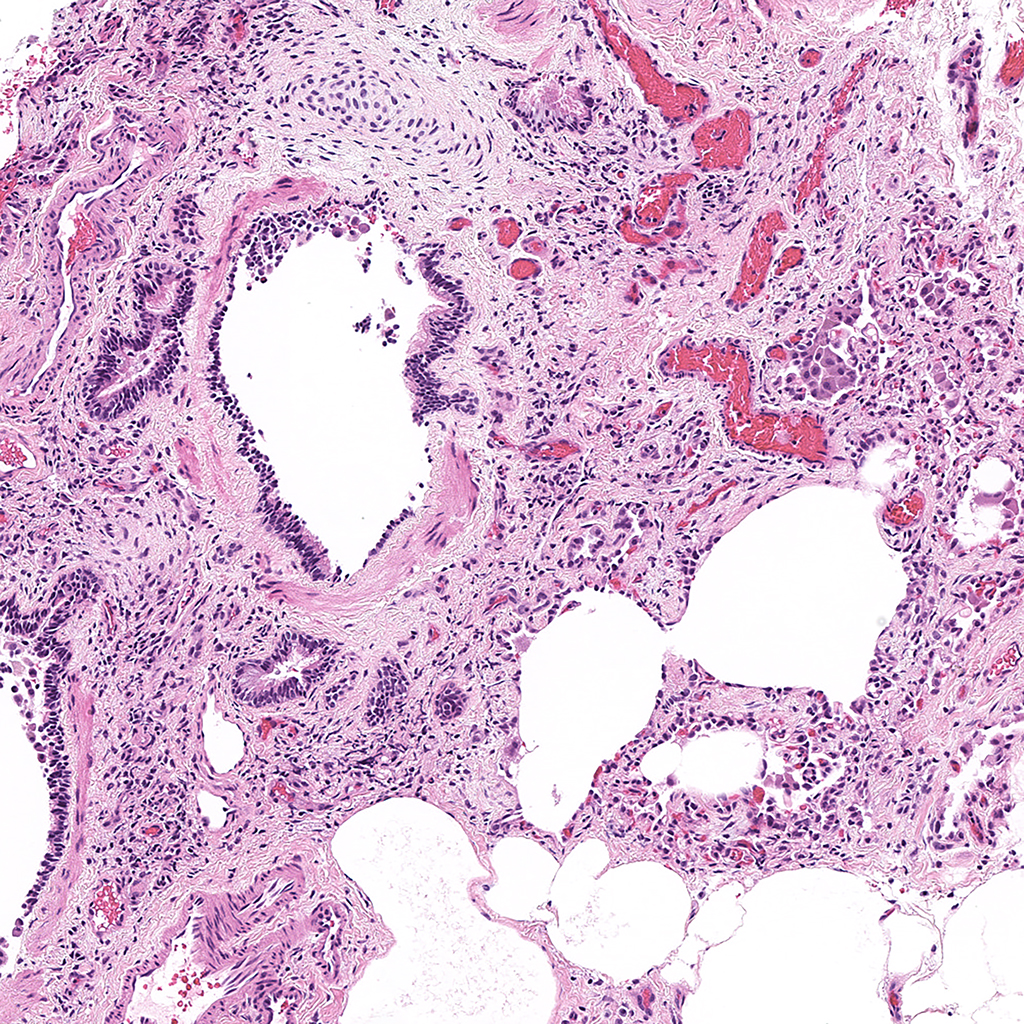
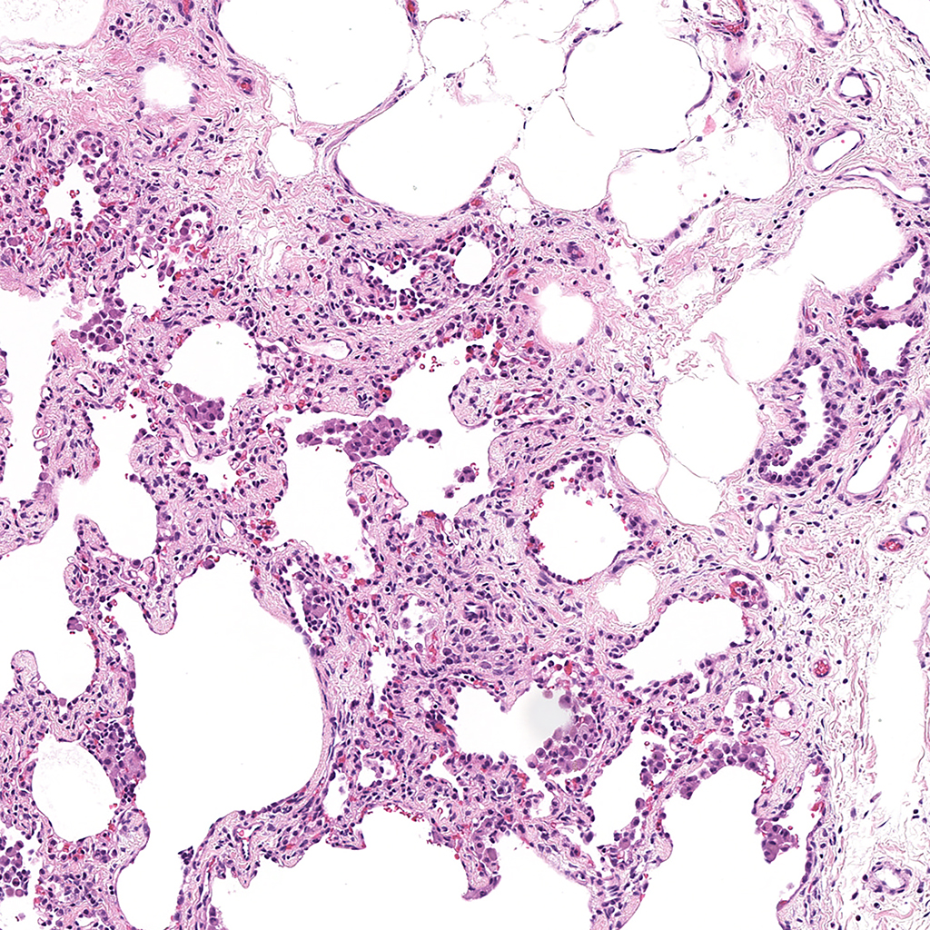
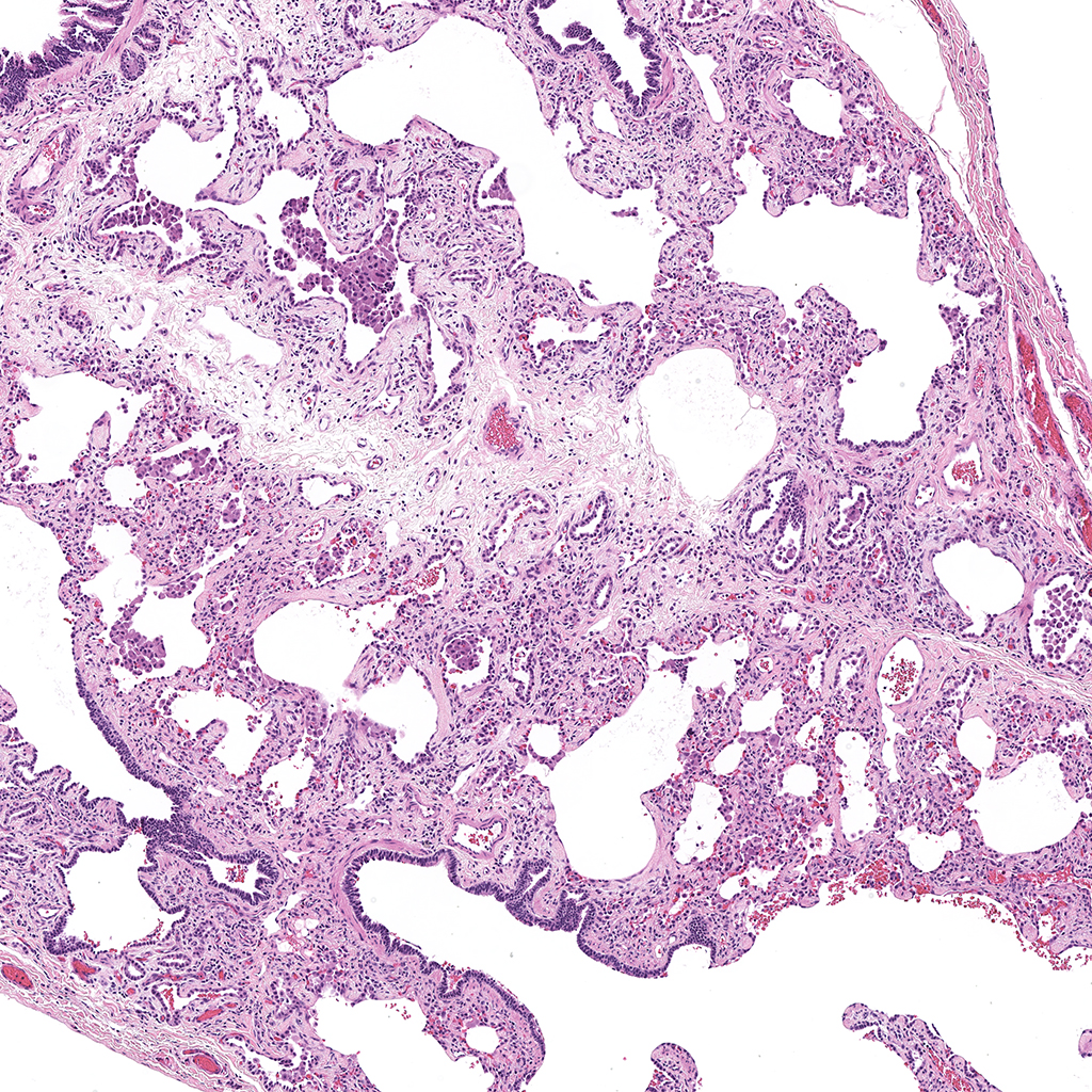
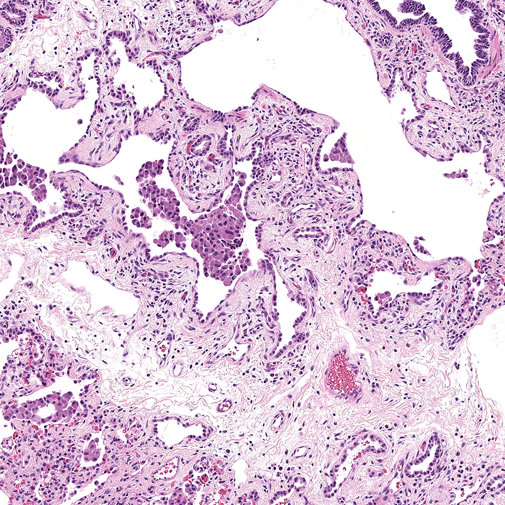
1 Day-Old Human Lung Confocal Imaging SOX9, SOX2 and ACTA2
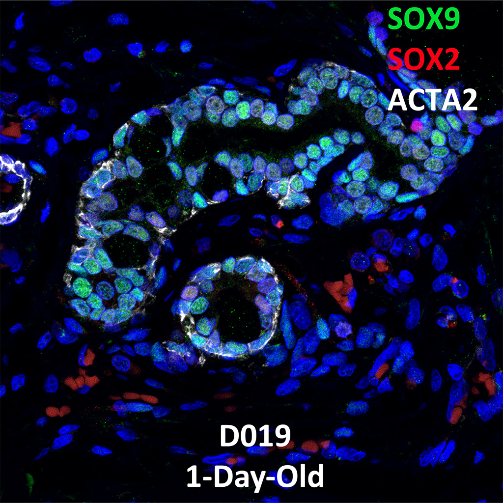

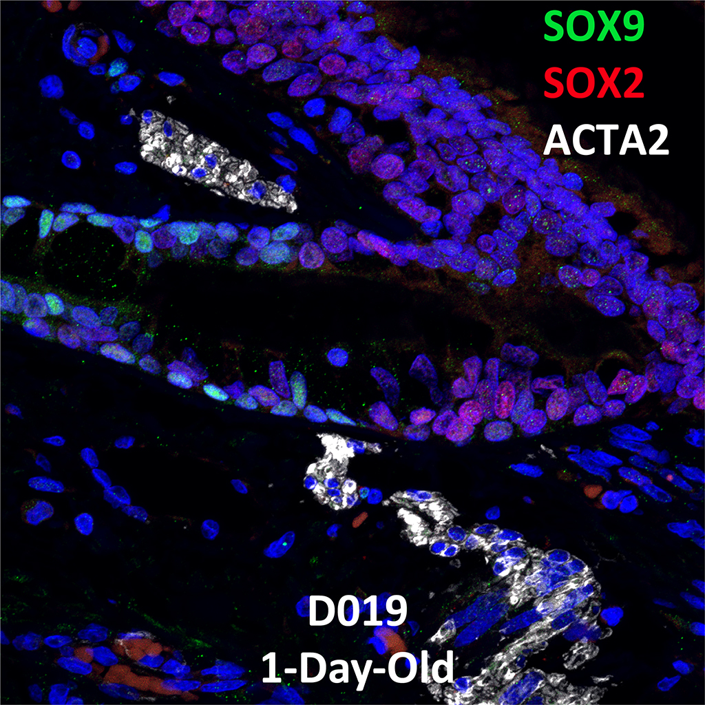
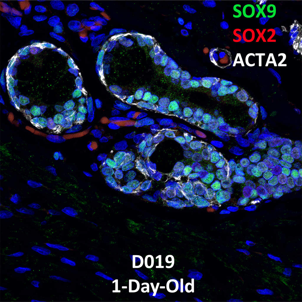
Donor Tissue Kindly Provided by Dr. Gloria Pryhuber from the University of Rochester Medical Center
![]()
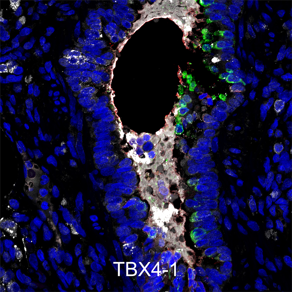
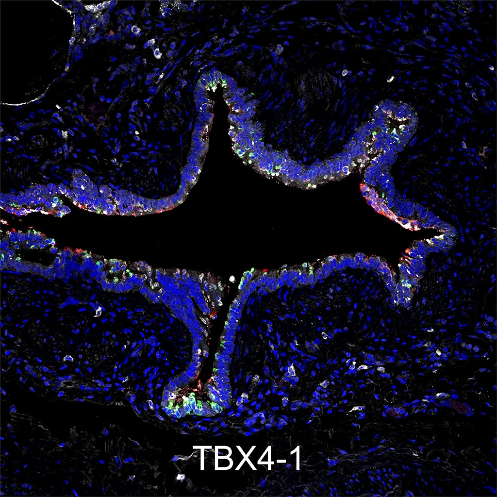
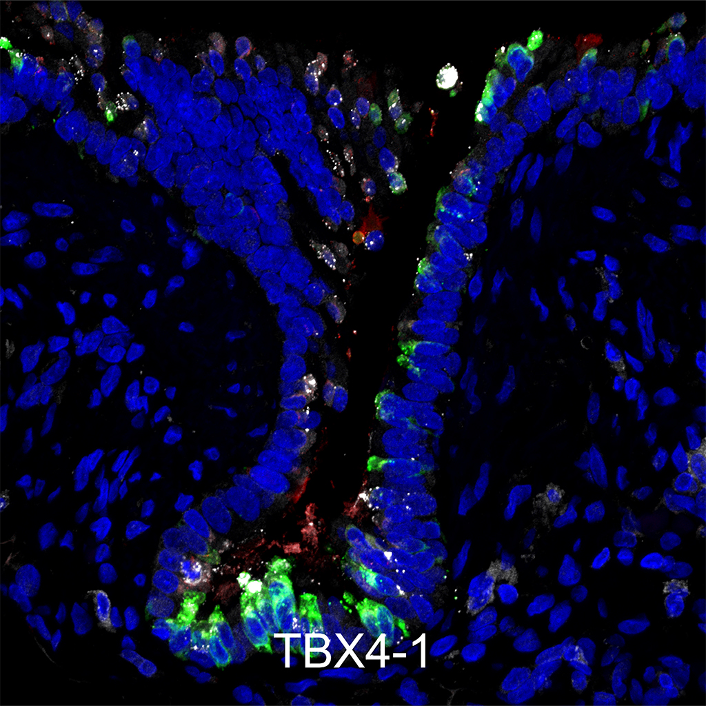
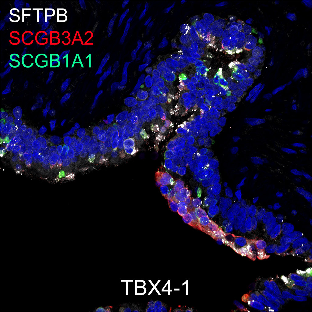
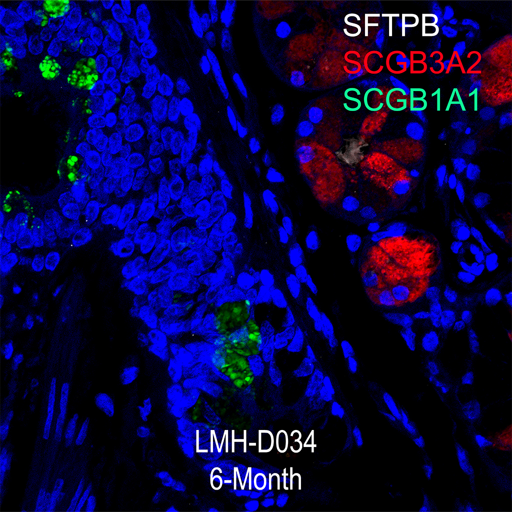
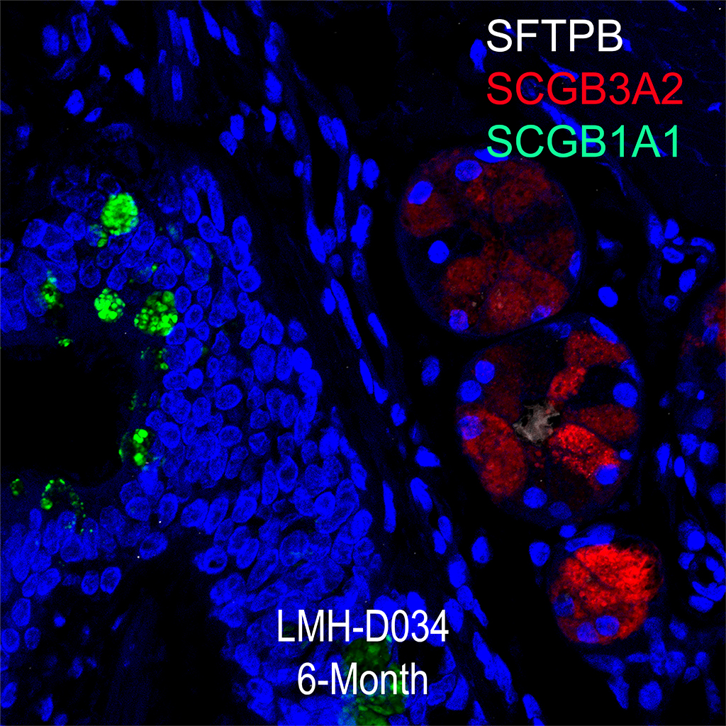
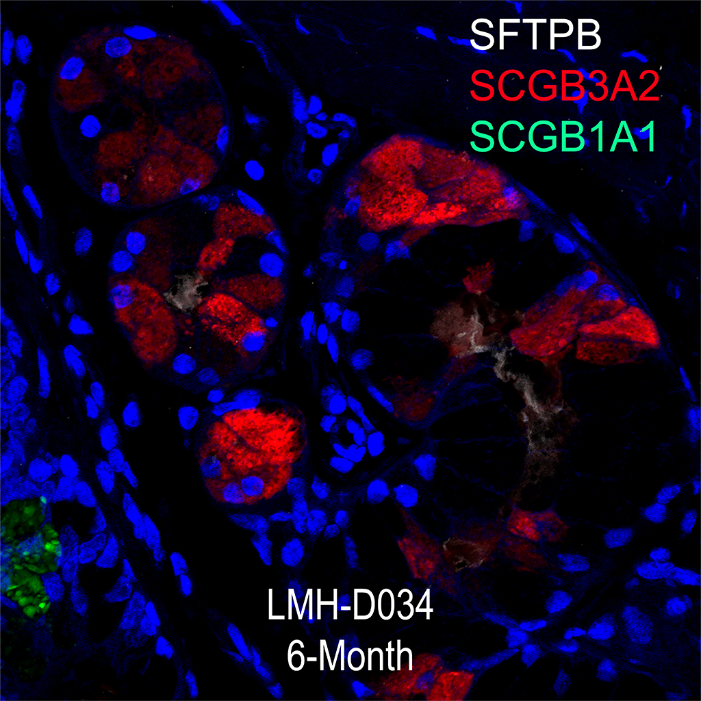
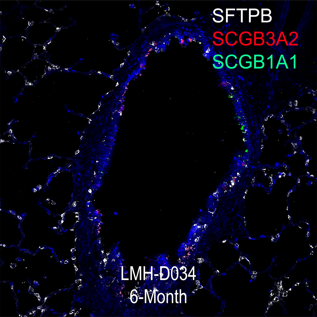
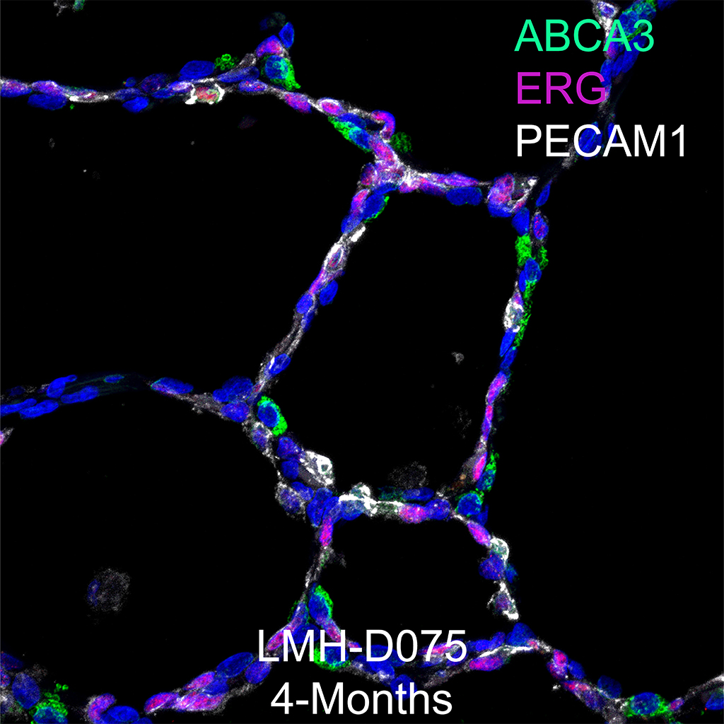
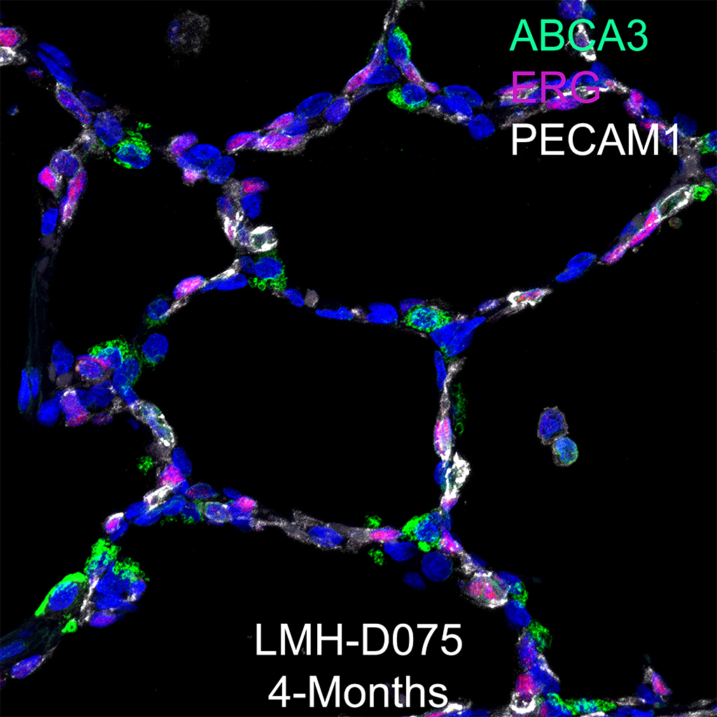
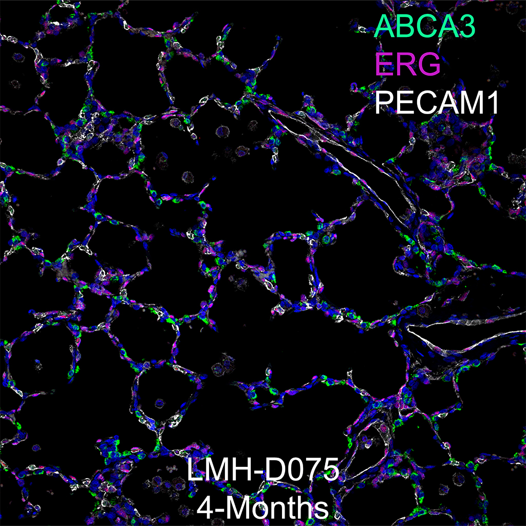
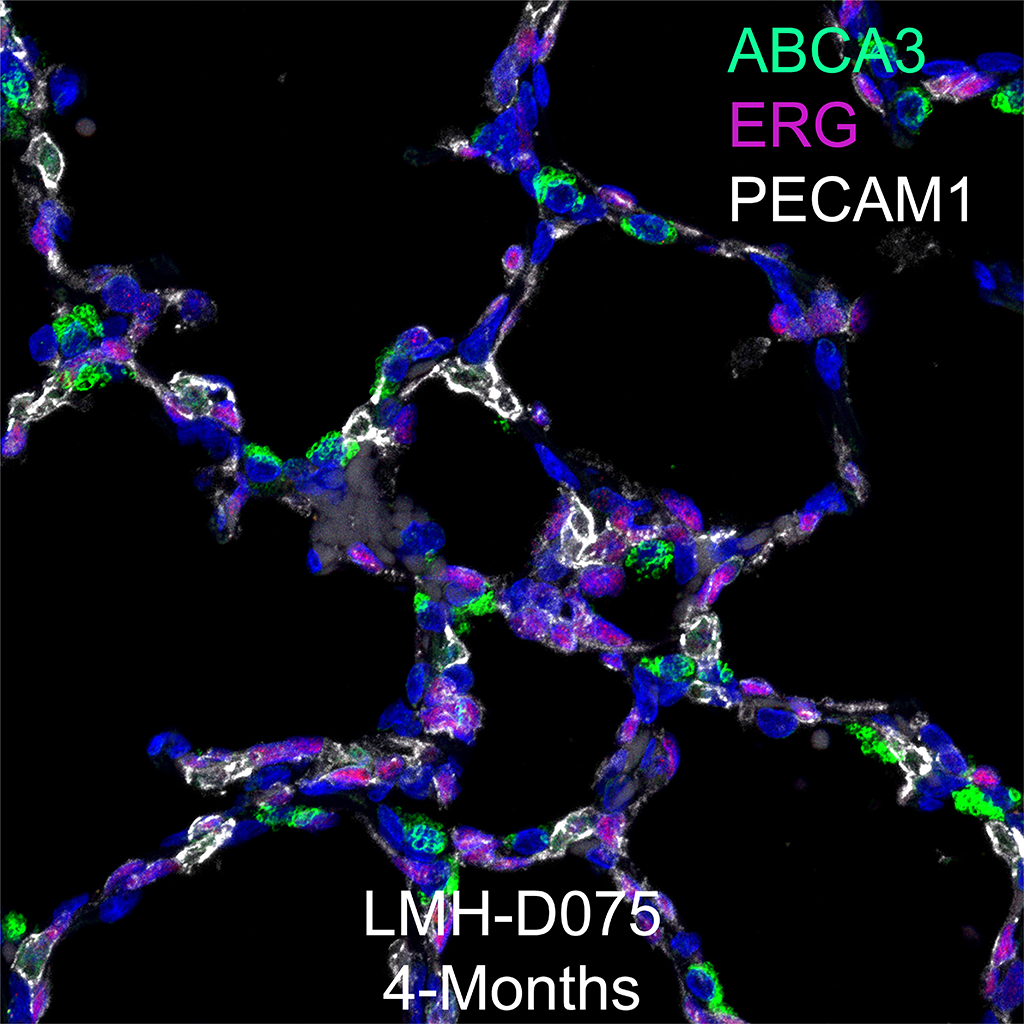
Immunofluorescence: SOX9, SOX2, and ACTA2
Purpose: Stain slides of 4µm frozen sections of human lung from HTC and from Pathology for SOX9, SOX2, and ACTA2 with antigen retrieval.
Day 1
- Paraffin sections are placed at 60oC for 2 hours to overnight to melt paraffin.
- Paraffin sections are then placed in xylene 3X for 10 minutes each, followed by 3X in 100% ETOH for 3 minutes each, 95% ETOH for 3 minutes each, and 70% ETOH for 3 minutes each. Slides are then placed in 1X PBS for 5 minutes to completely rehydrate tissue.
- Antigen retrieval, pH 6.0 (times will vary according to microwave).
- 10 mM sodium citrate, pH 6.0, and heat in a microwave at 96oC. *We usually do a series of three runs (6-7 minutes each run) to equal the time/temp because of evaporation (refill coplin jars with dH2O).
- Microwave according to instructions on microwave.
- Cool on countertop, 15 min.
- Rinse with dH2O
- 1X PBS, 5 min.
- Block in 4% Donkey serum/PBS-T, 2 hours at RT.
- For Rabbit anti-SOX9 (AB–5535, Lot # 2167153, Millipore) dilute 1:100, for mouse anti-SOX2 (SC-365823, Santa Cruz, Lot# C0317) dilute 1:100, for mouse anti-ACTA2 (α Smooth muscle actin, A5228, Sigma) dilute 1:2000 in blocking buffer. Spin down in µfuge for 10 minutes and incubate on tissue overnight @ 4oC.
Day 2
- Rinse slides in PBS-T 3X, 5 min.
- Apply secondary antibody , Donkey Alexa Fluor 488 anti-rabbit IgG (A21206, Lot# 1608521, for anti-SOX9) at 1:200, Goat Alexa Fluor 568 anti-mouse IgG1 (A21124, Lot# 1600878, for anti-SOX2) at 1:200, Donkey Alexa Fluor 633 anti-mouse IgG2A A21136, Lot# 1345055, ACTA2) at 1:200, in blocking buffer. Spin down in µfuge for 10 min, apply to tissue and incubated at room temperature for 1 hour.
- Rinse in PBS-T 3X, 5 min.
- Dilute DAPI 1:2000 and apply to slides for 10 min.
- Wash in PBS-T 3X, 5 min.
- Rinse slides in 0.1M PB or TB, 3X, 5 min.
- Add 1 drop of Prolong Gold anti-fade mounting medium (P36930).
- Coverslip with Gold Seal Coverslip (Cat# 3422 Electron Microscopy Sciences, 22 X 22 mm).
- Allow Prolong Gold to cure overnight at room temp in light sealed box.
- Store slides in light sealed box @ 4oC
Tissue Used:
LMH-D019-RLL-5B1.22_SOX9_SOX2_ACTA2
Gender: Male
Age: 1 Day
Appendix
Antigen Retrieval Solution
18ml Solution A- 0.1M Citric Acid (Sigma, C1909) pH 2.5
82ml Reagent B- 0.1M Sodium Citrate (Sigma, S4641 ) pH 8.2
1L dH2O
pH 6.0


