
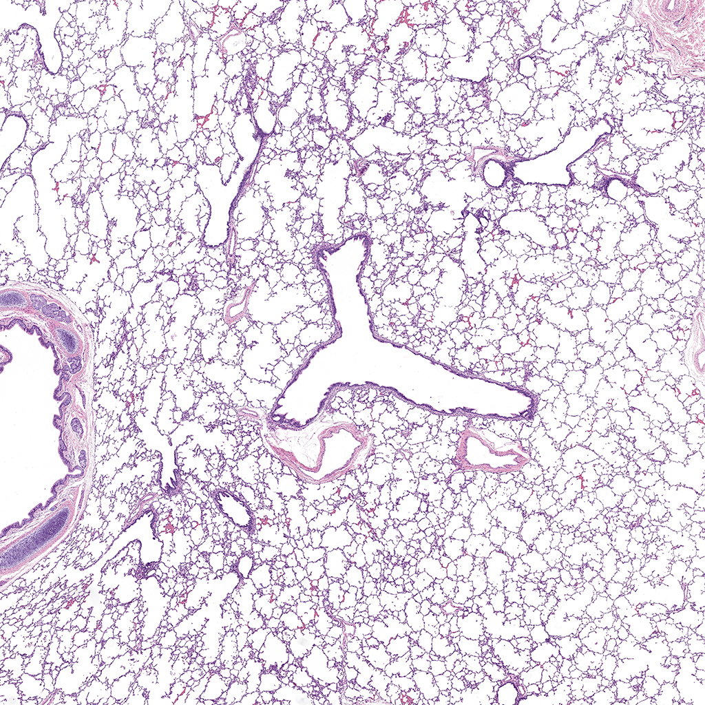
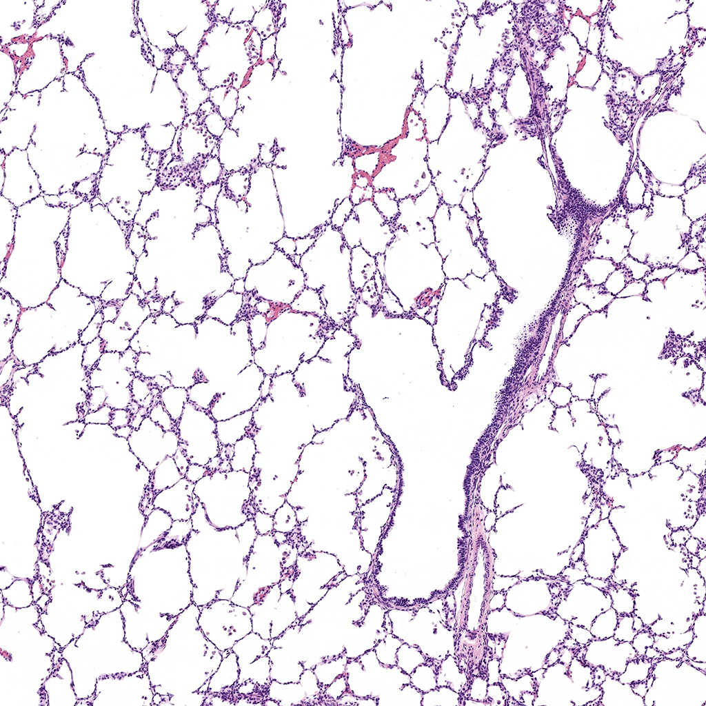
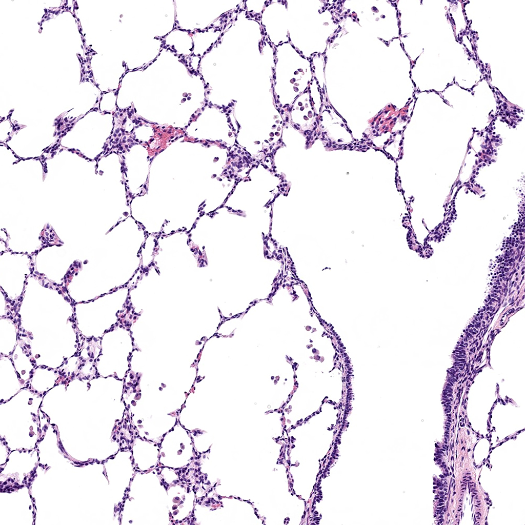
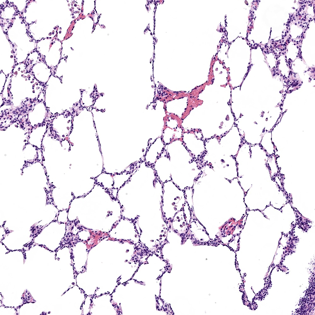
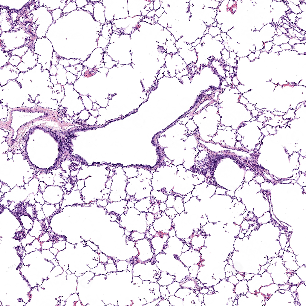
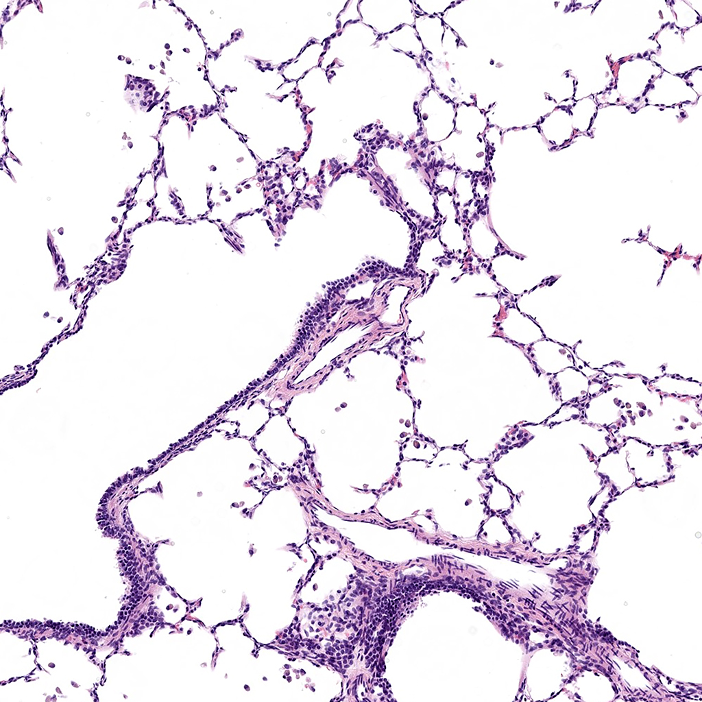
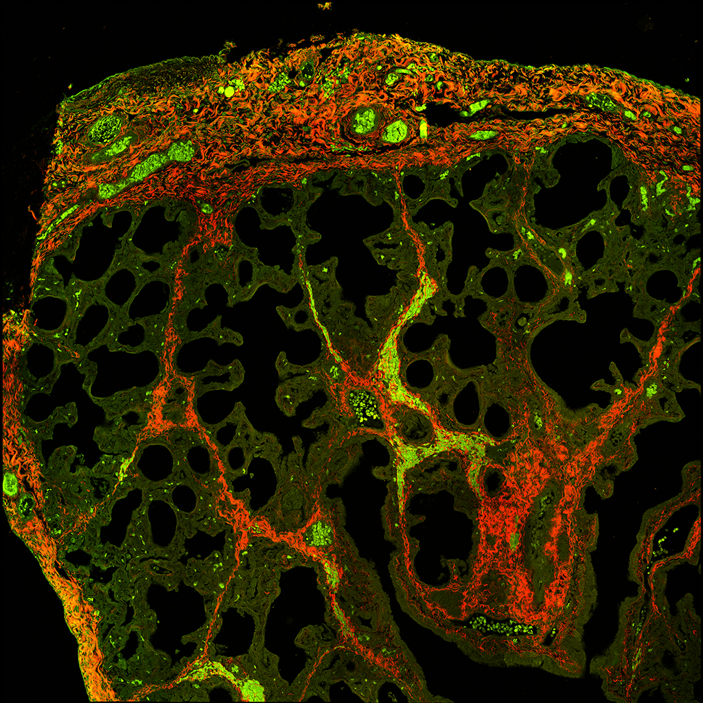
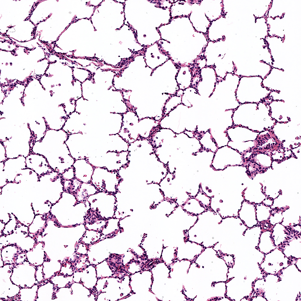
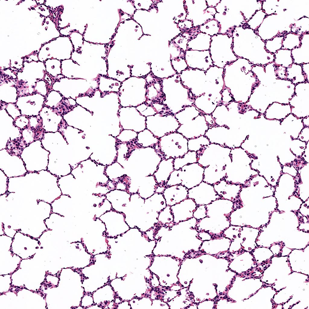
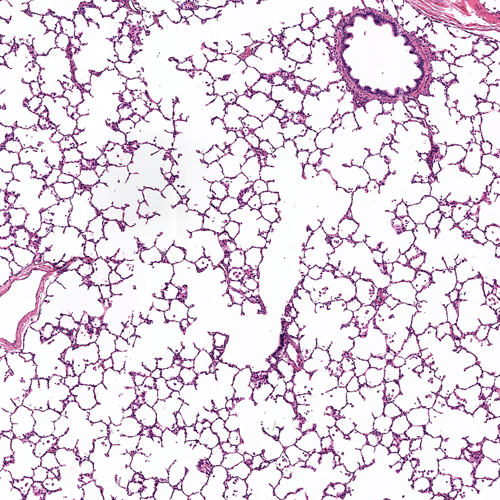
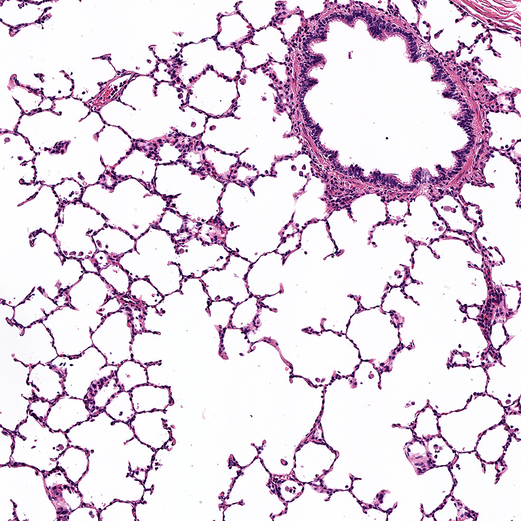
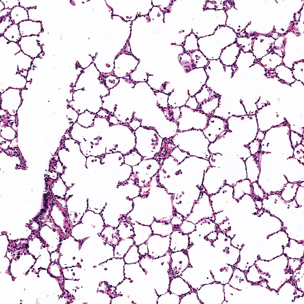
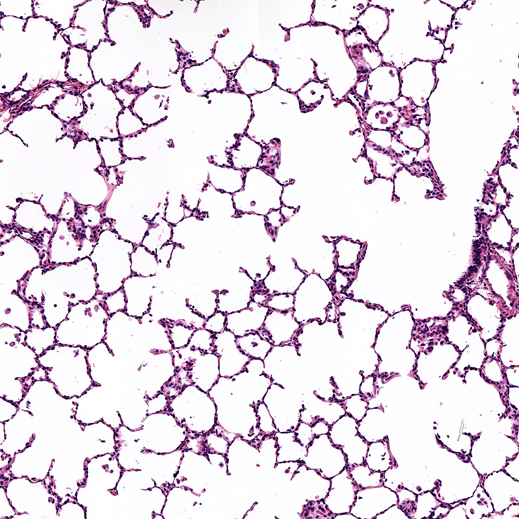
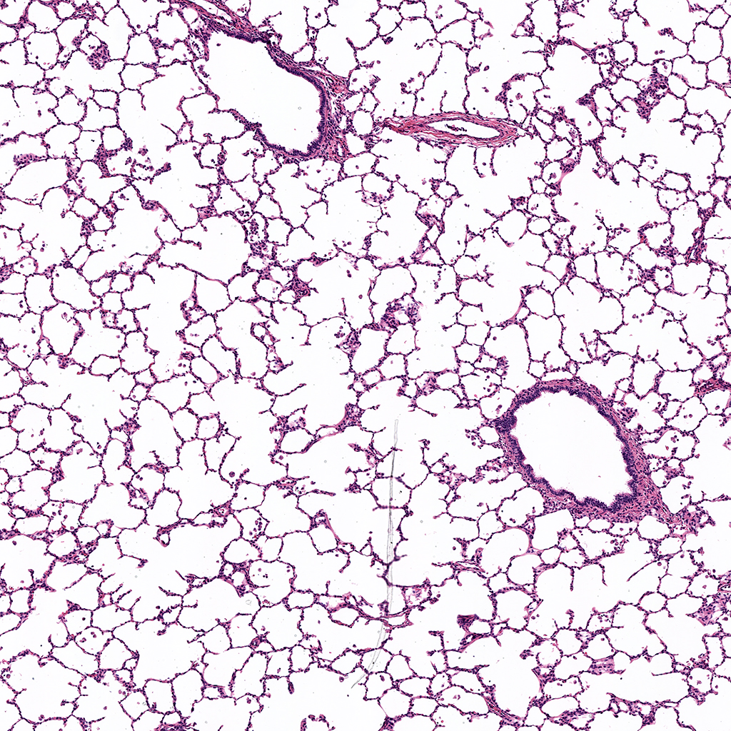
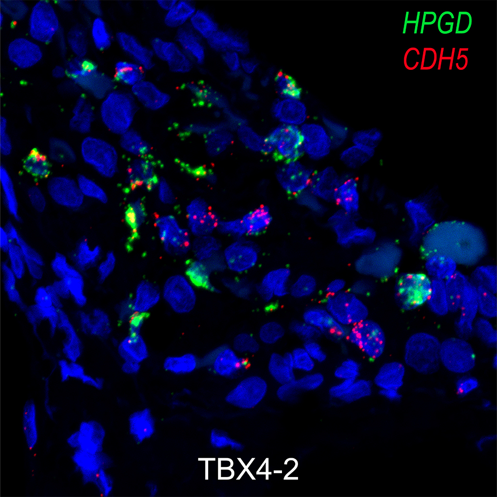
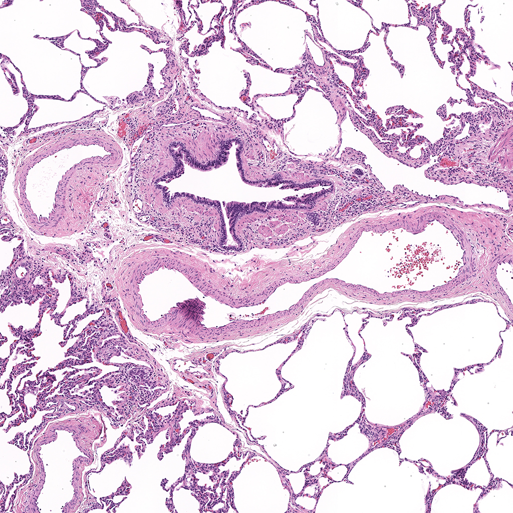
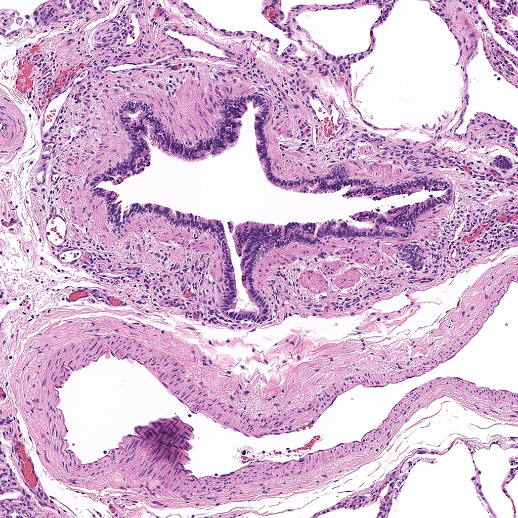
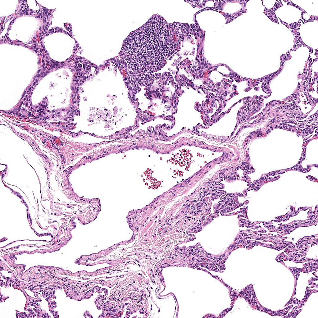
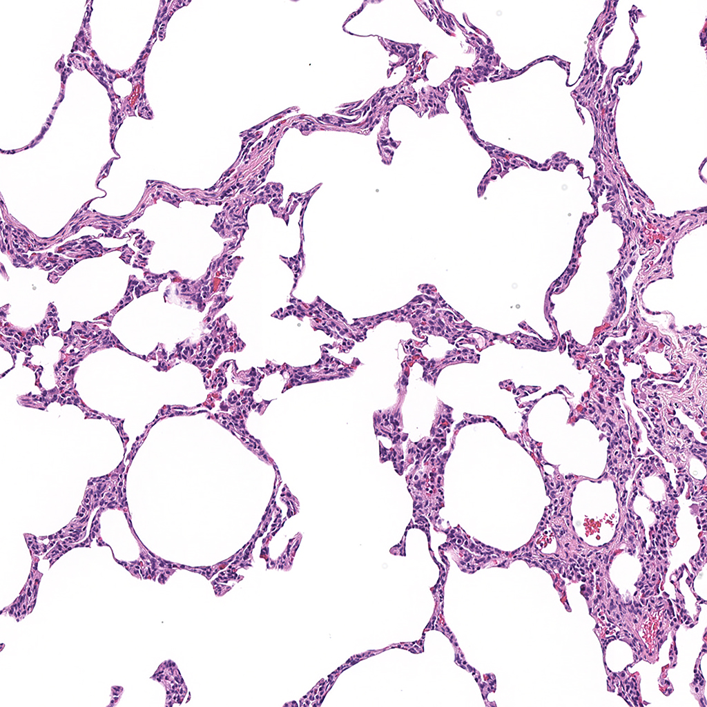
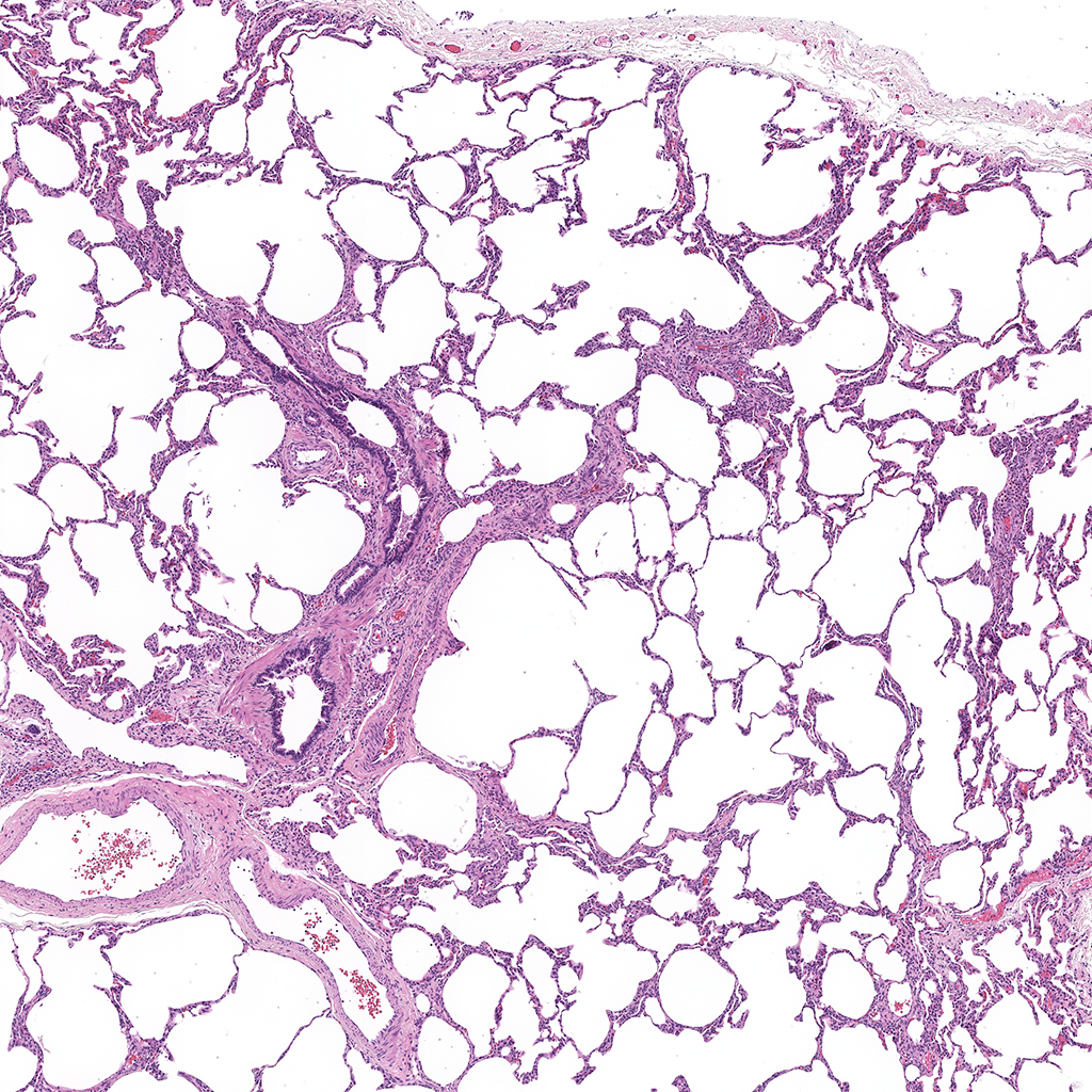
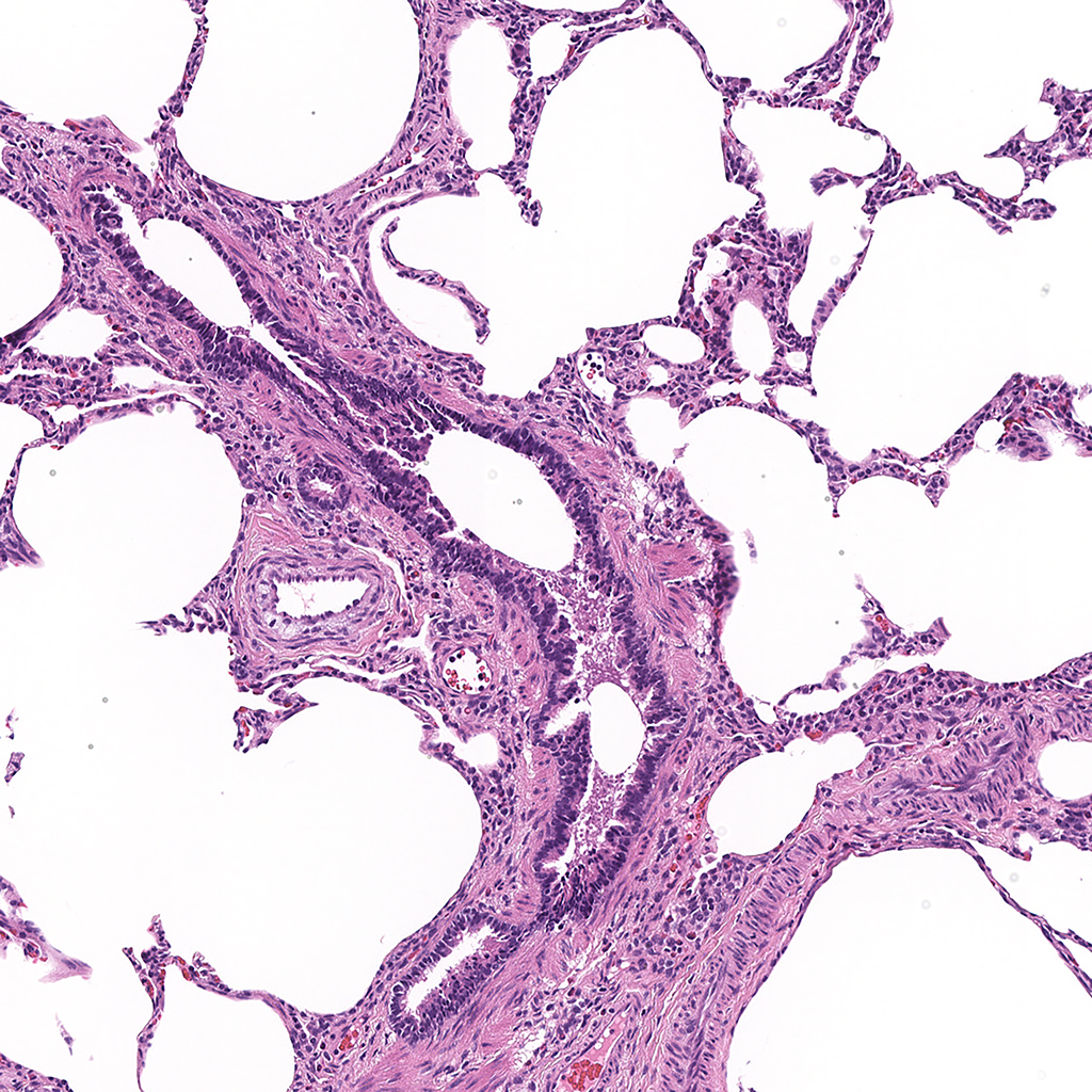
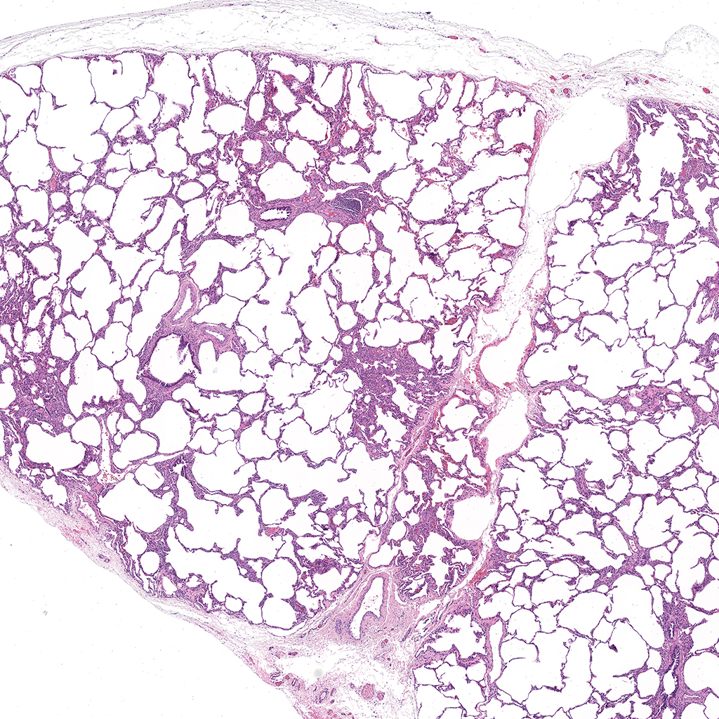
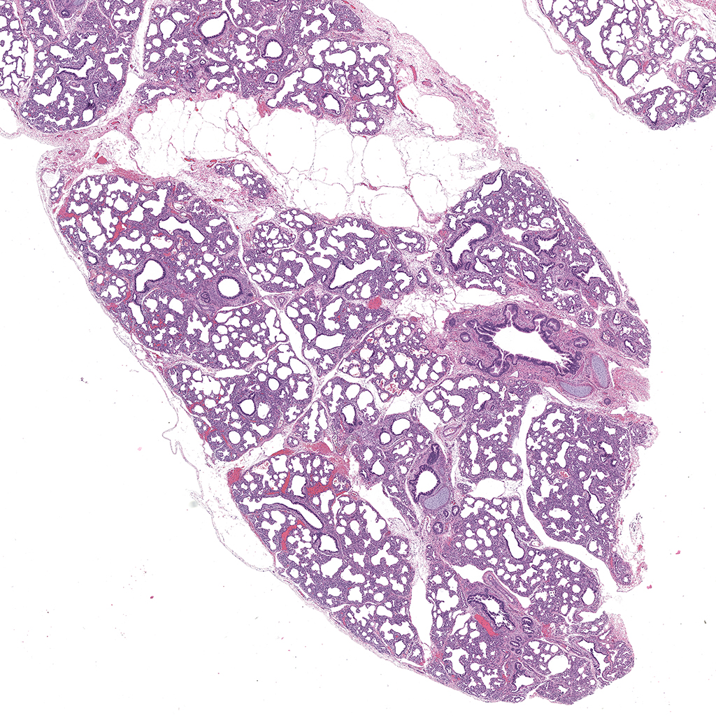
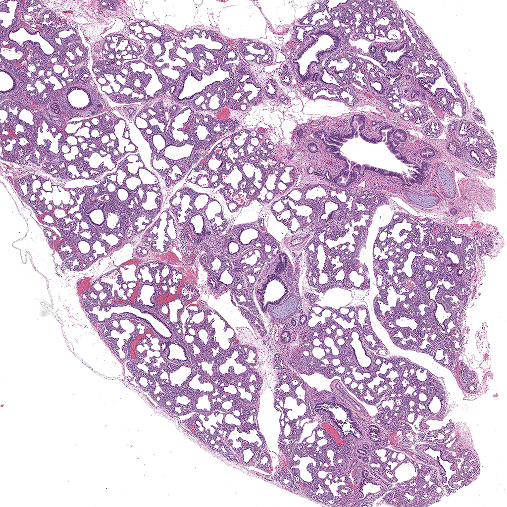
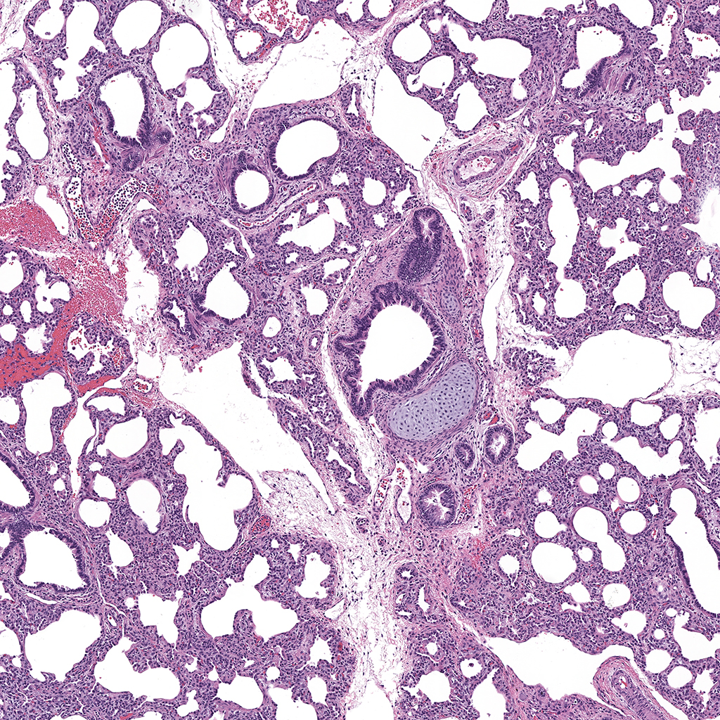
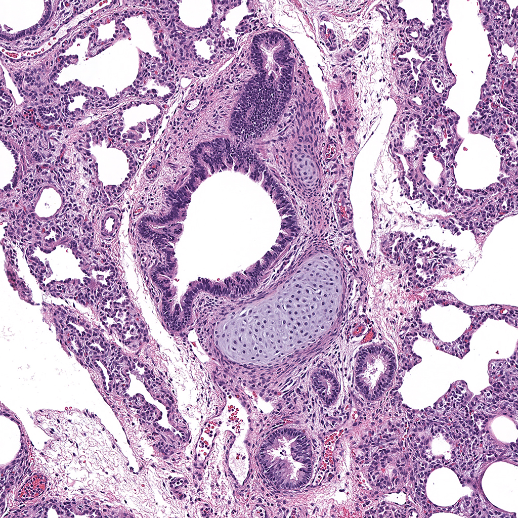
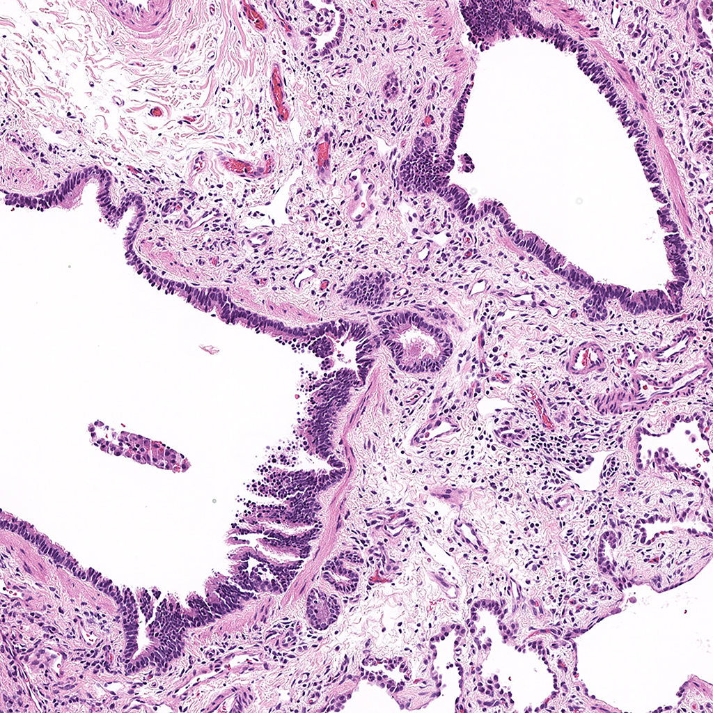
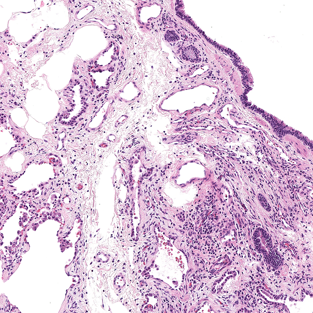
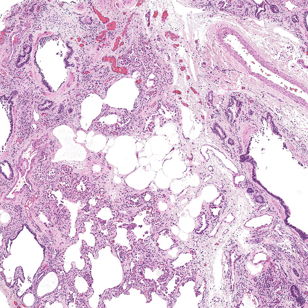
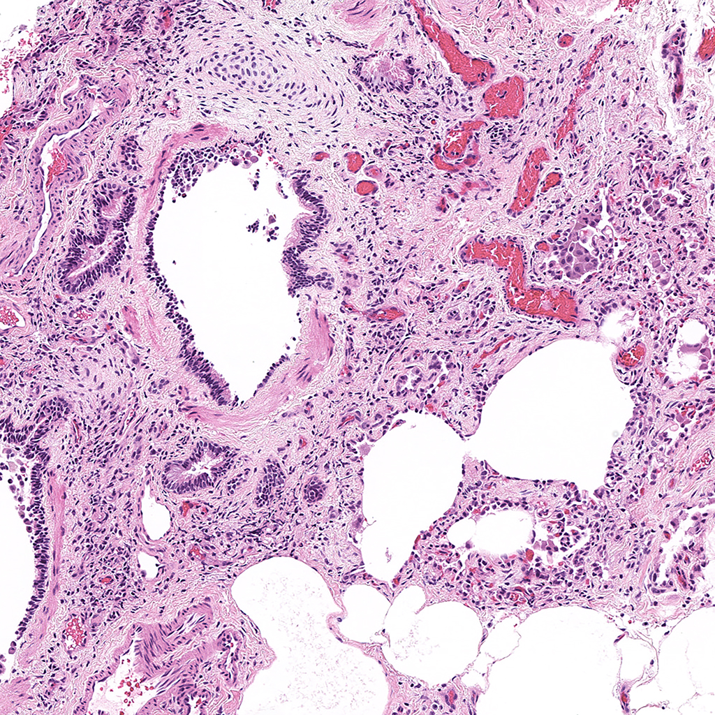
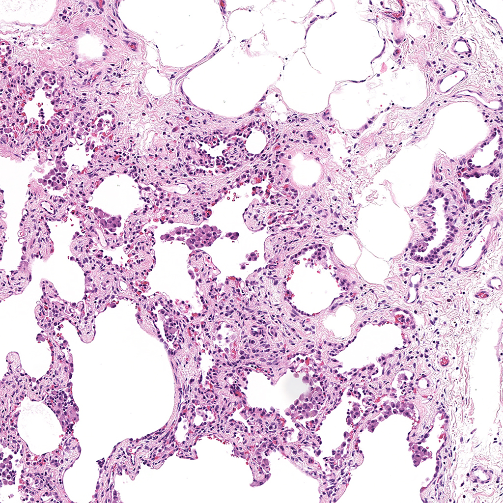
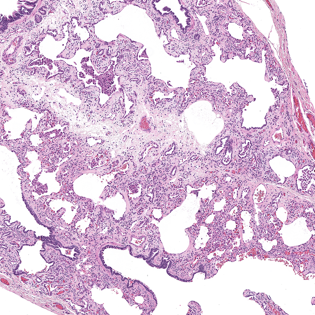
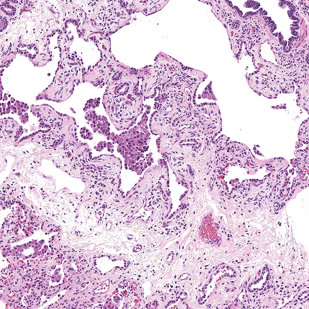
Confocal Imaging for C57BL6
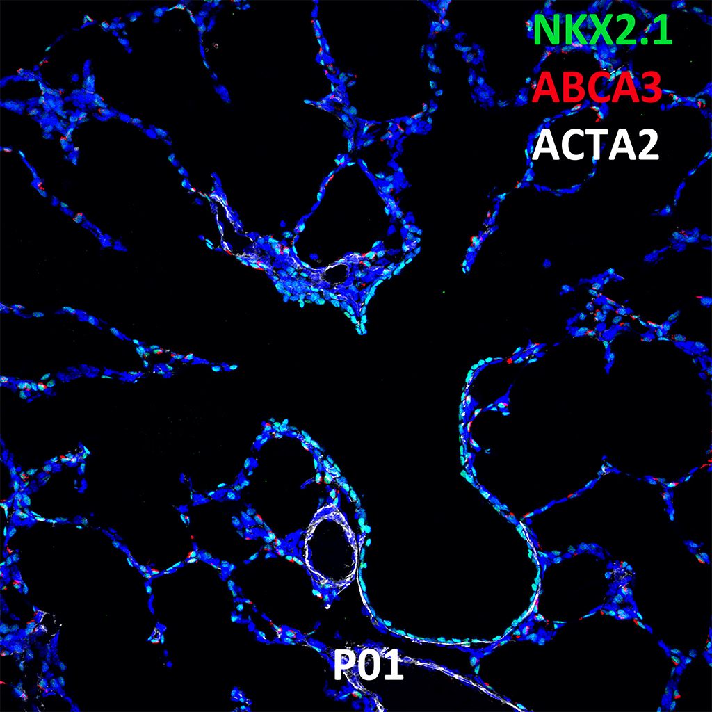
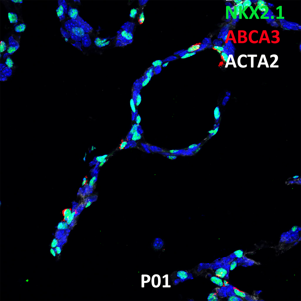
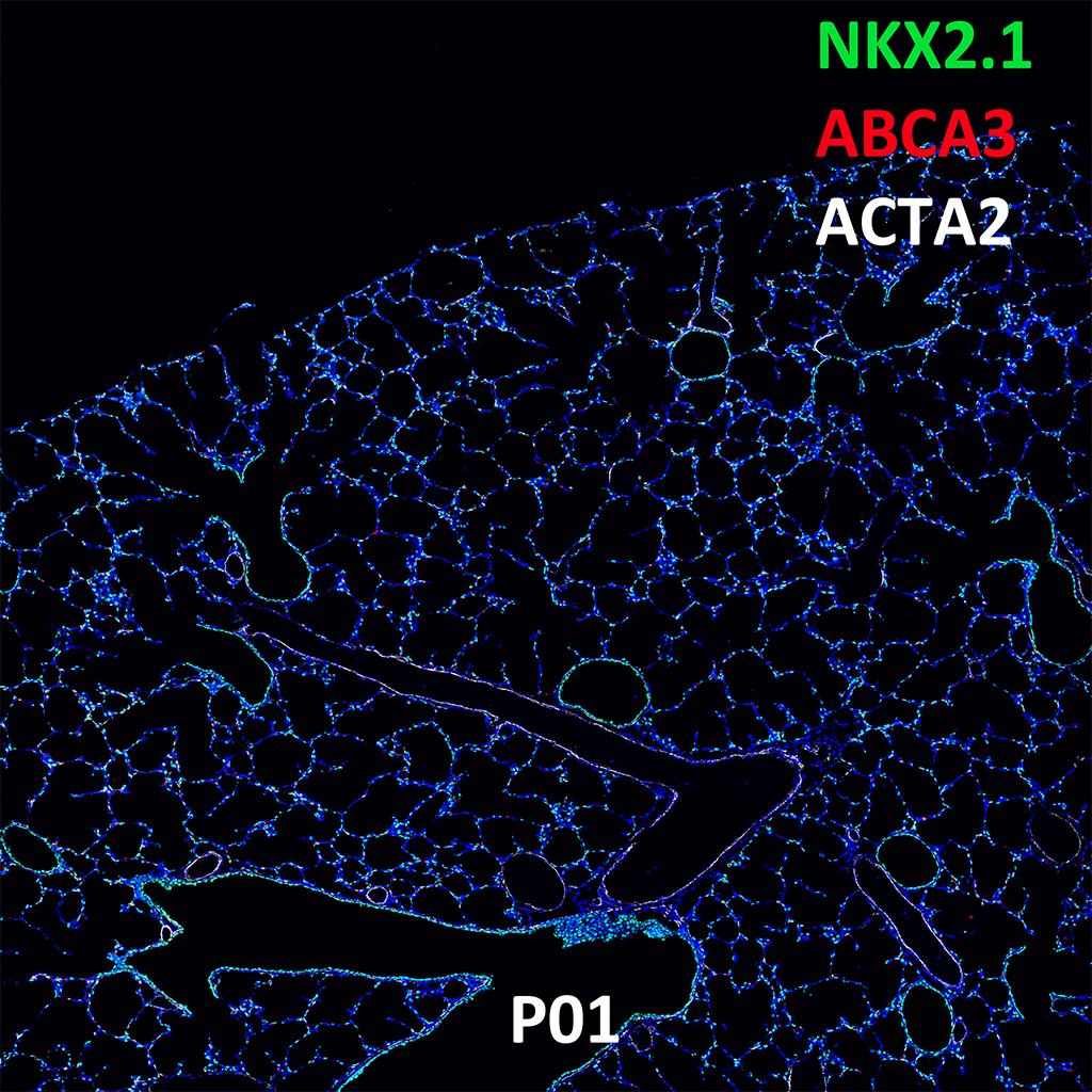
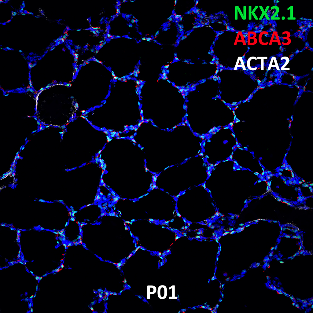
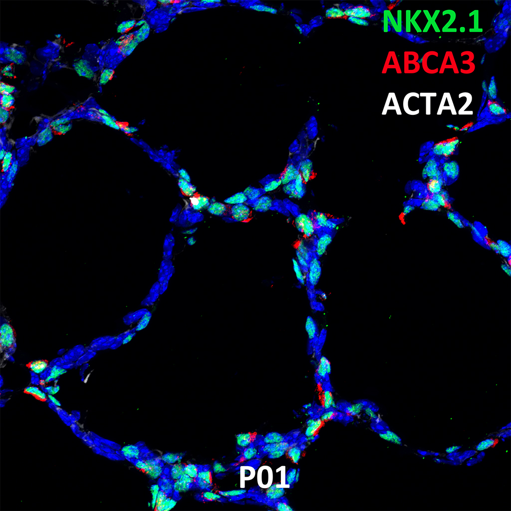
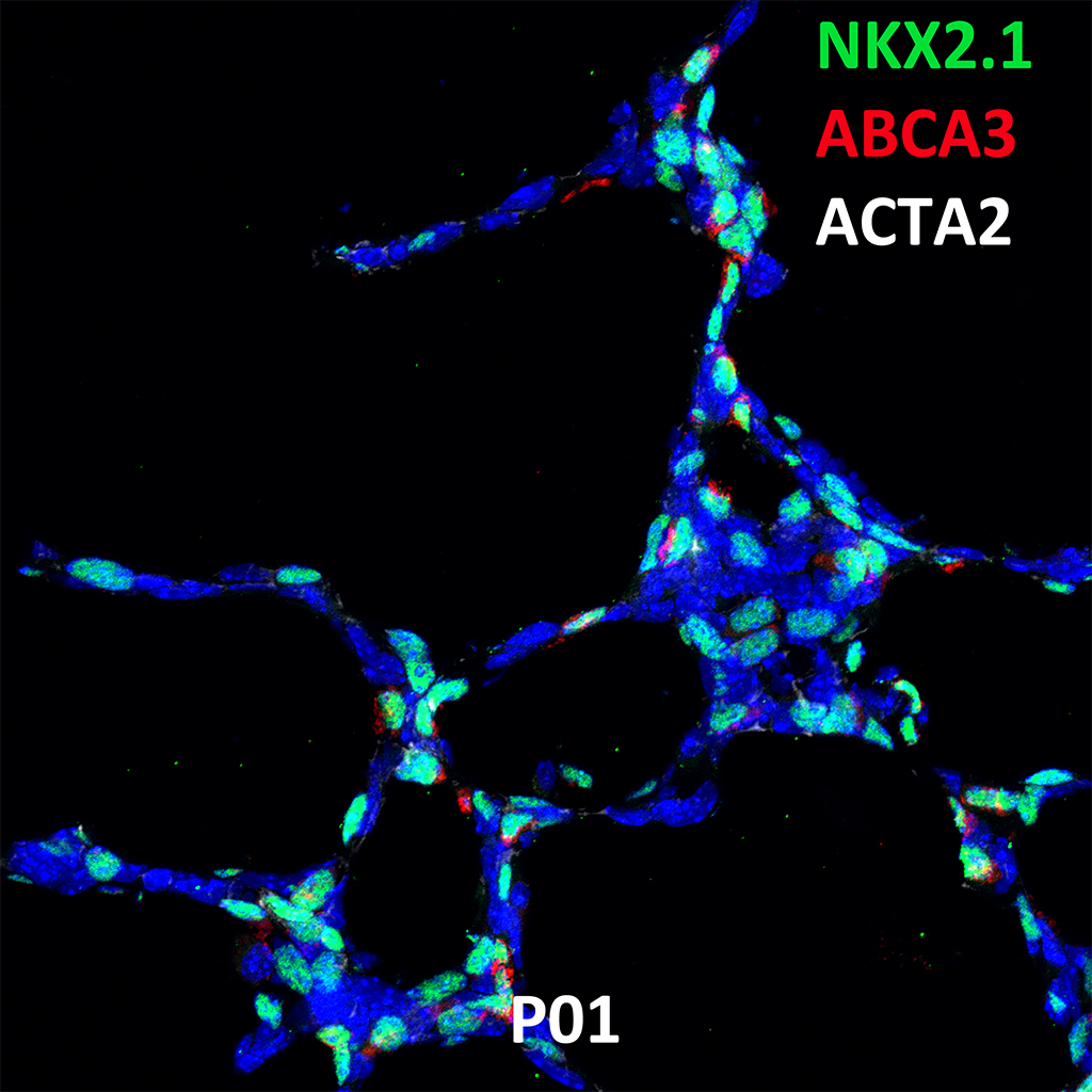
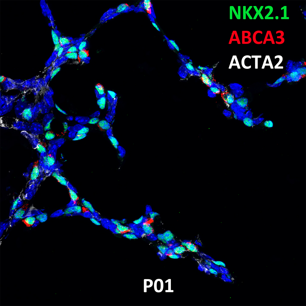
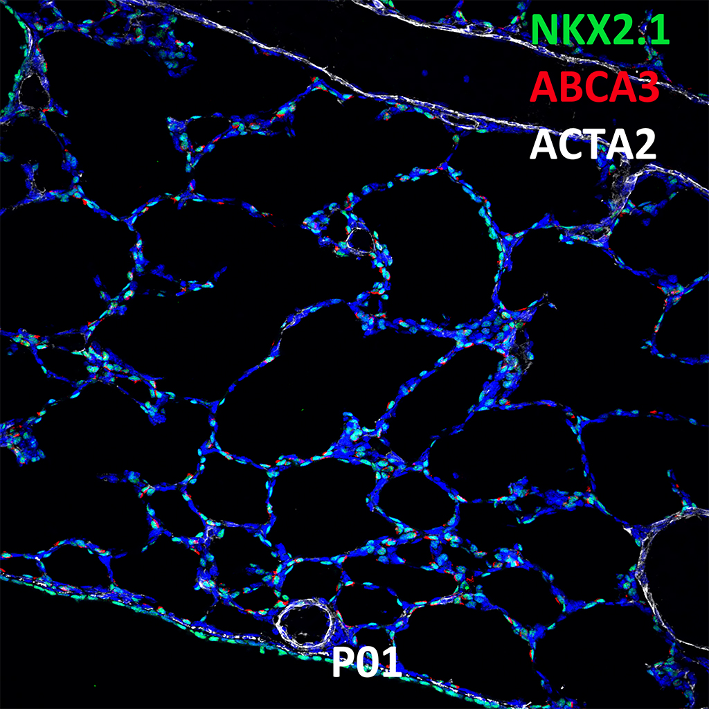
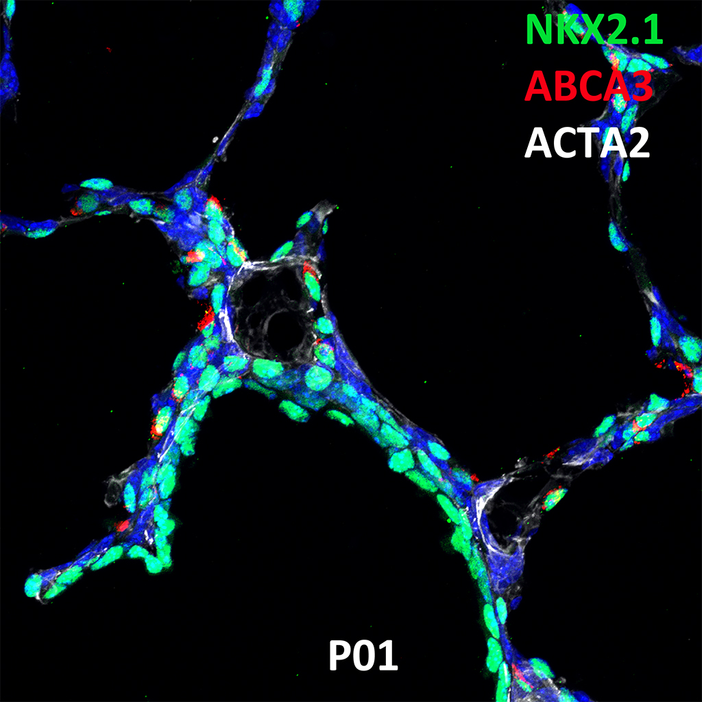
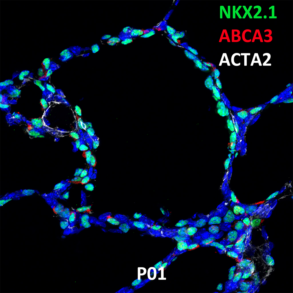
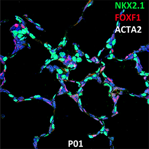
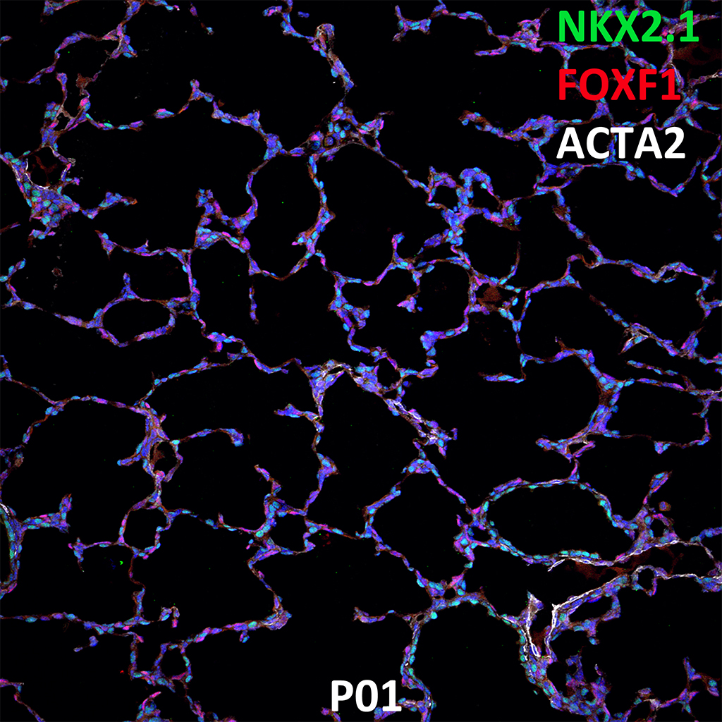
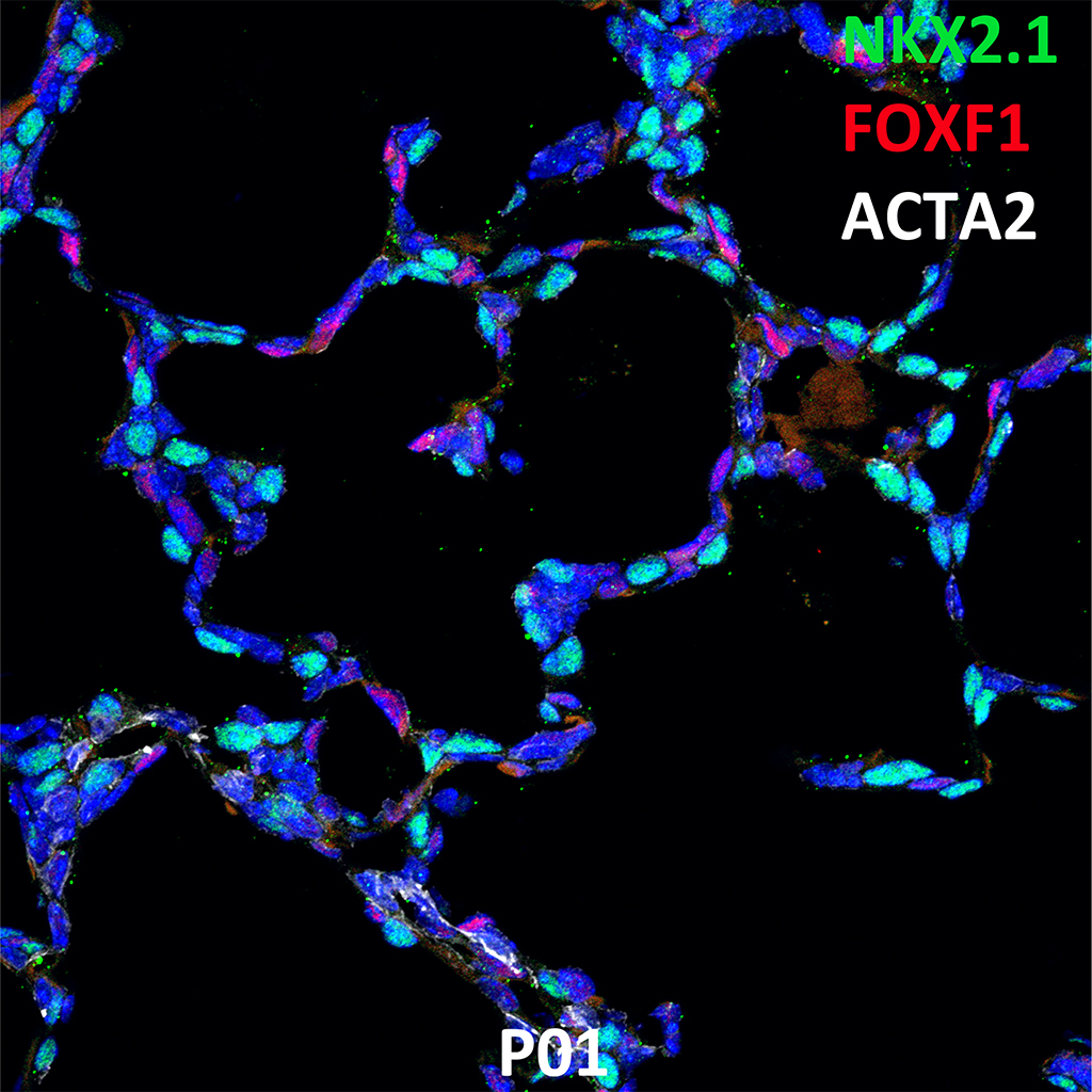
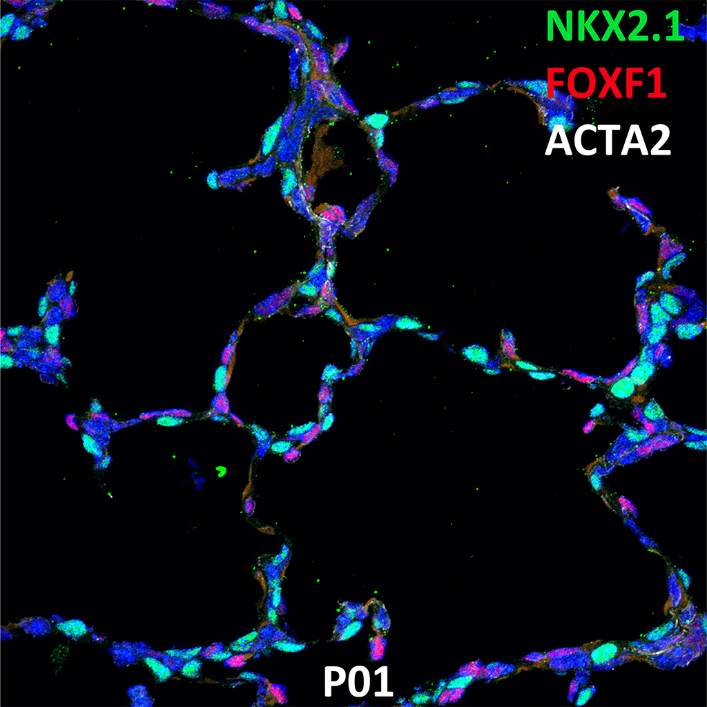
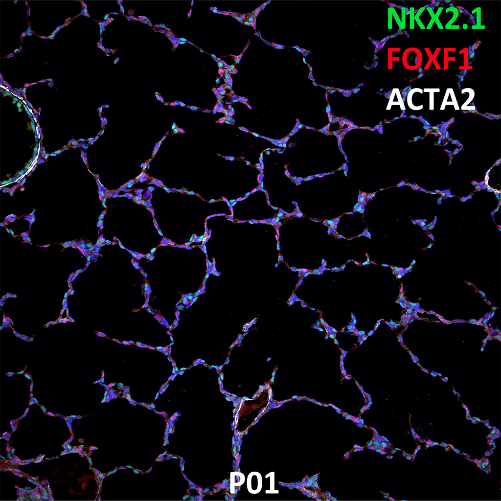
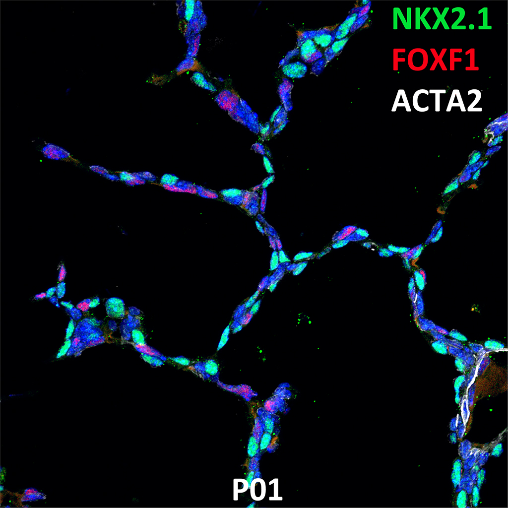
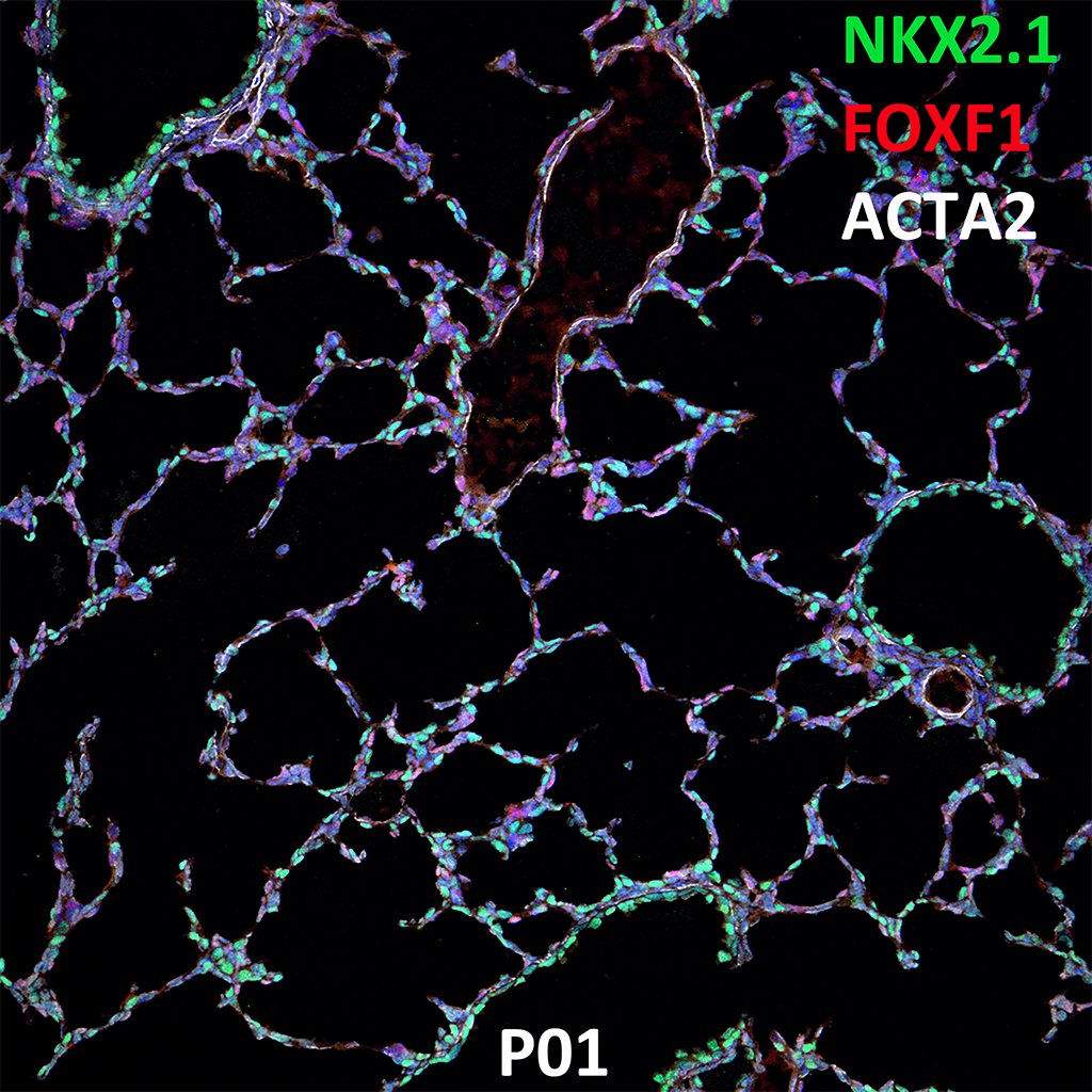
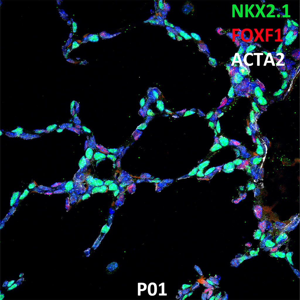
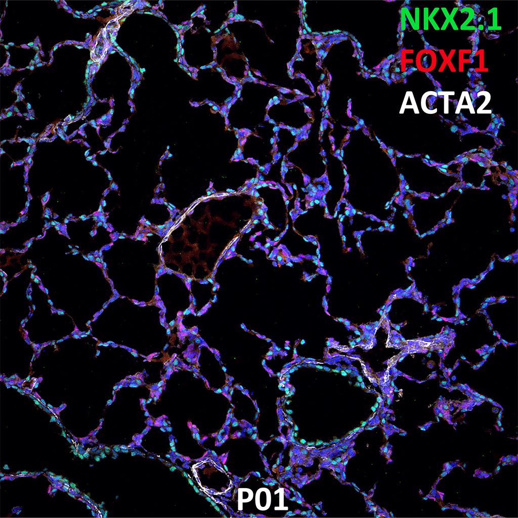
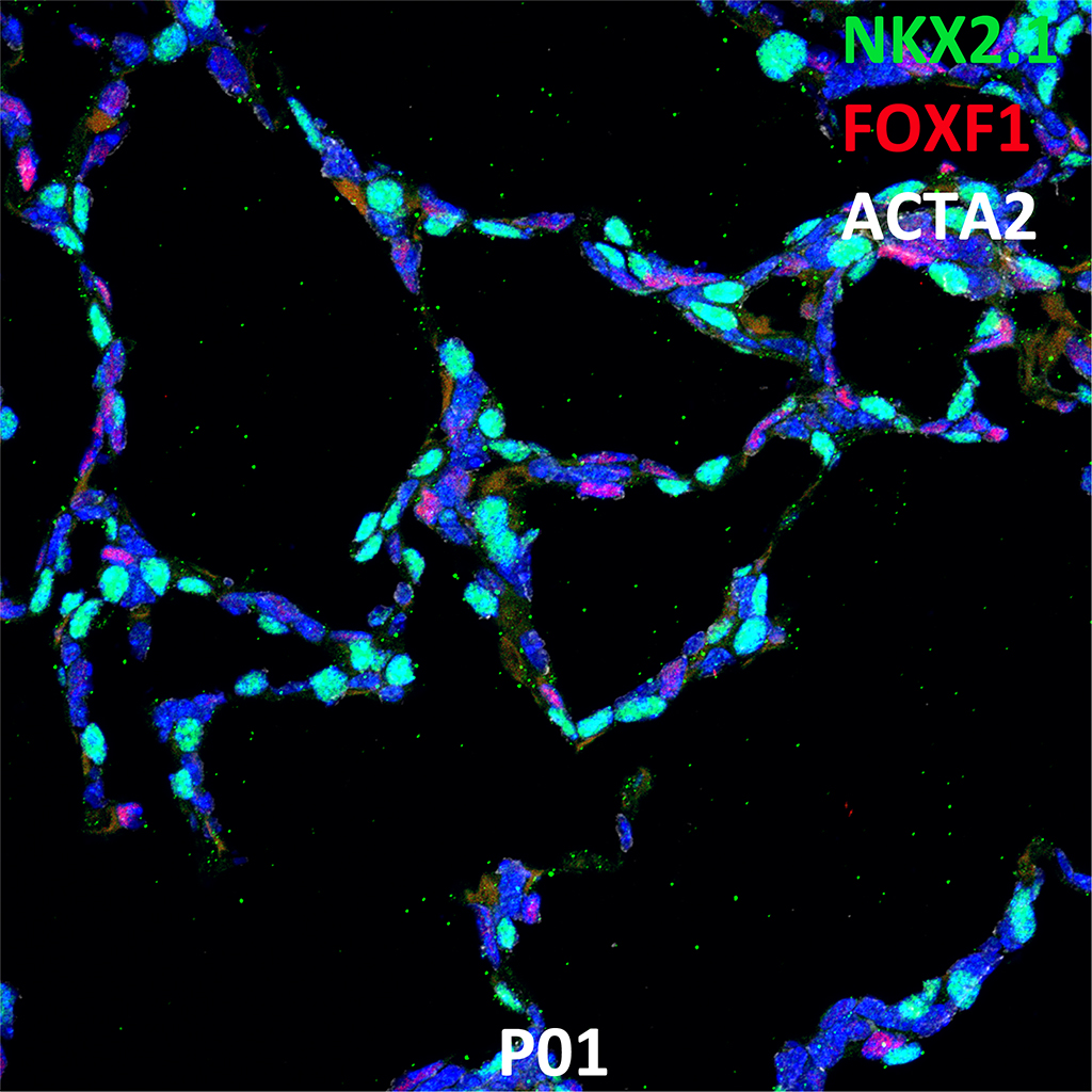
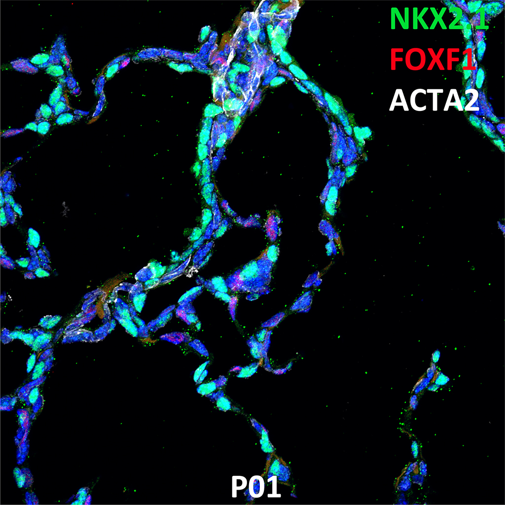
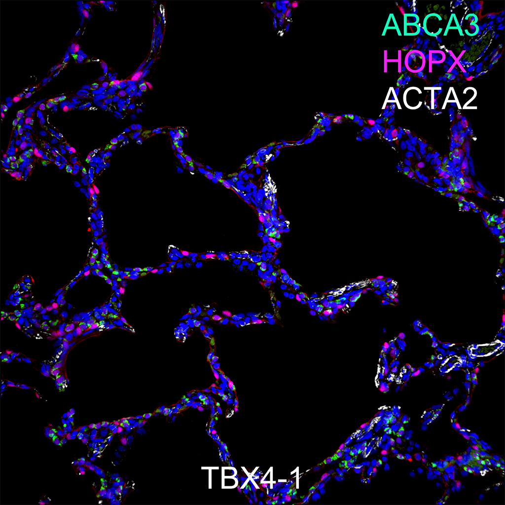
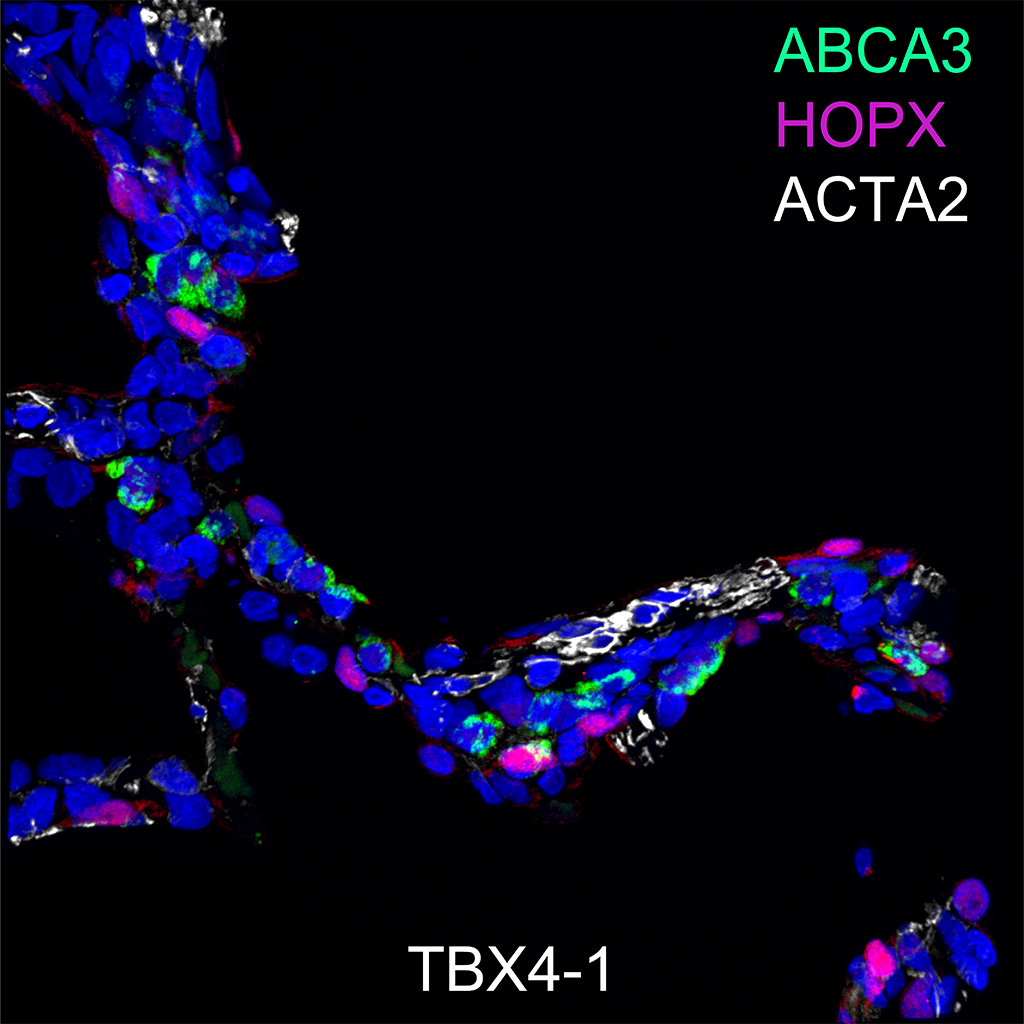
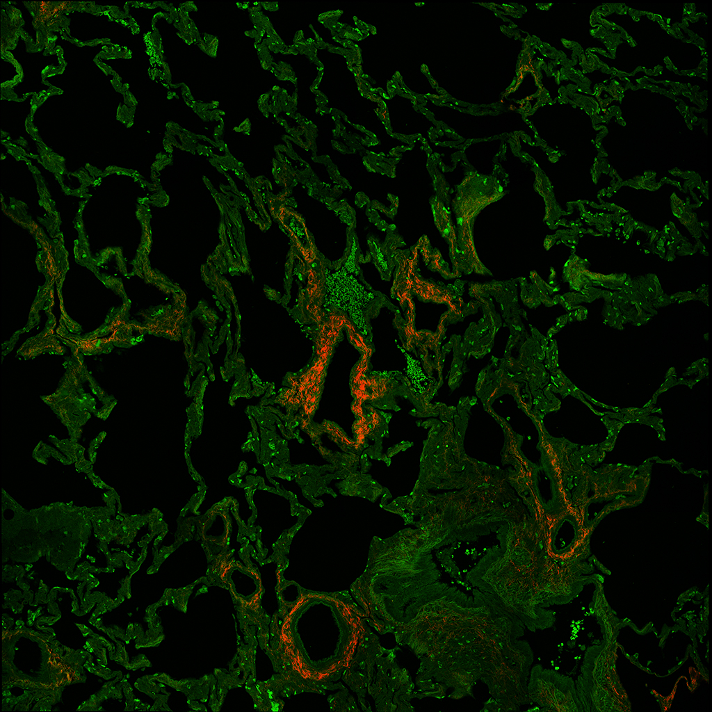
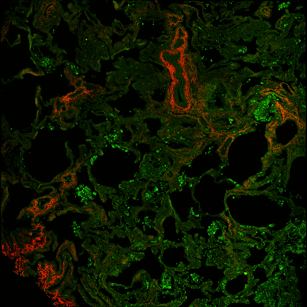
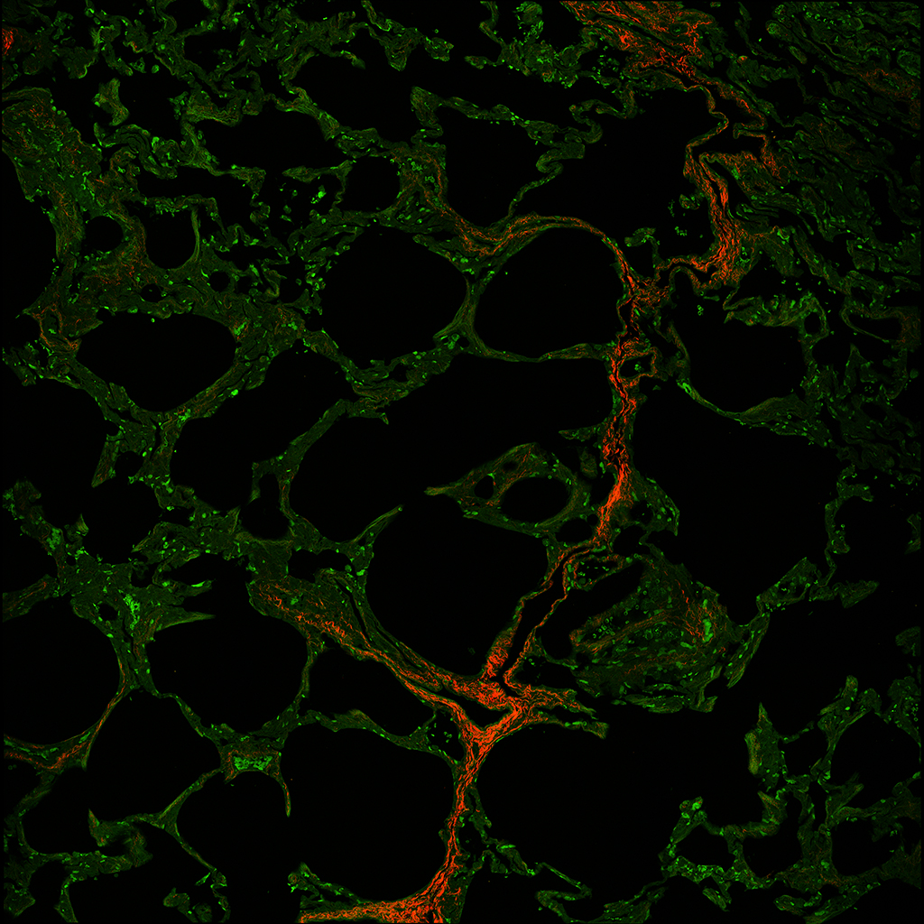
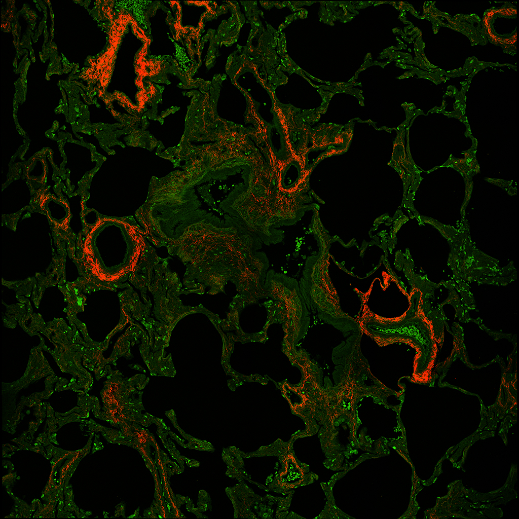
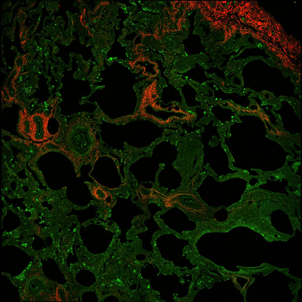
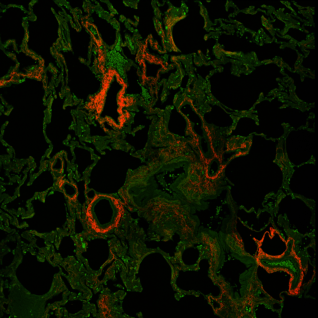
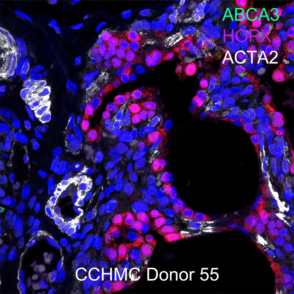
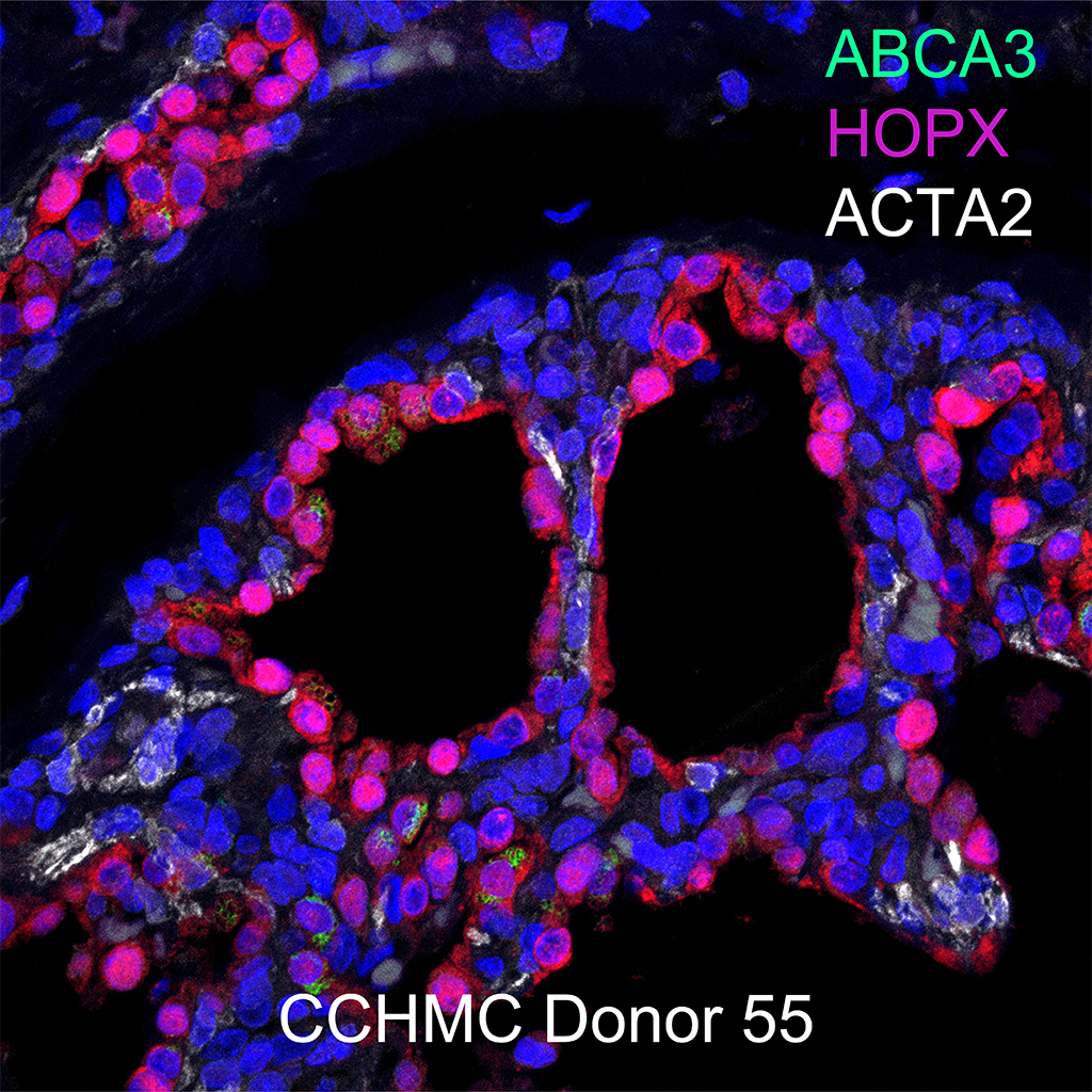
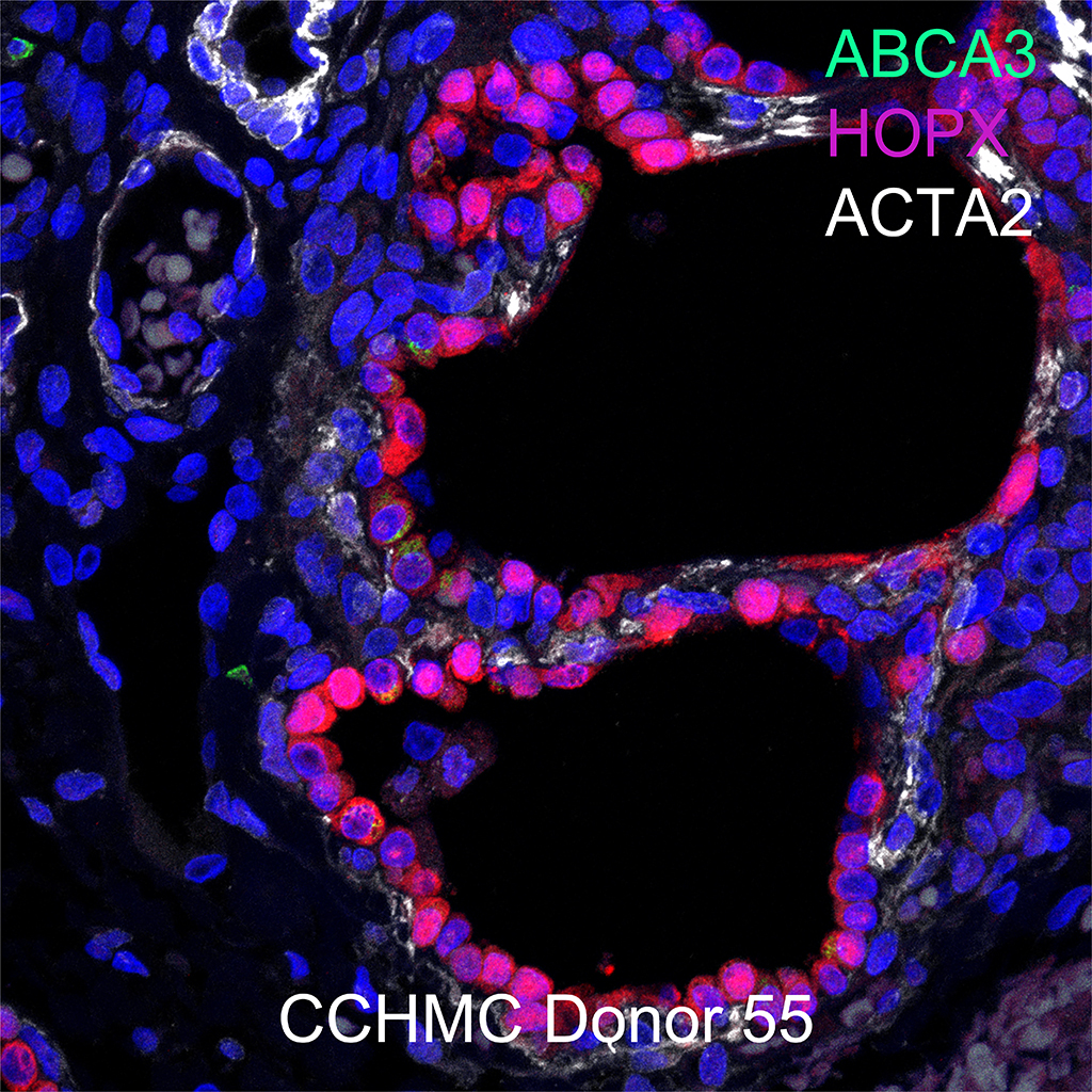
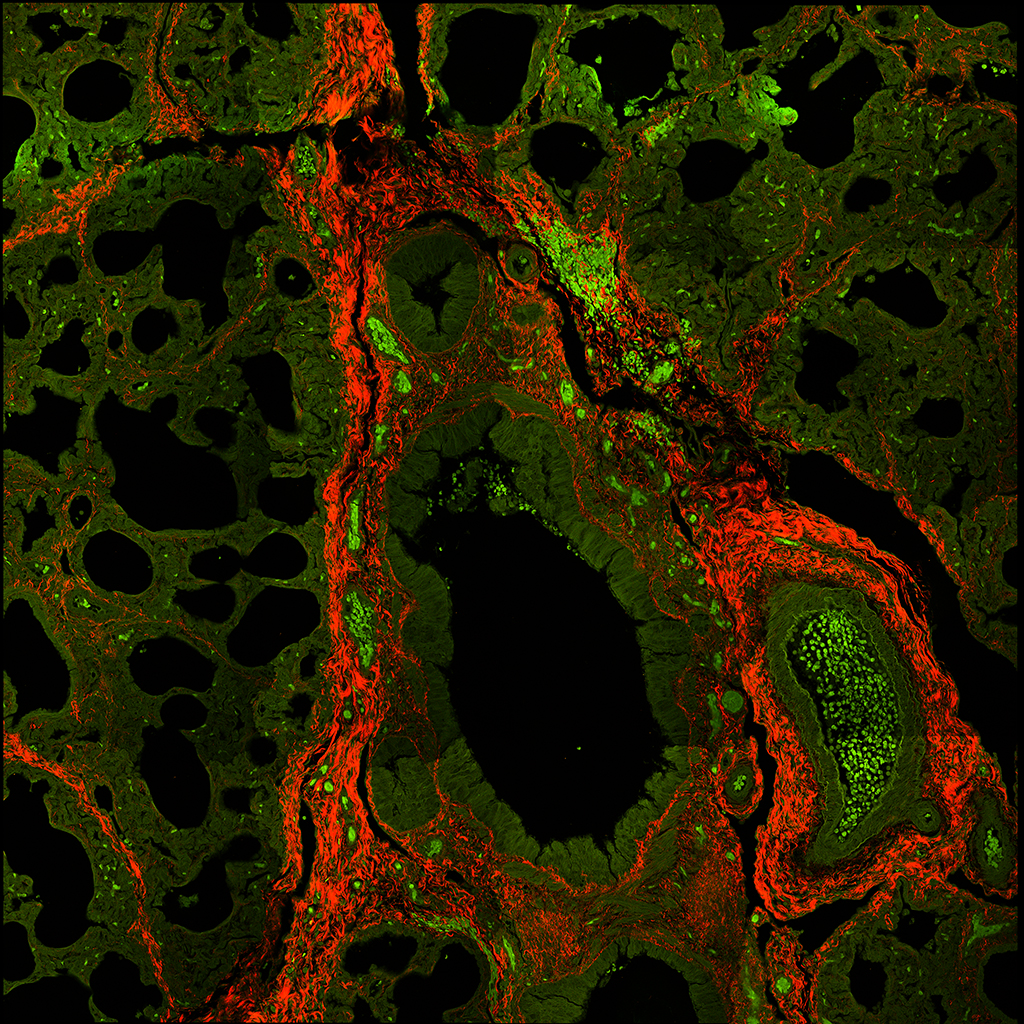
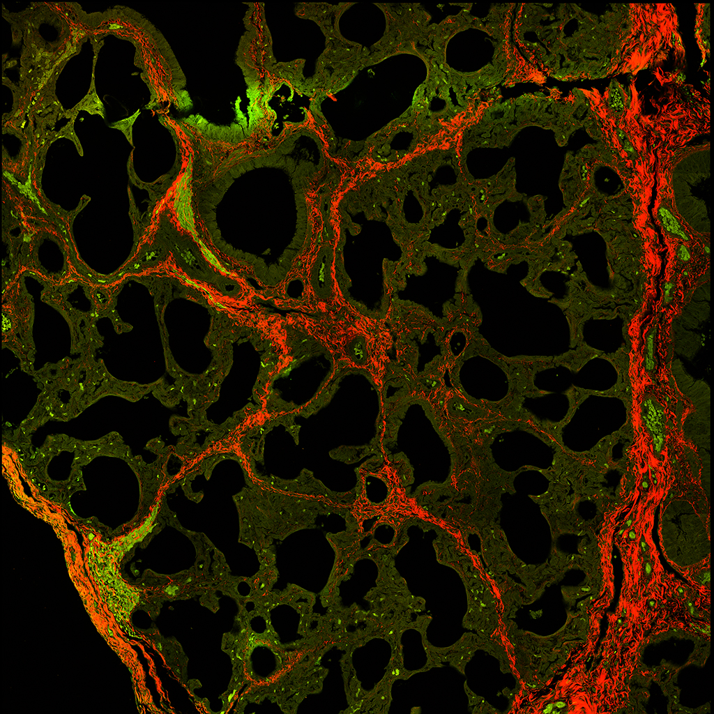
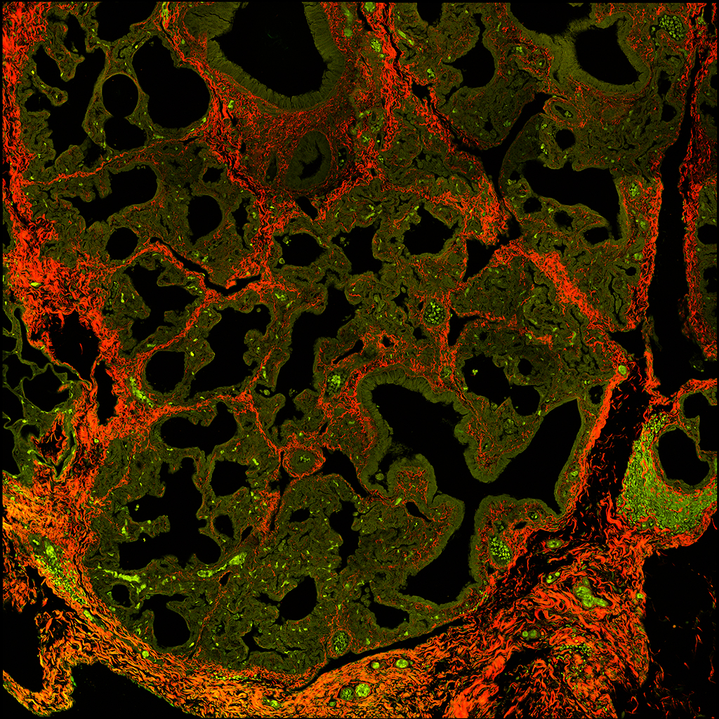
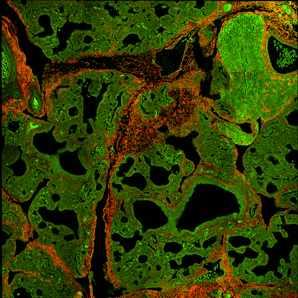
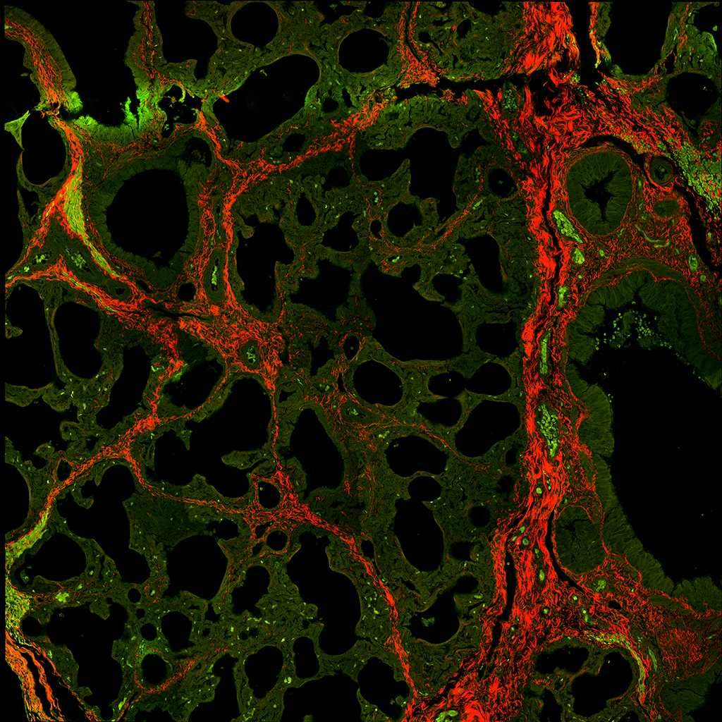
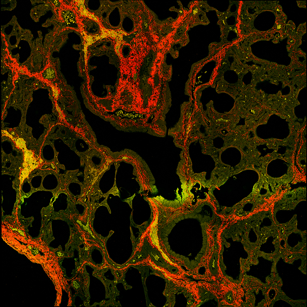
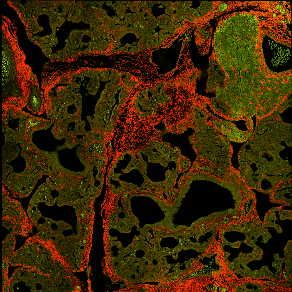
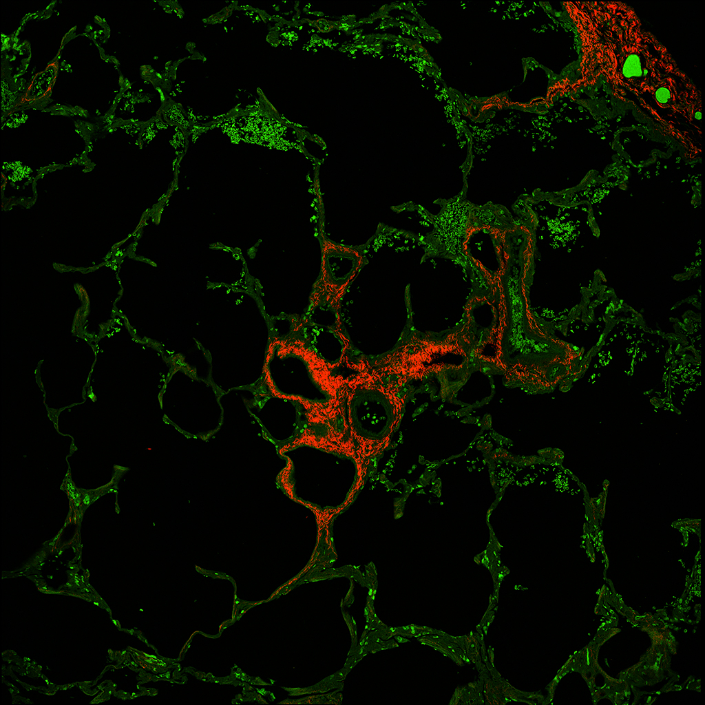
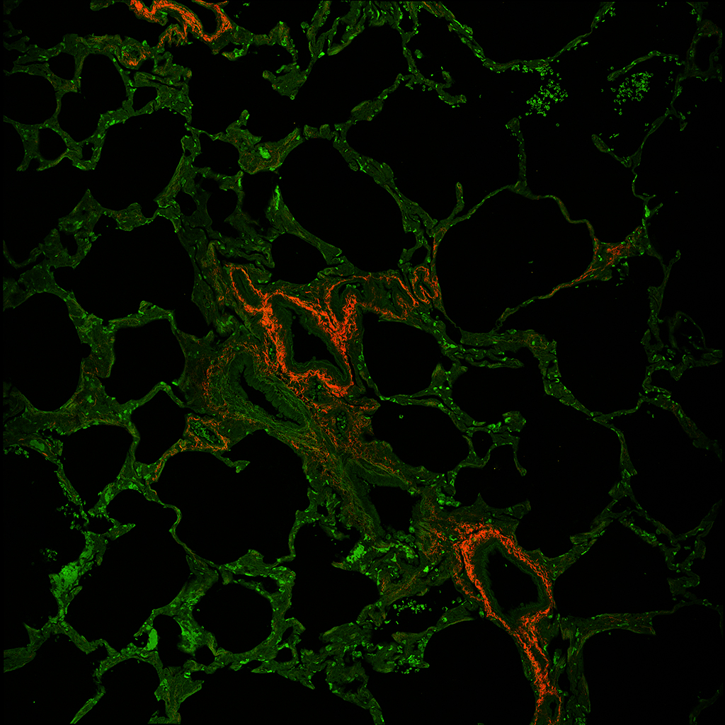
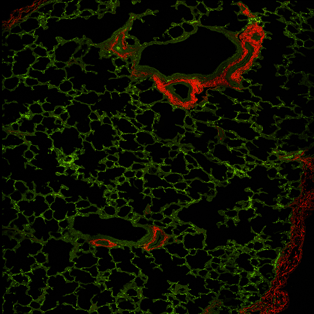
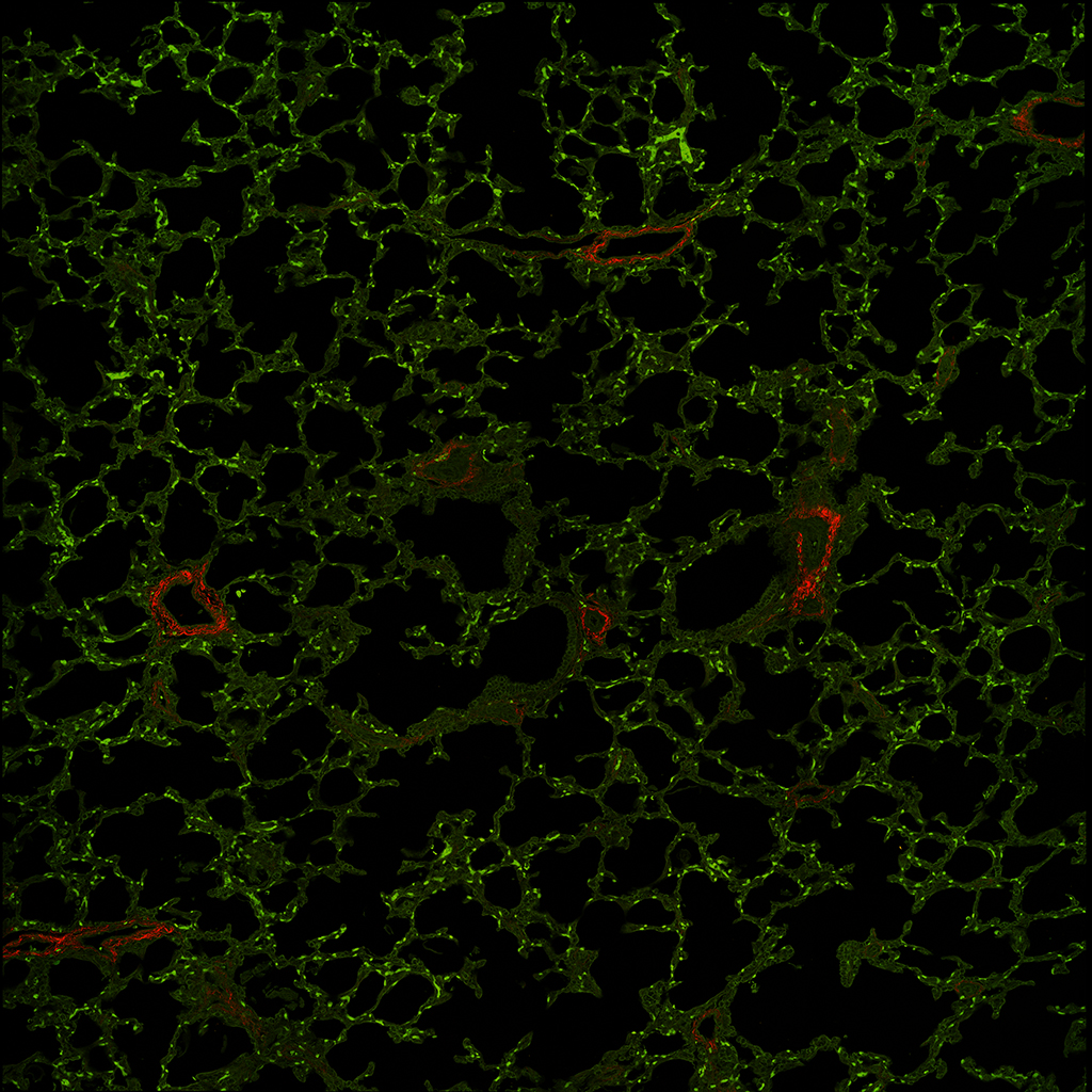
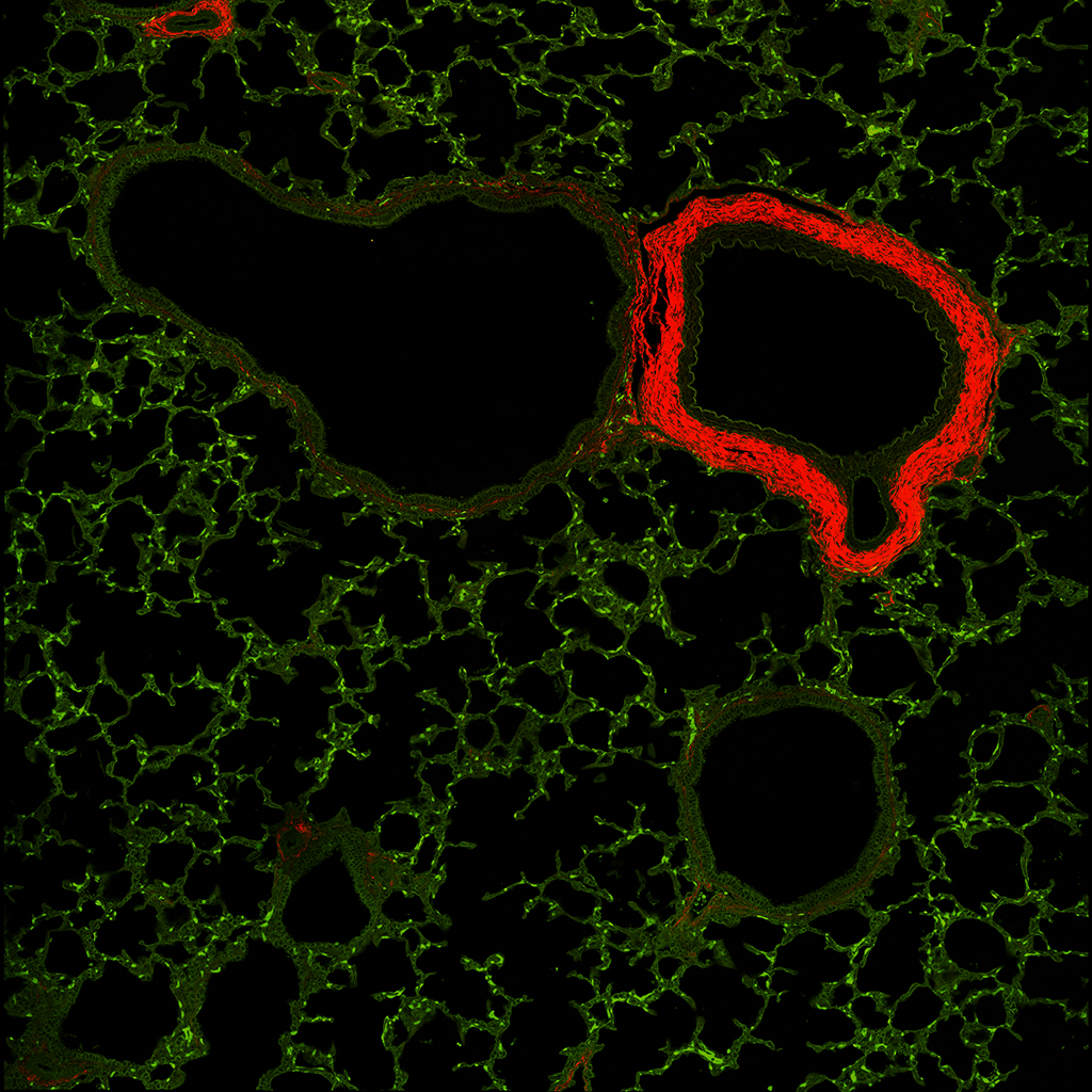
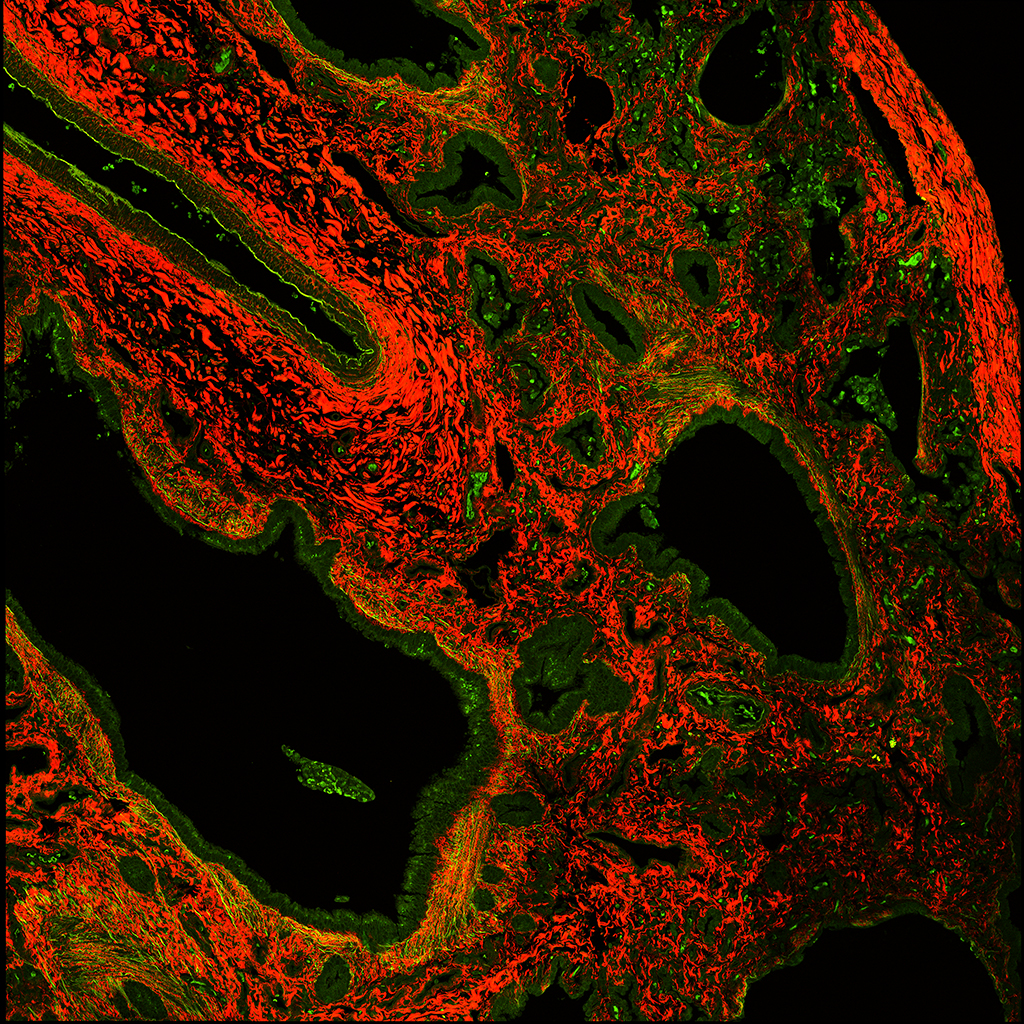
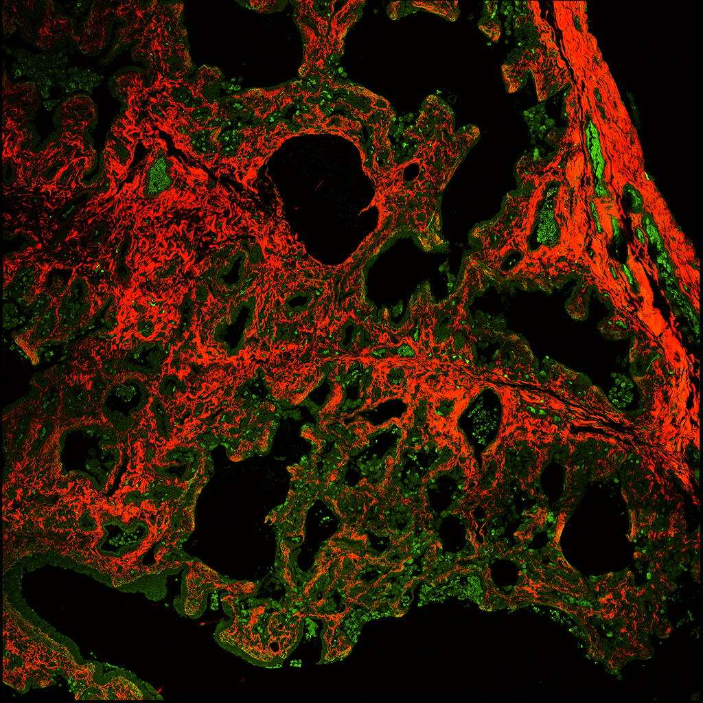
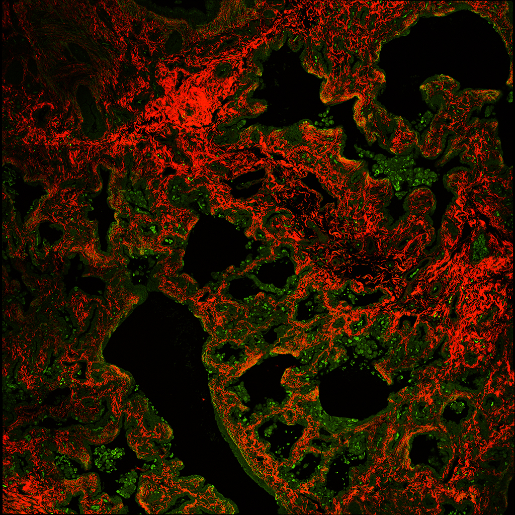
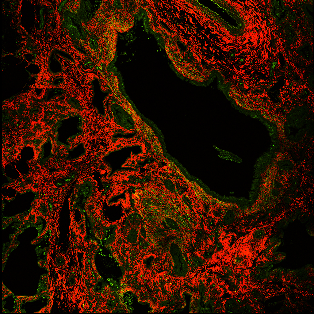
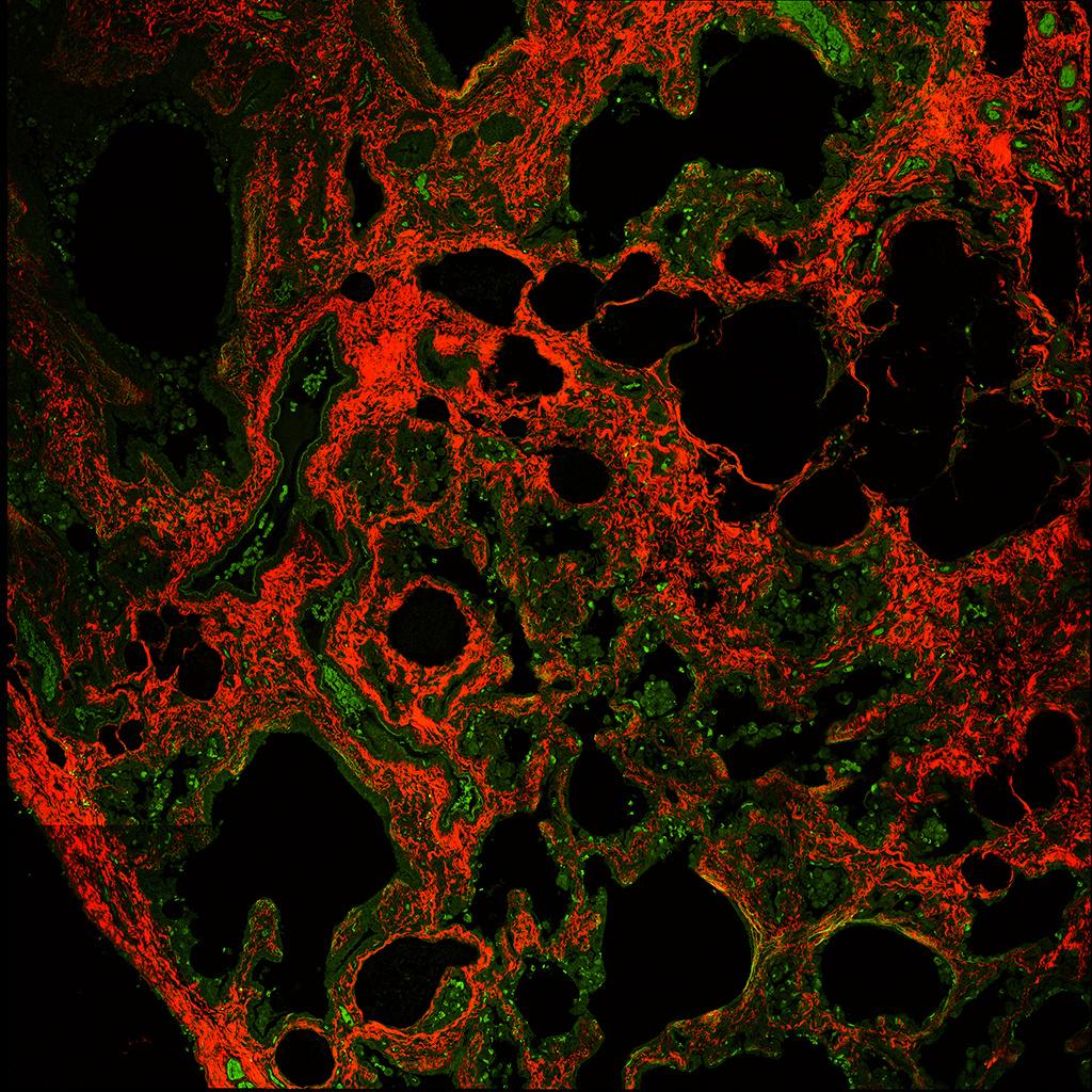
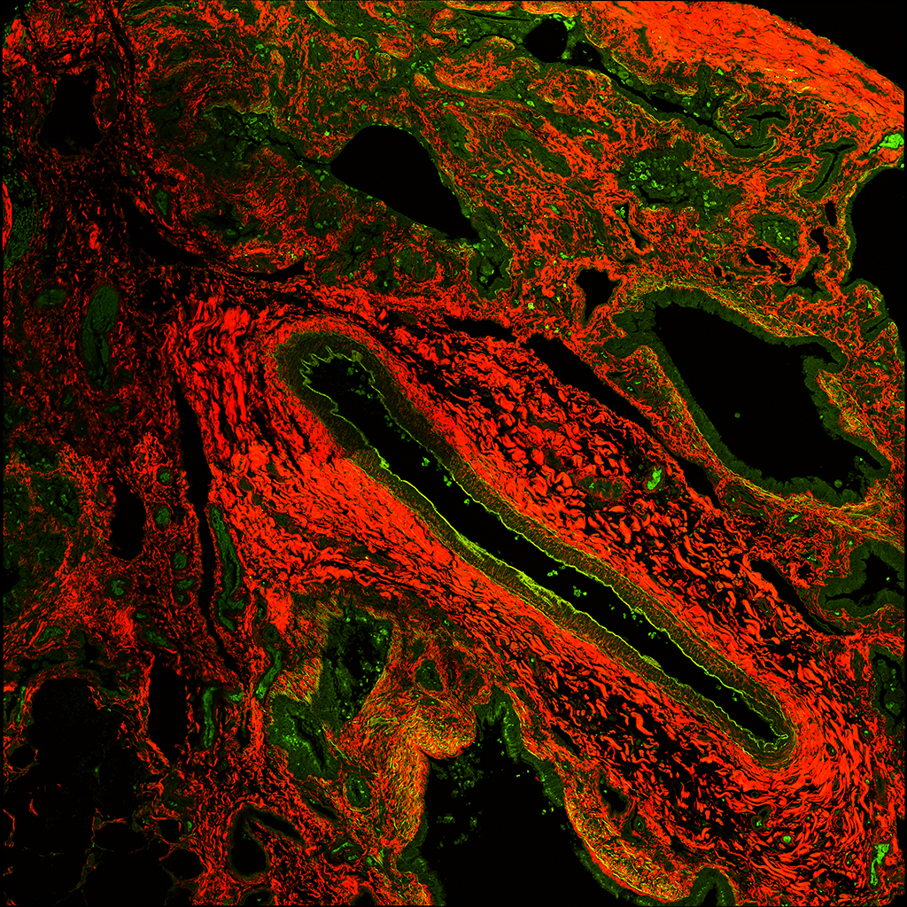
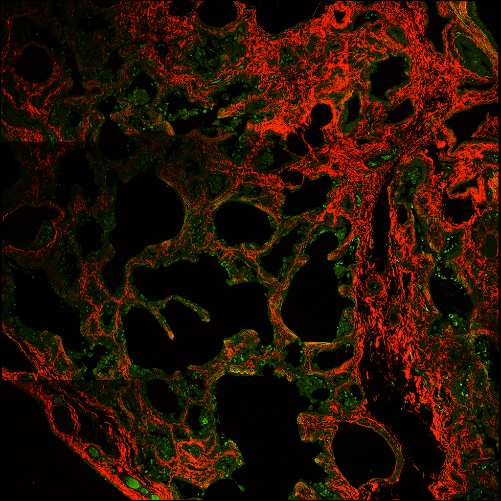
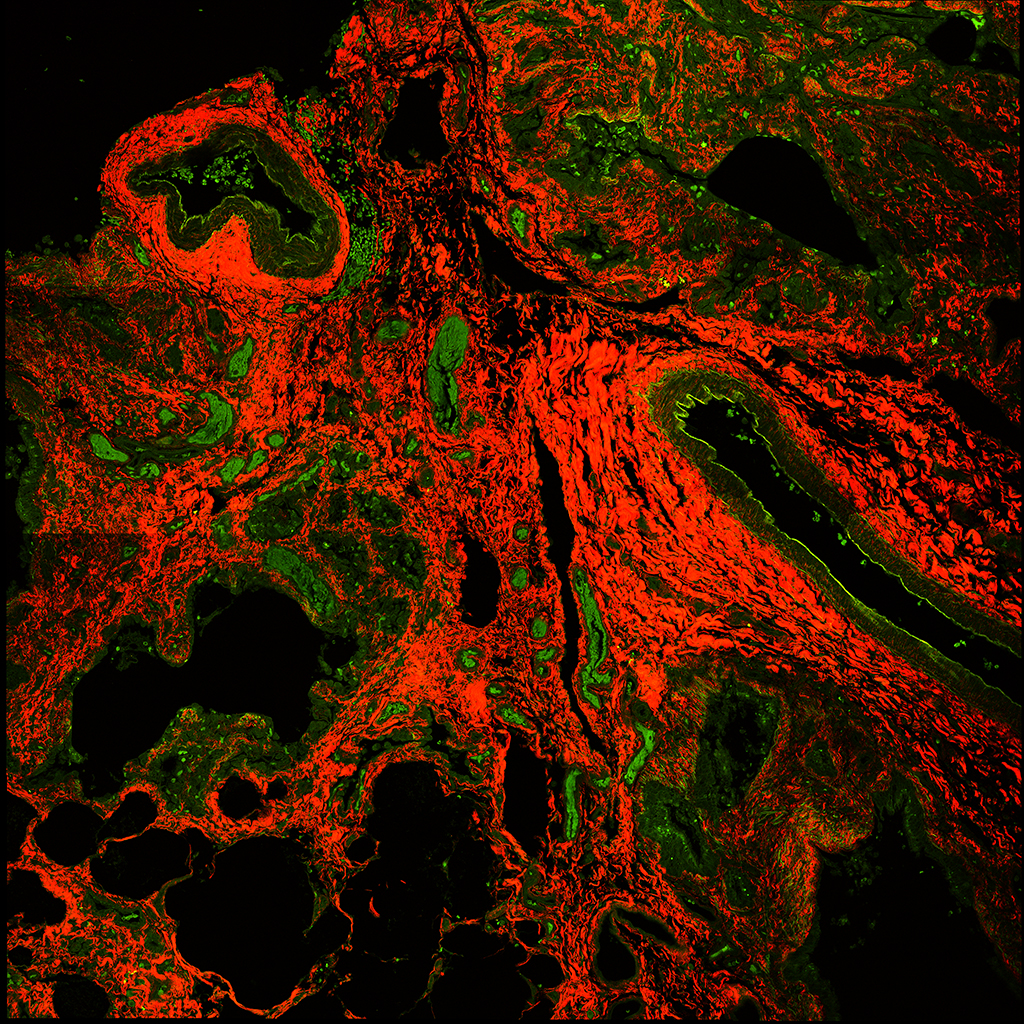
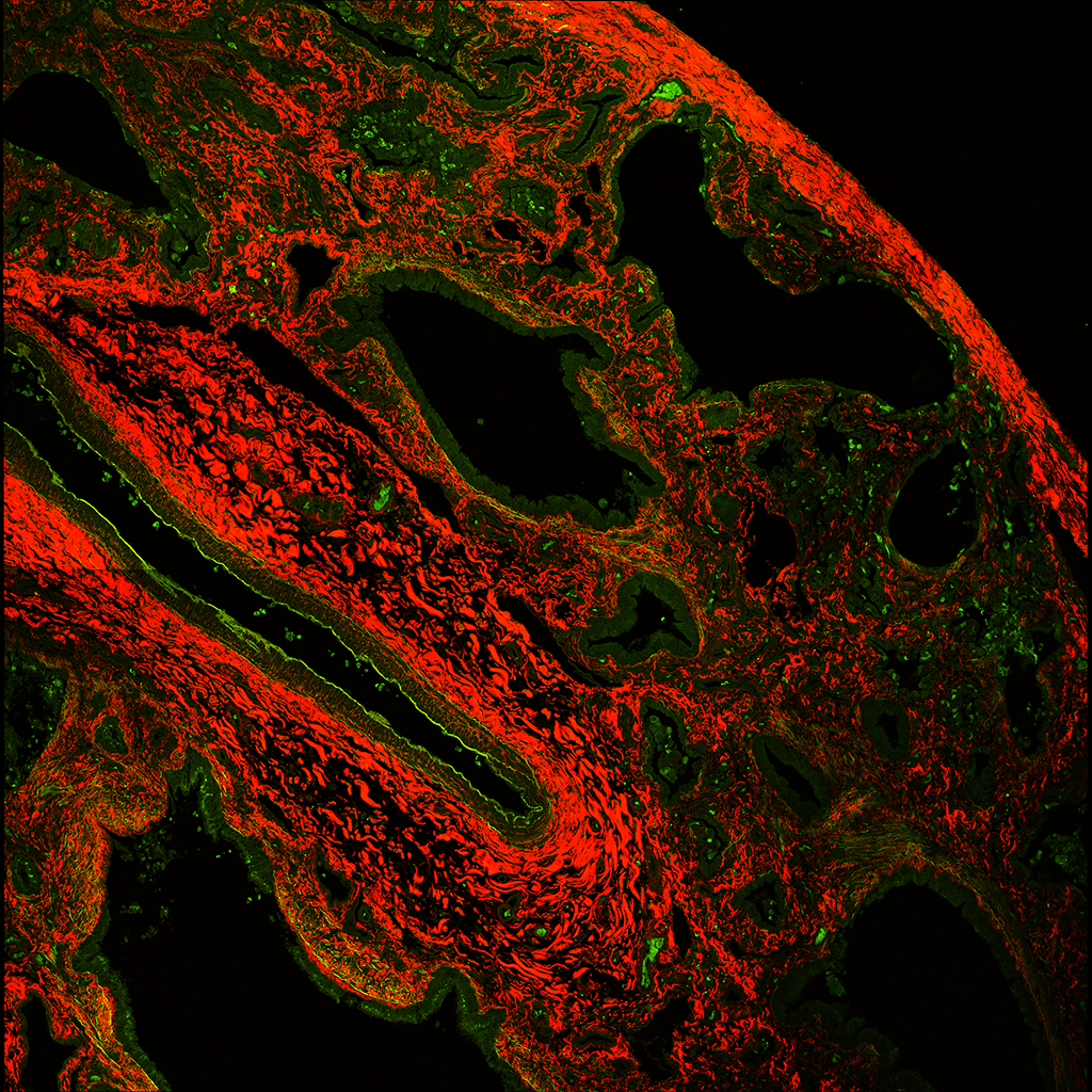
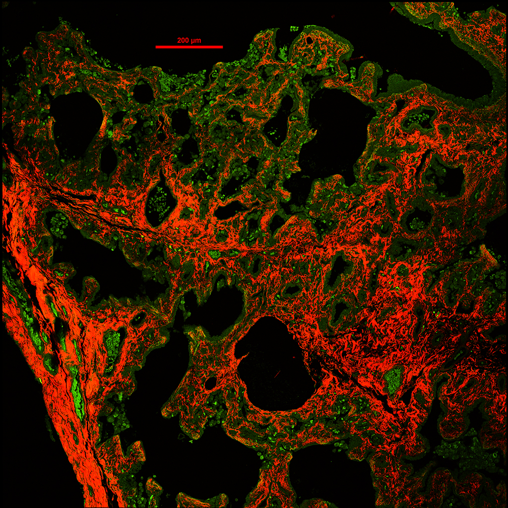
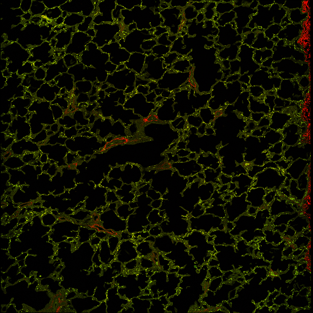
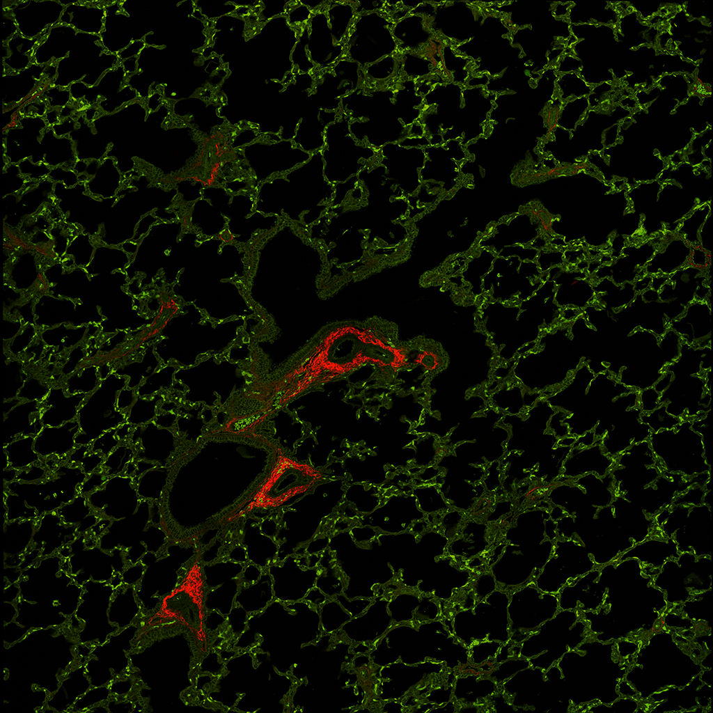
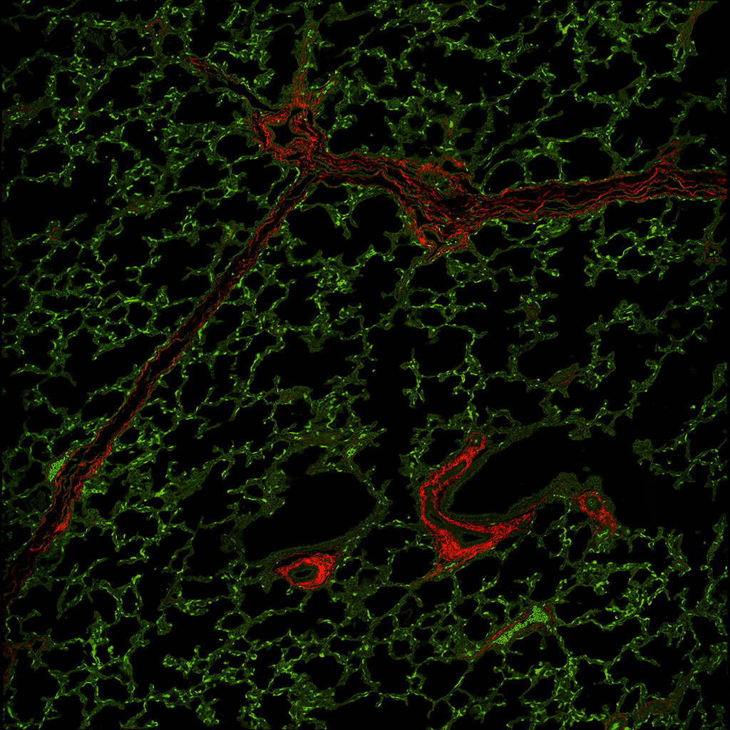
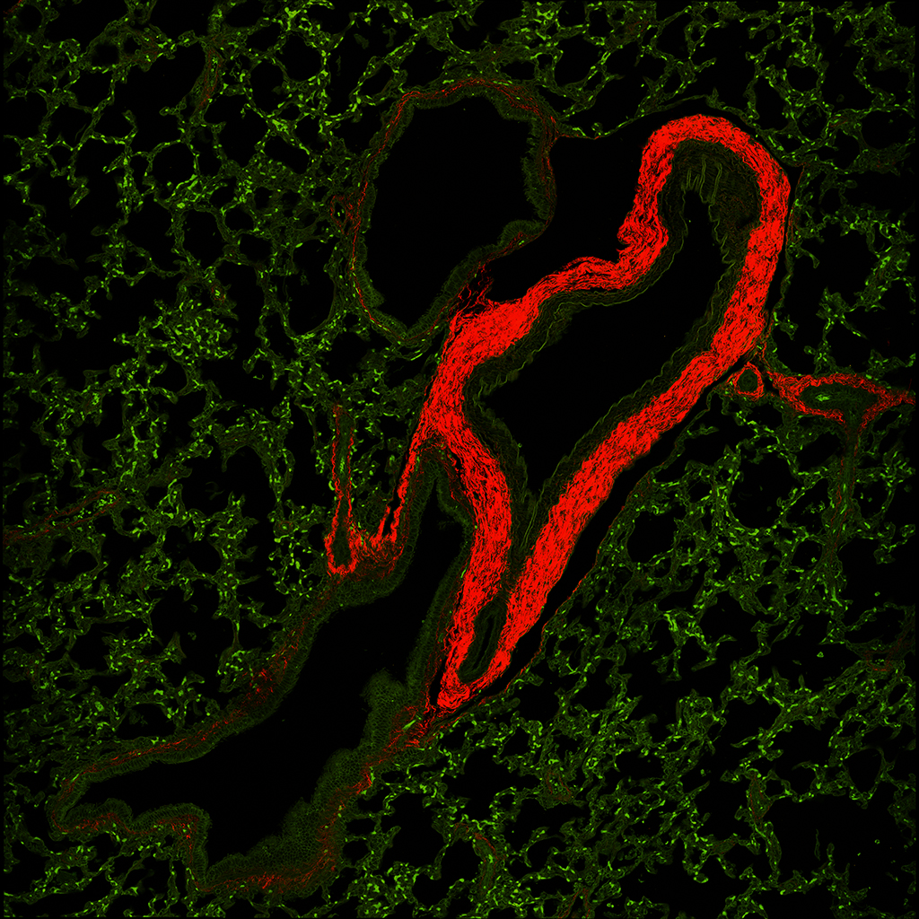
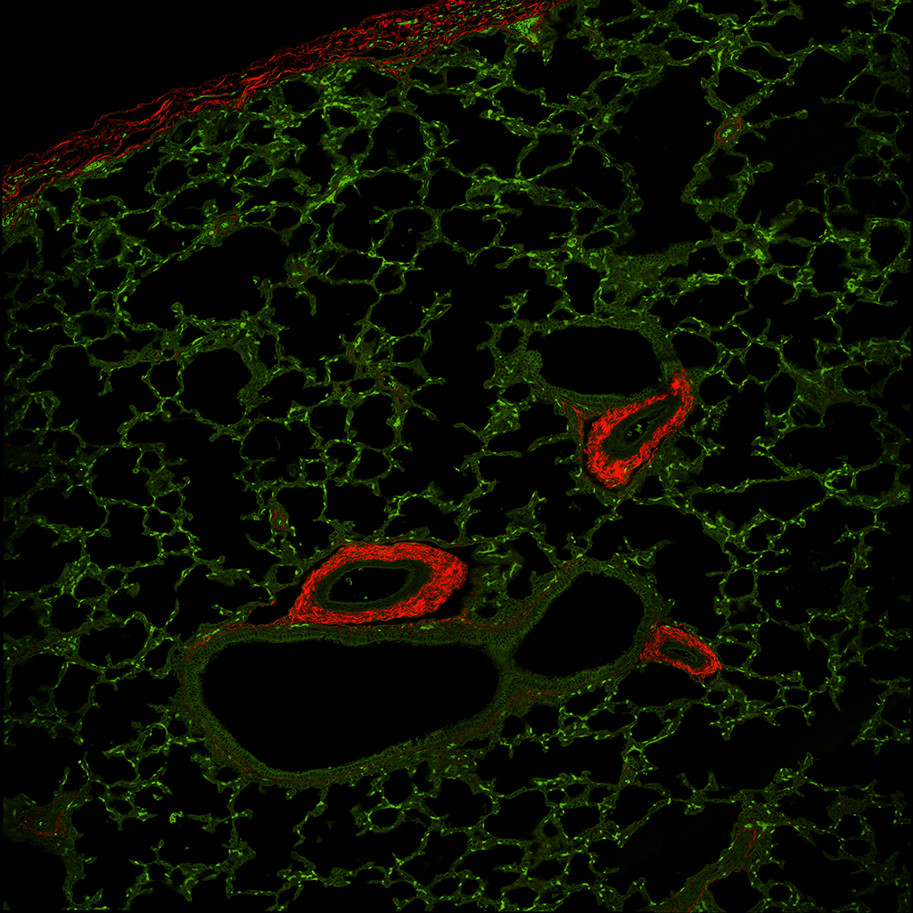
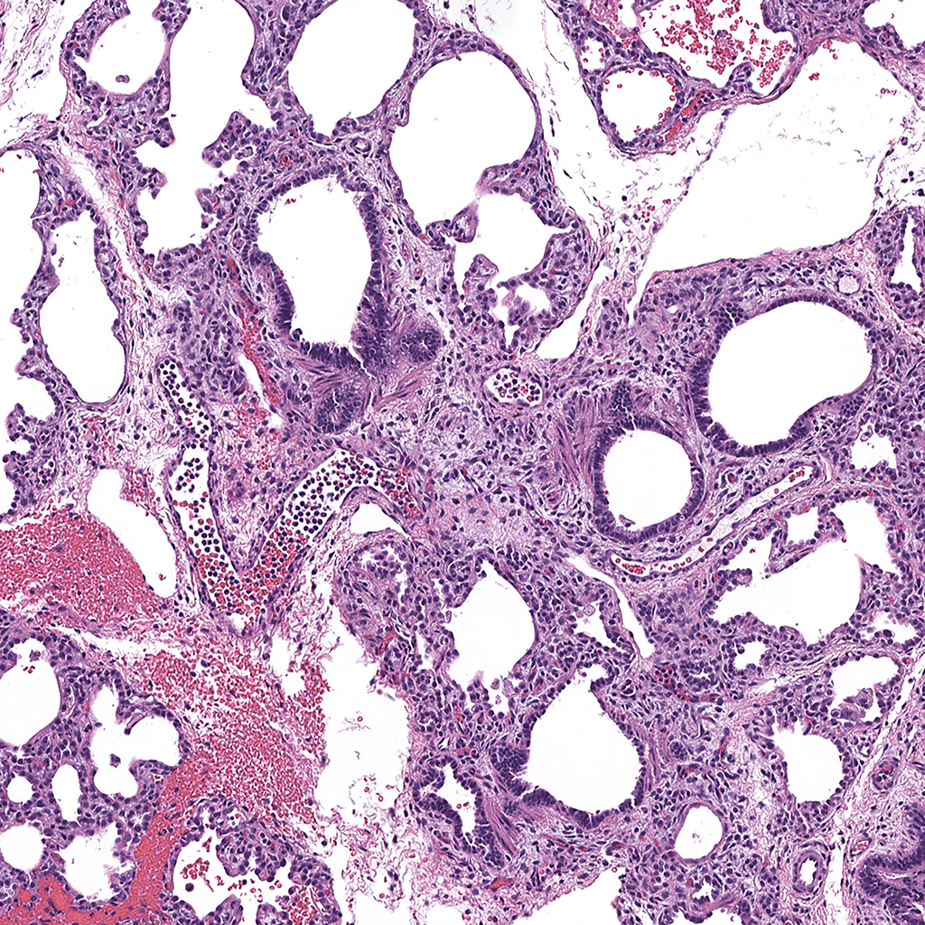
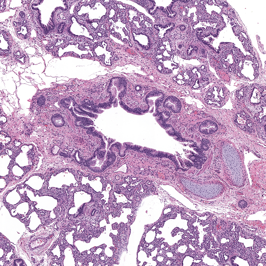
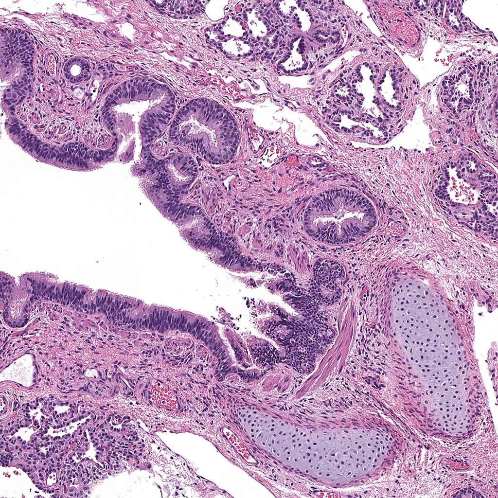
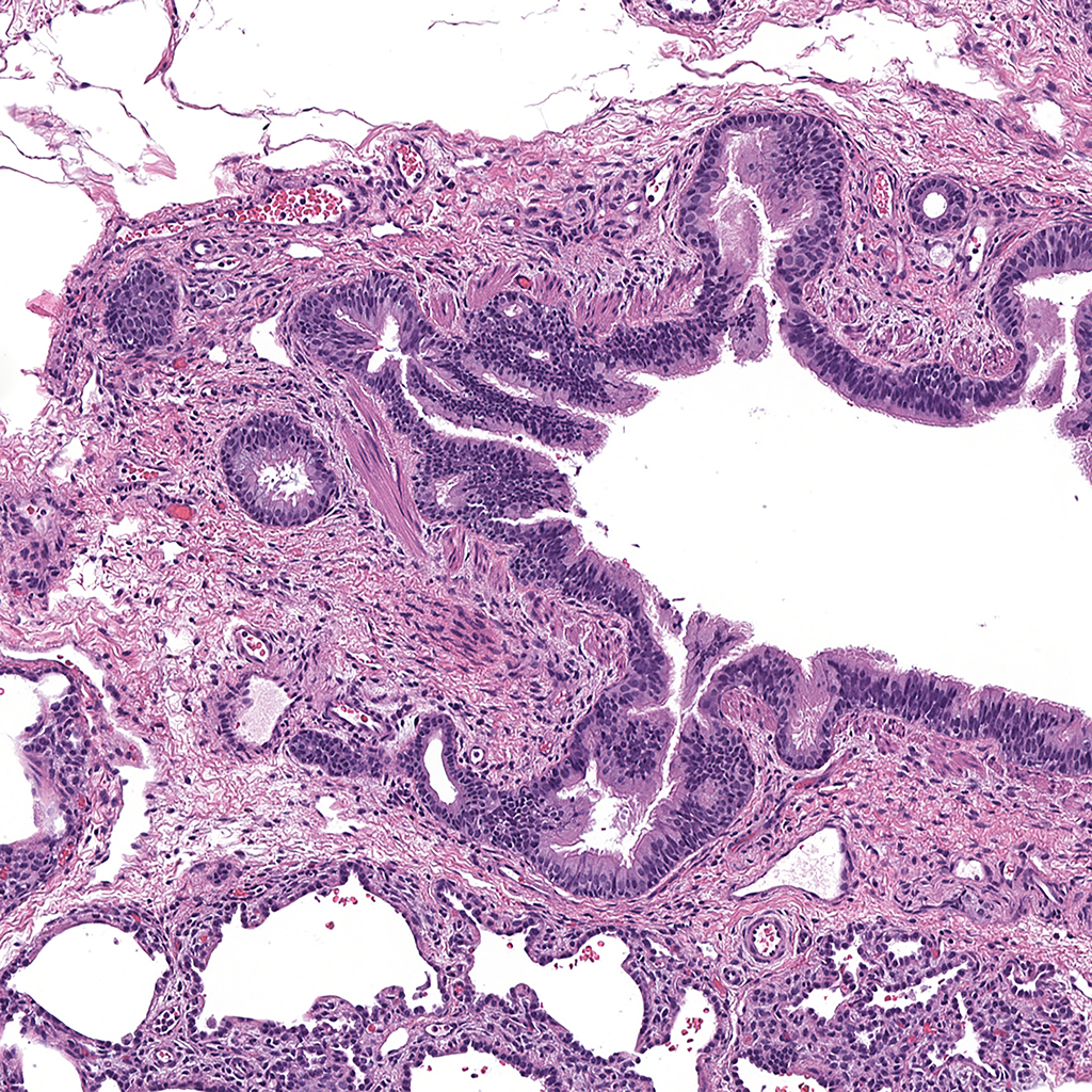
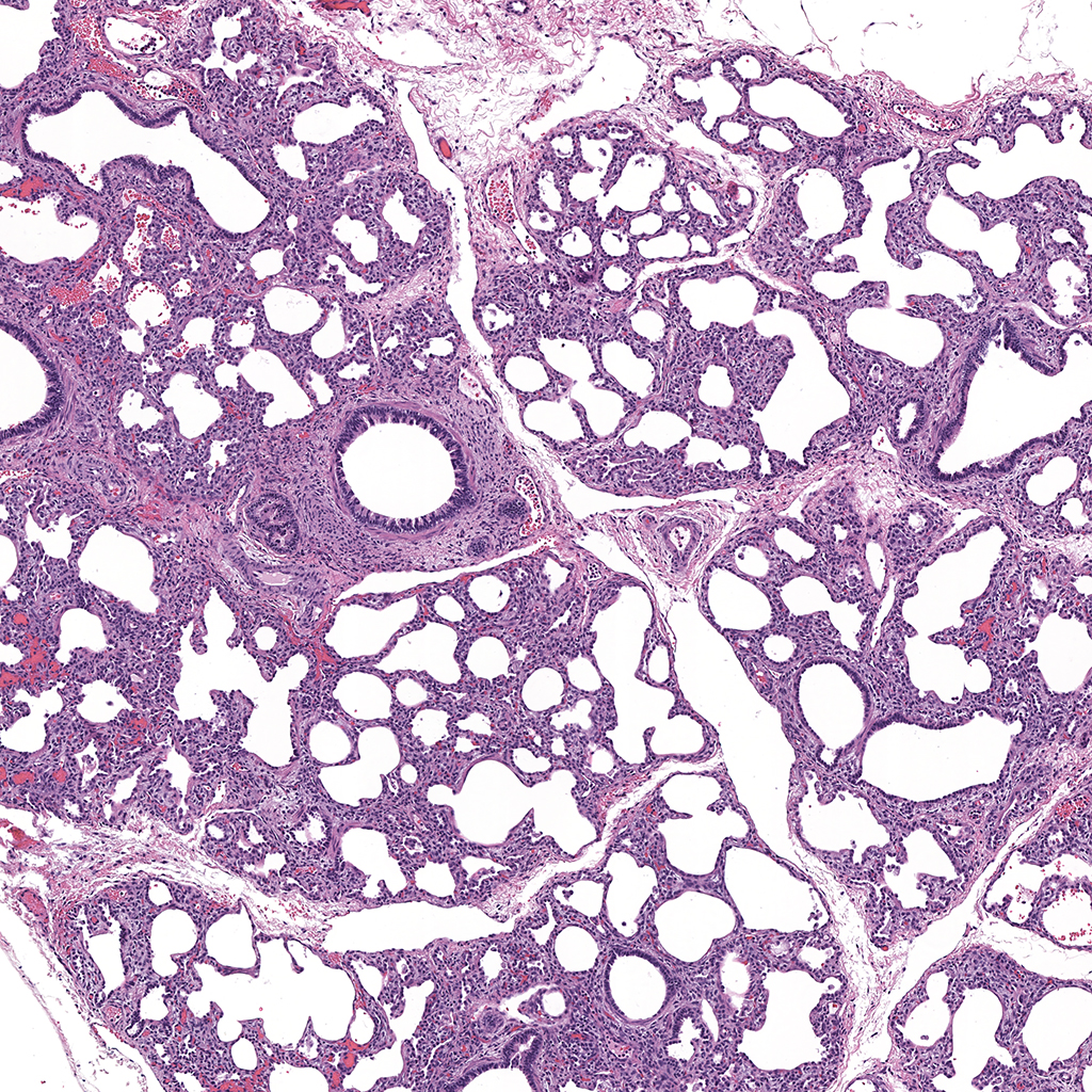
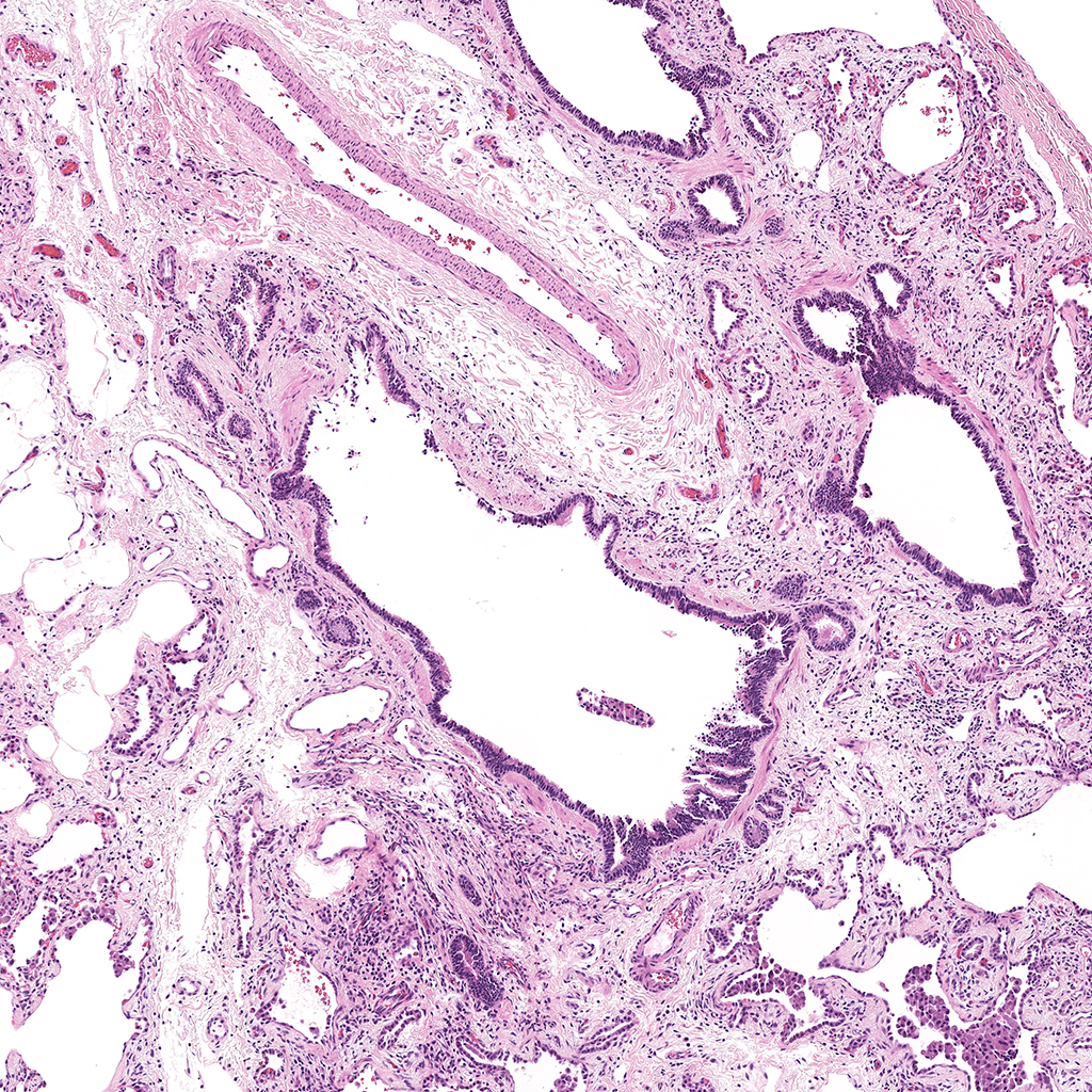
PND01 NKX2.1, ABCA3, and ACTA2 Confocal Imaging


| Gene |
|||||
| Nkx2.1 | LungGens | Ensemble | Genecards | NCBI | The Human Protein Atlas |
| FOXF1 | LungGens | Ensemble | Genecards | NCBI | The Human Protein Atlas |
| Acta2 | LungGens | Ensemble | Genecards | NCBI | The Human Protein Atlas |
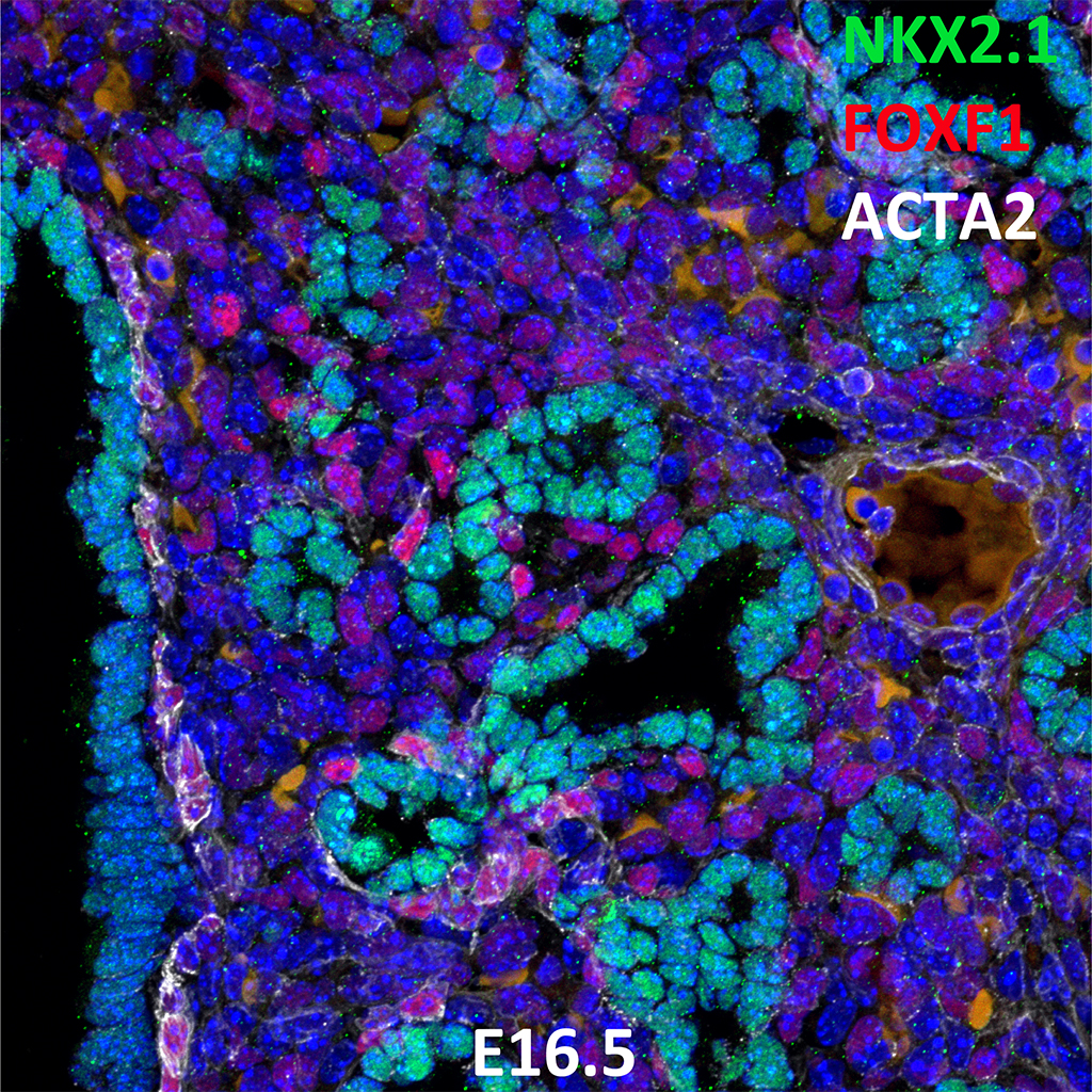
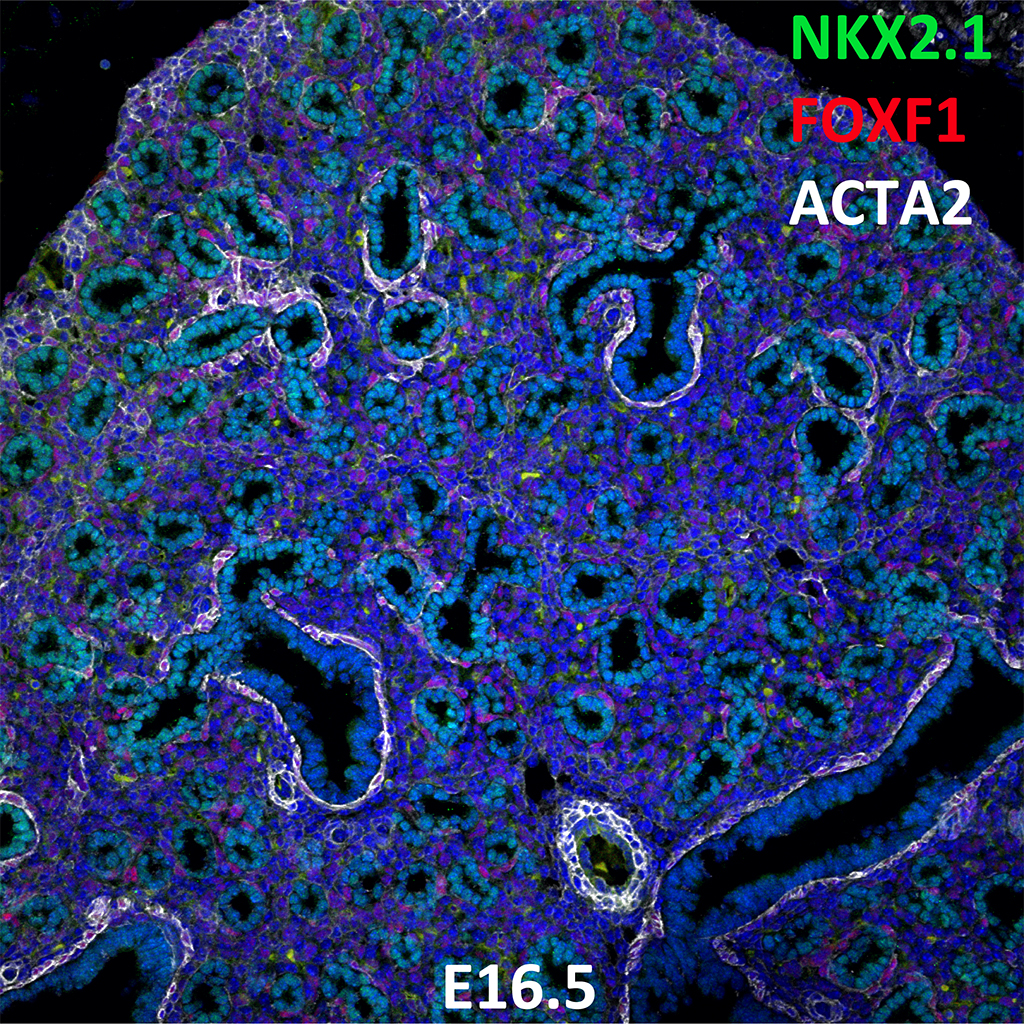
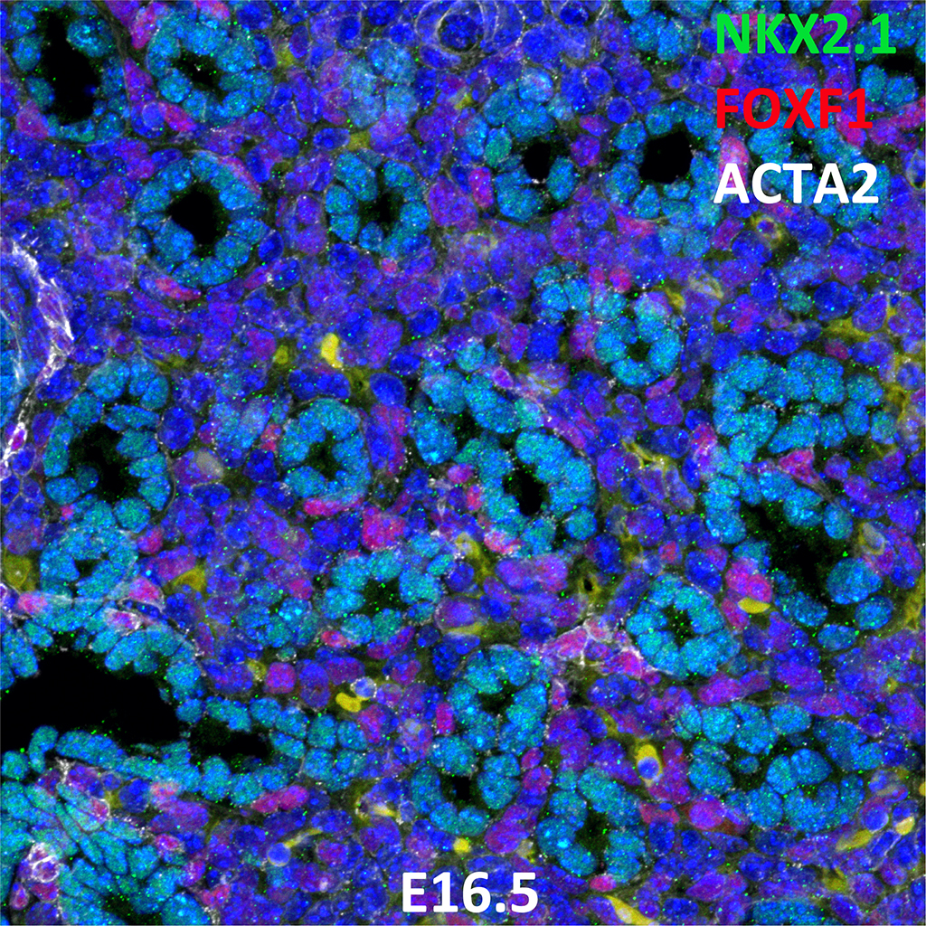
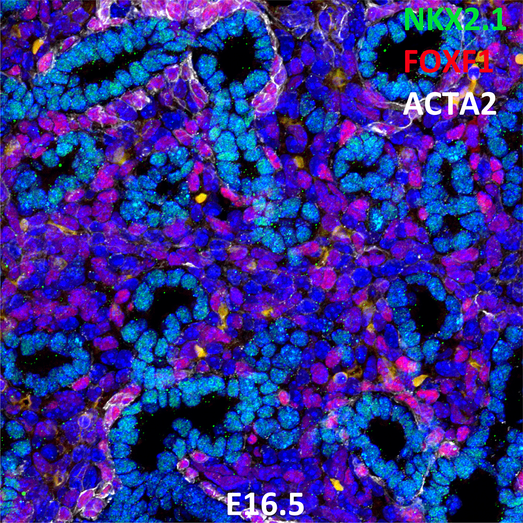
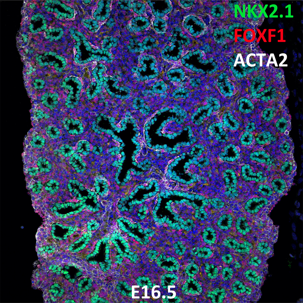
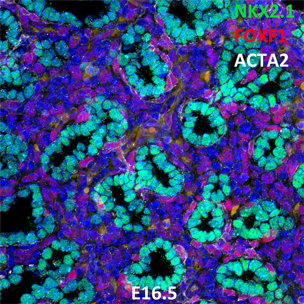
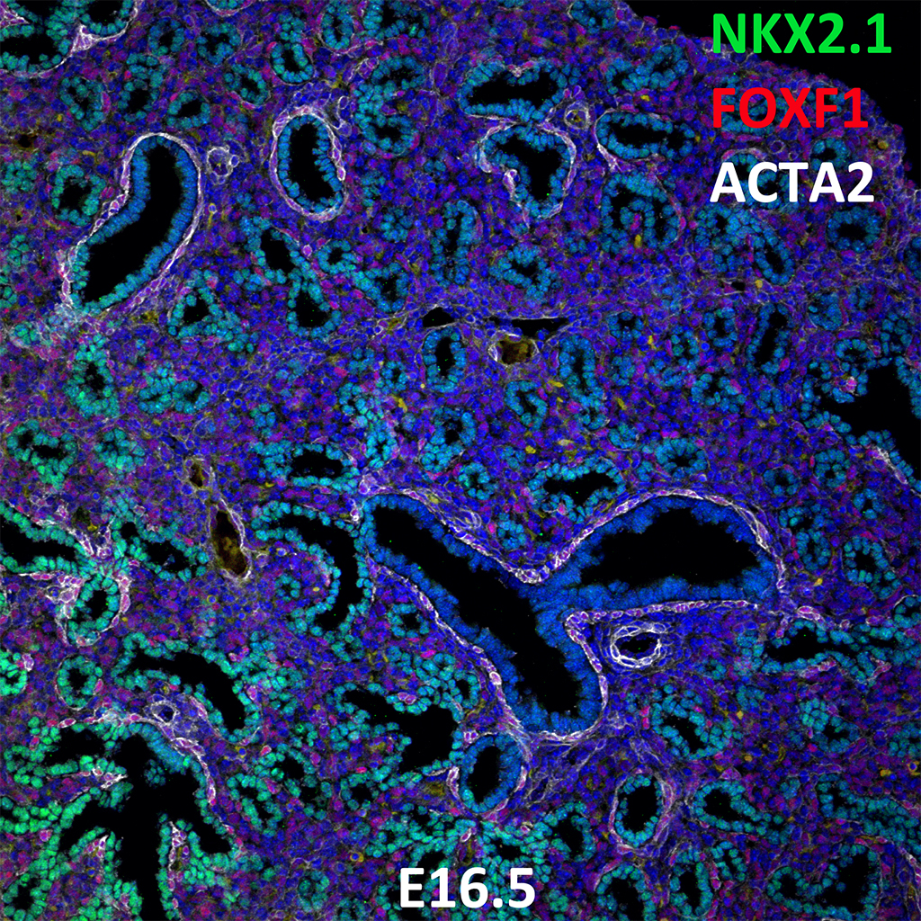
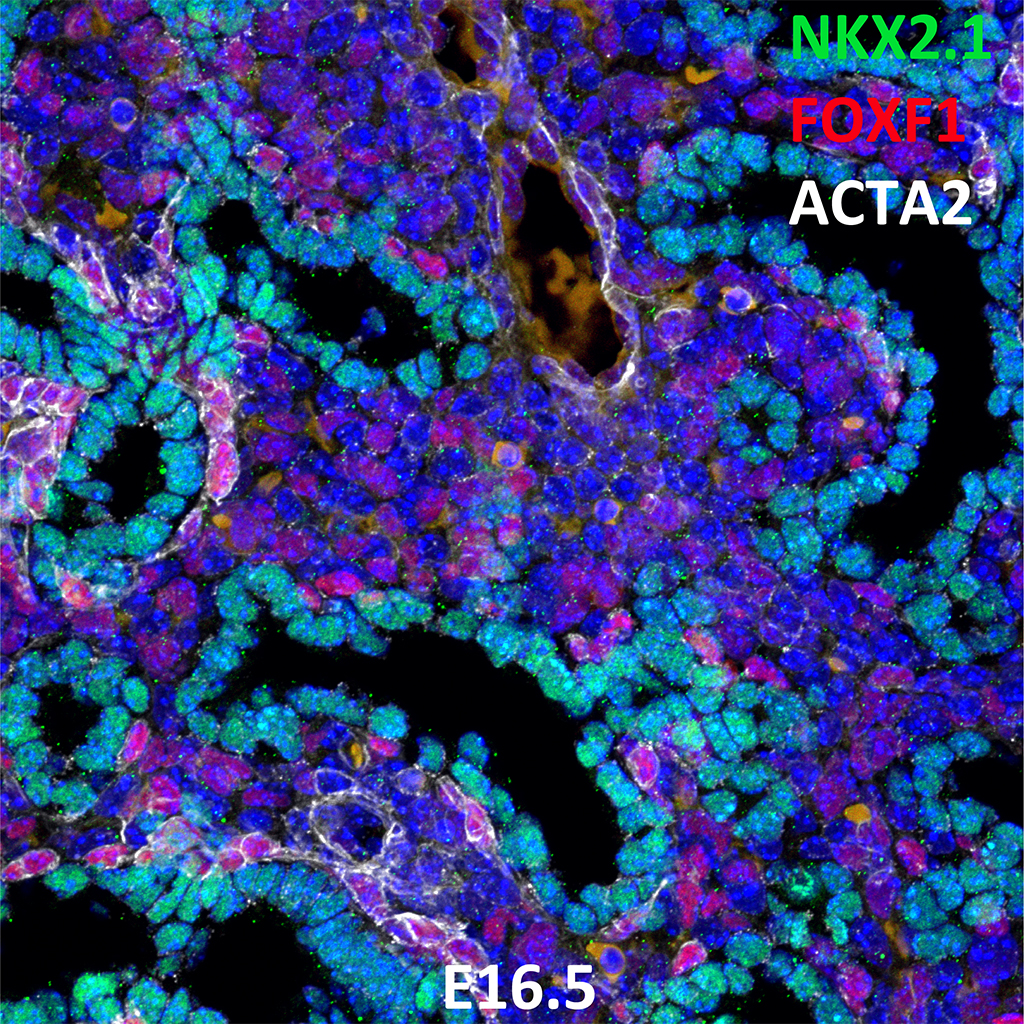
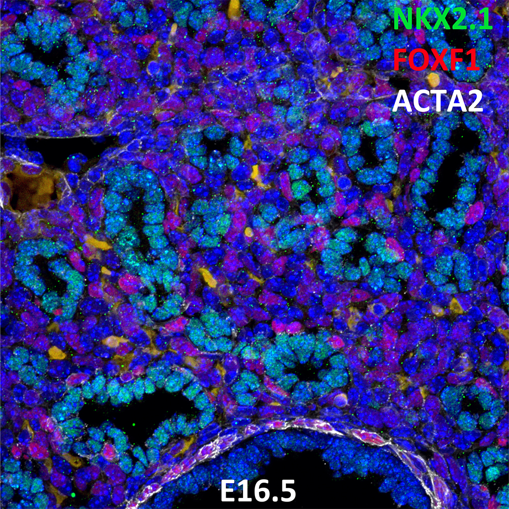
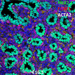
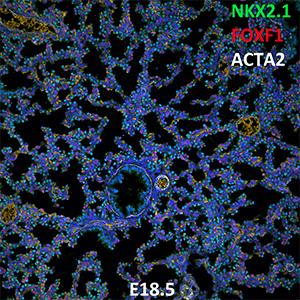
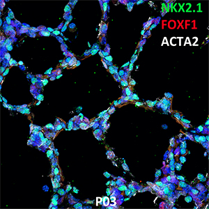
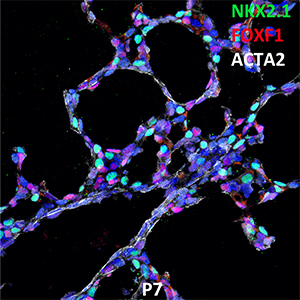
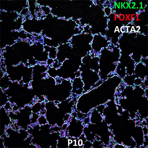
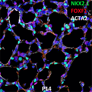
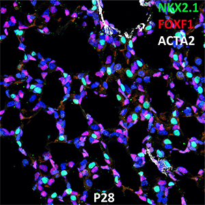
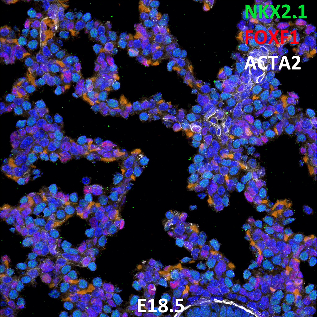
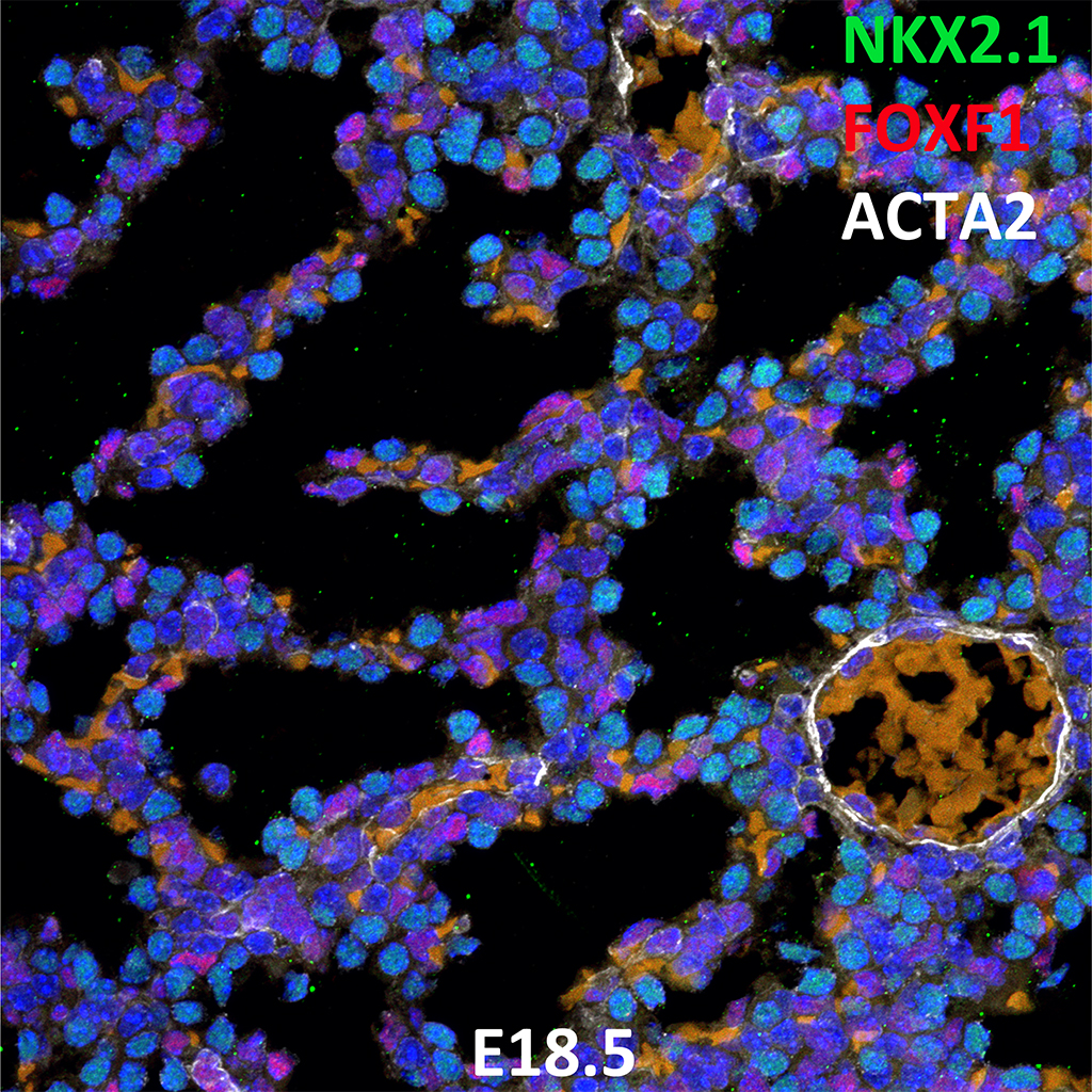
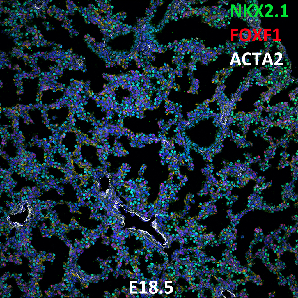
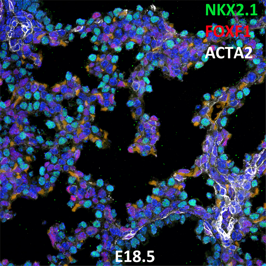
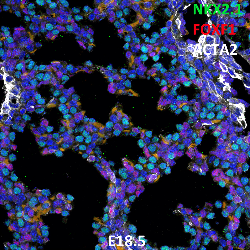
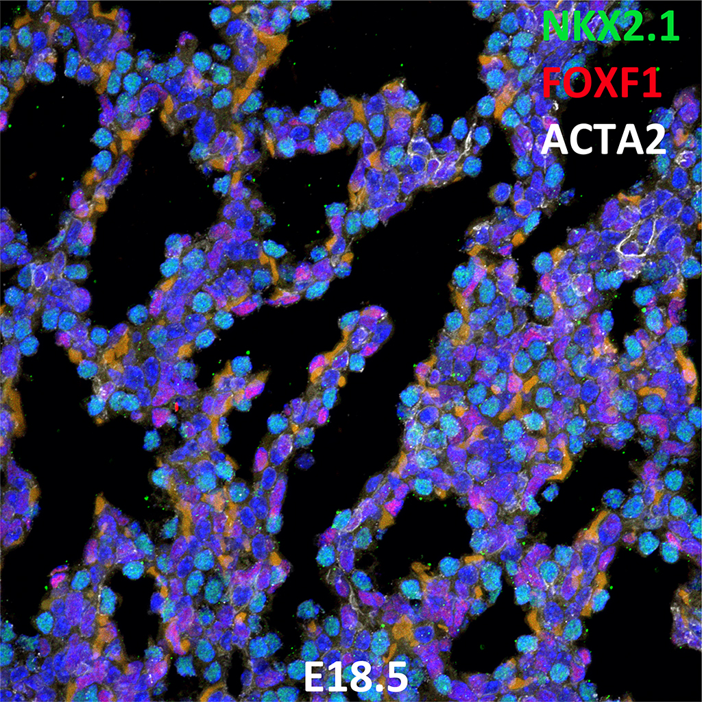
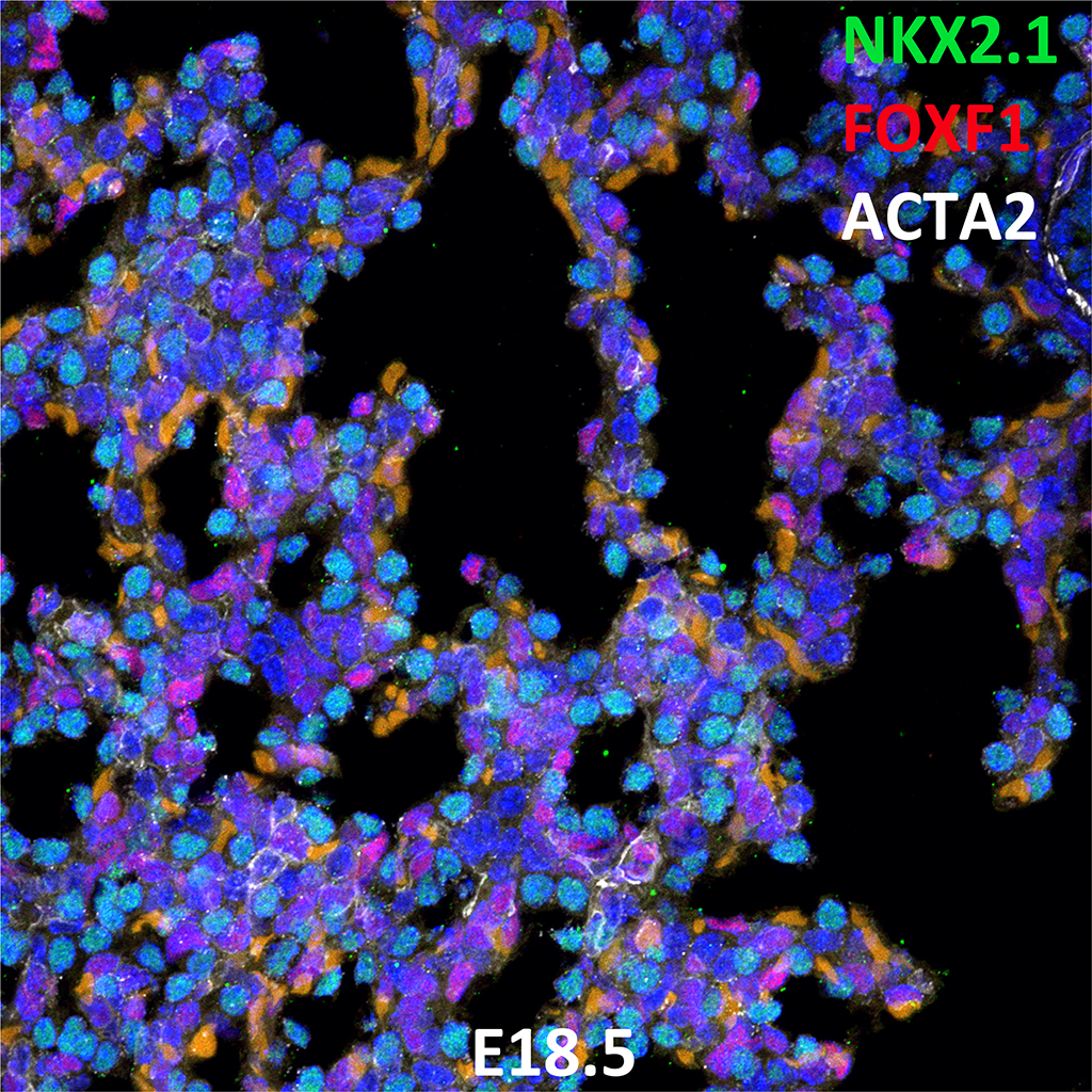
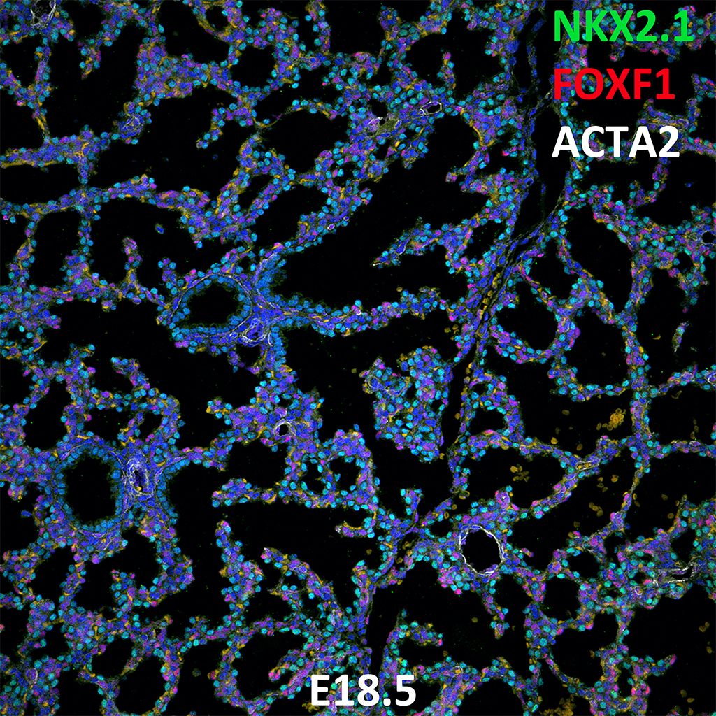
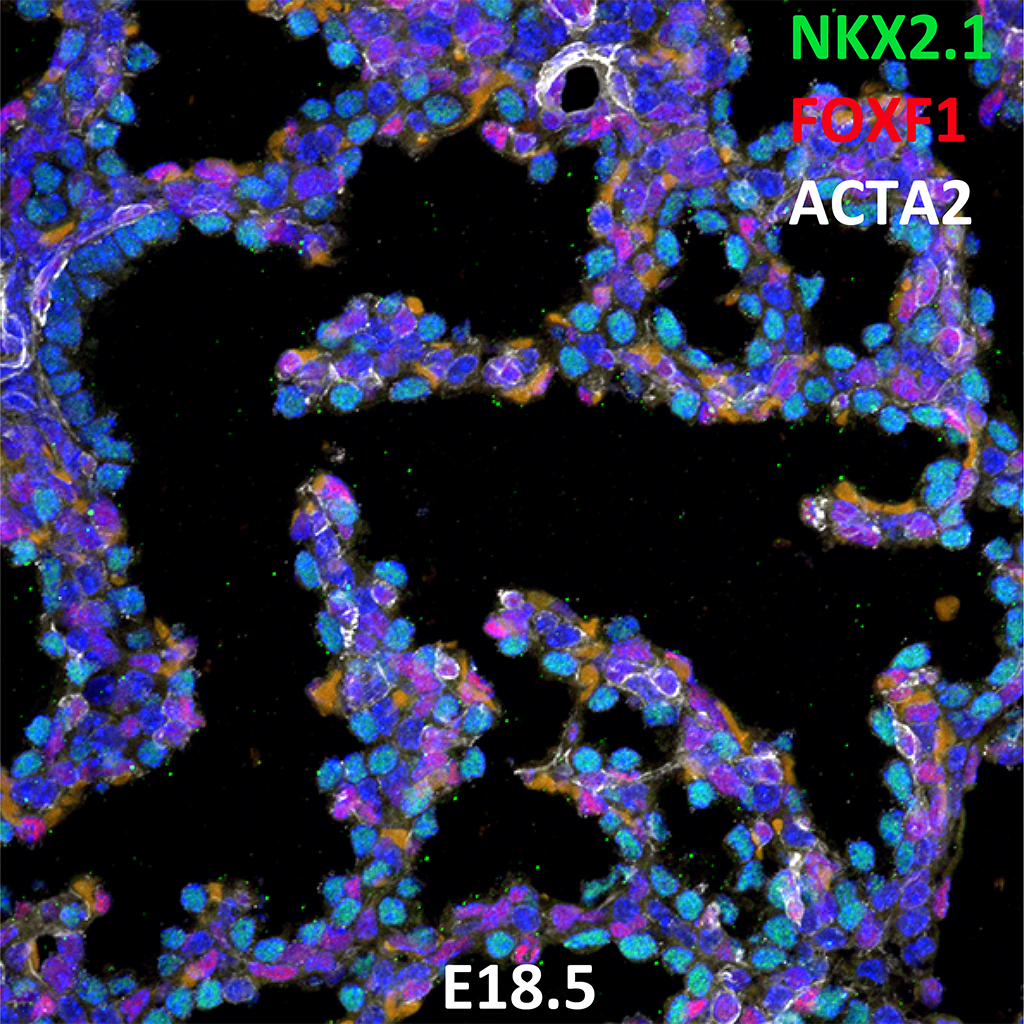
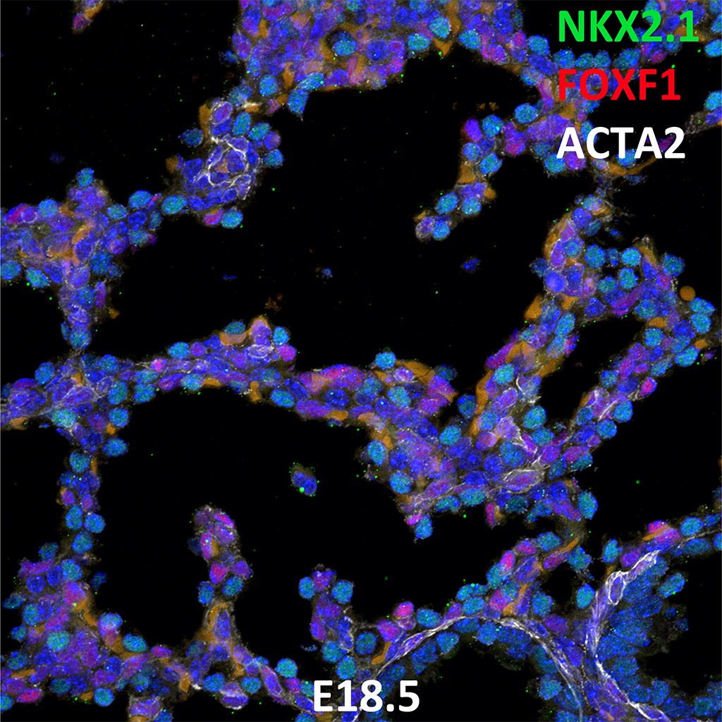
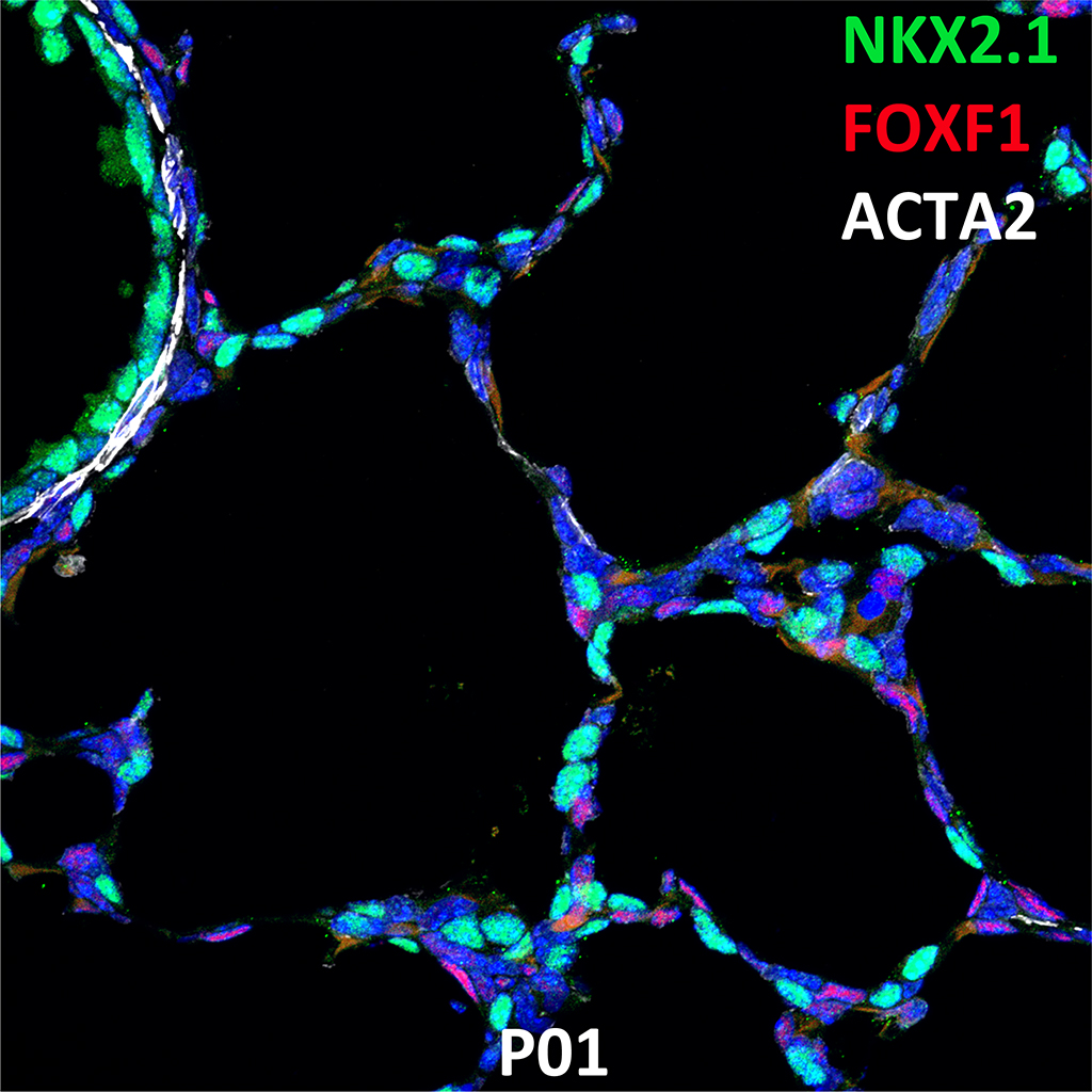
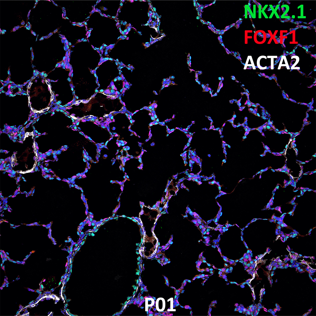
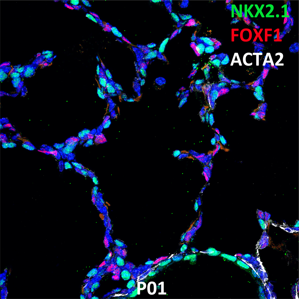
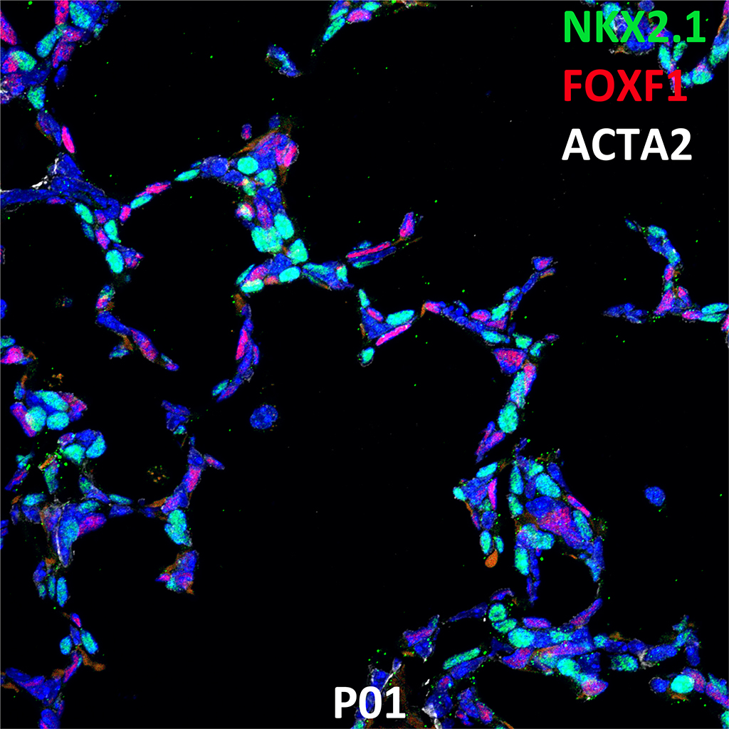
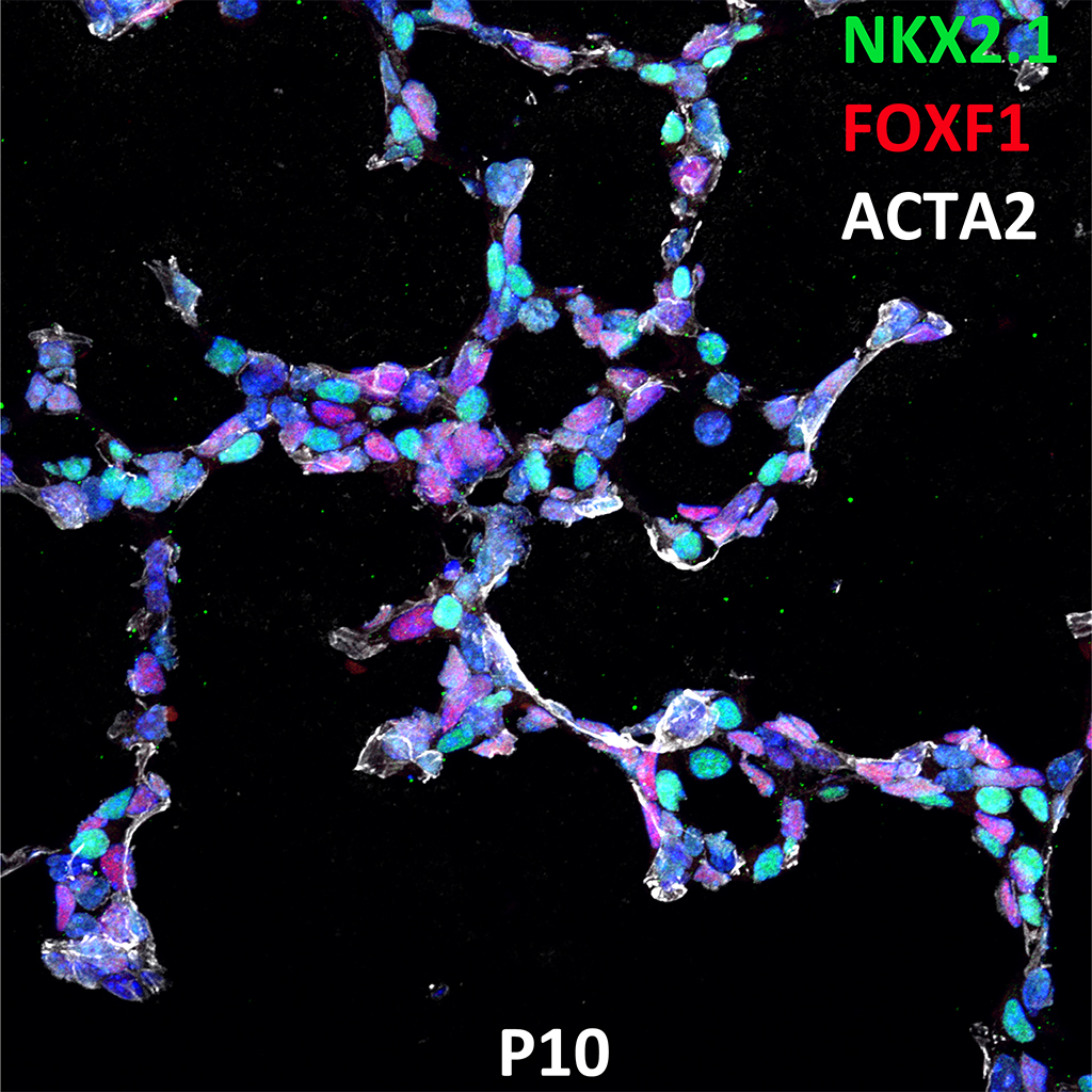
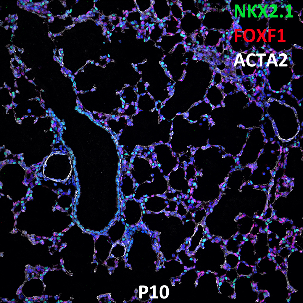
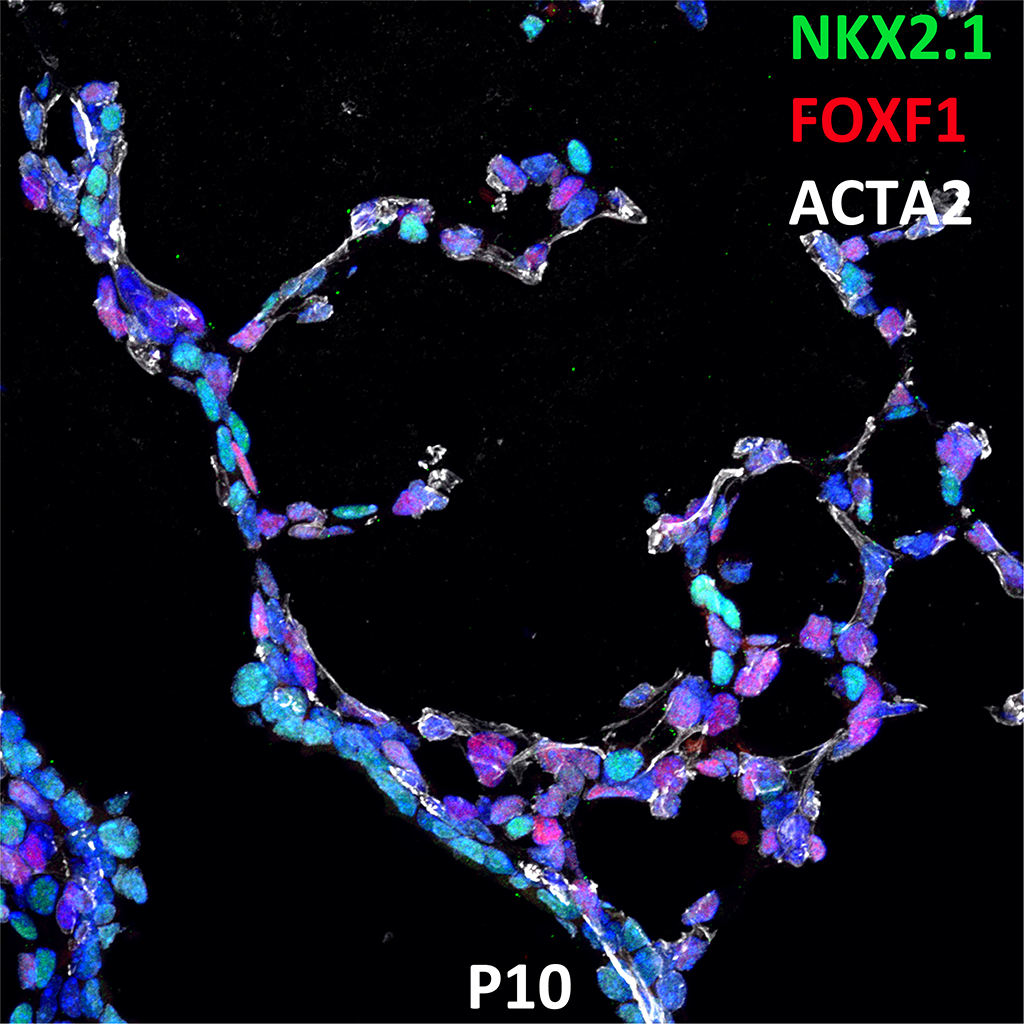
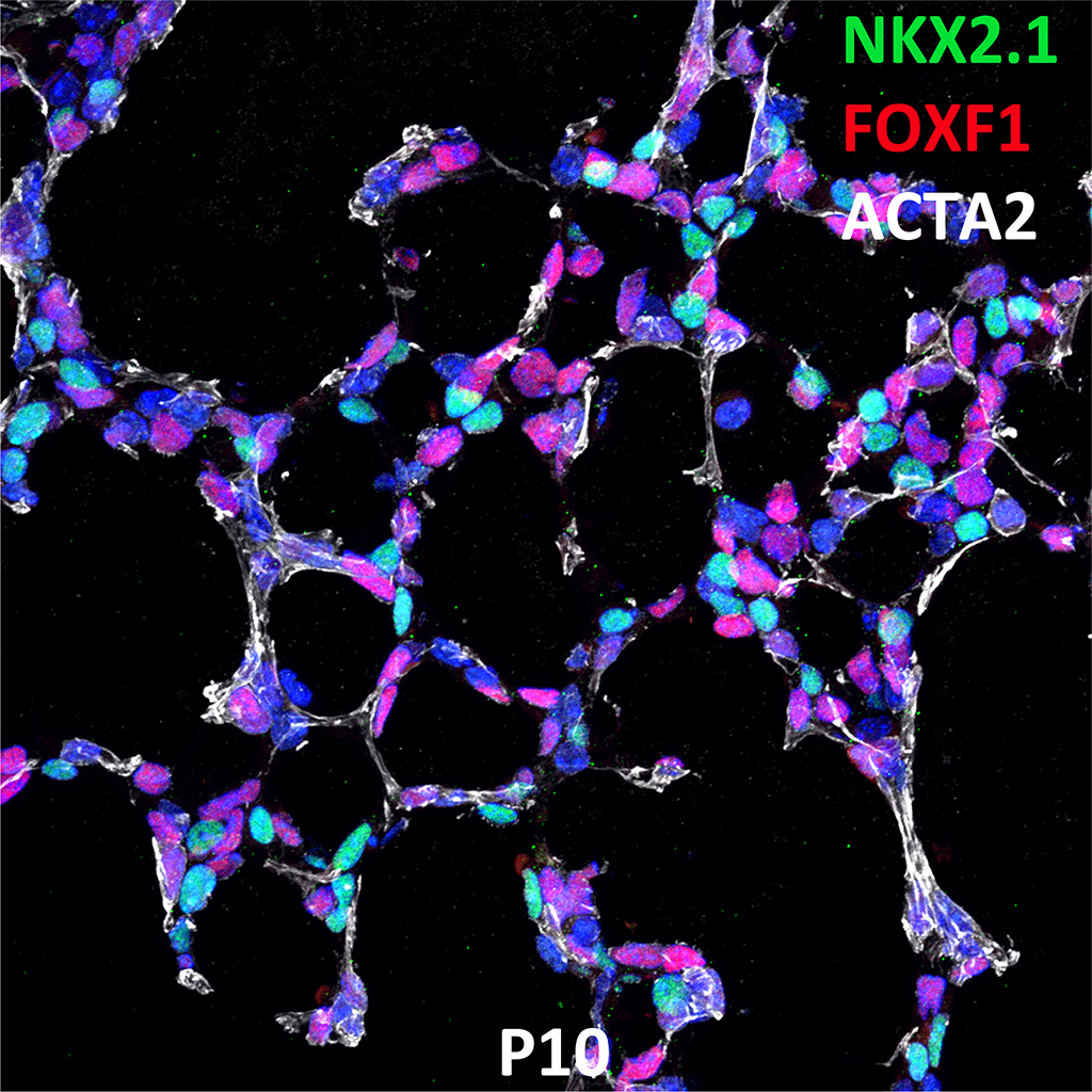
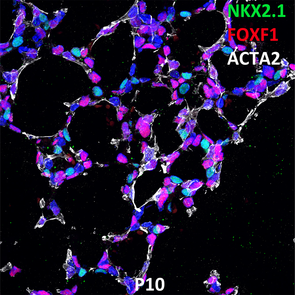
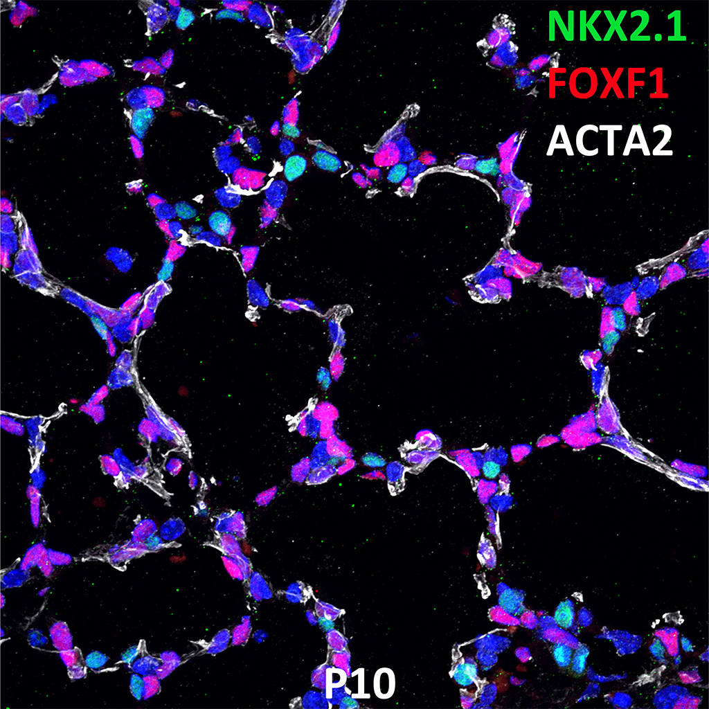
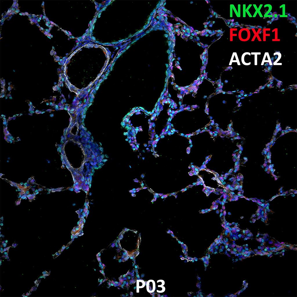
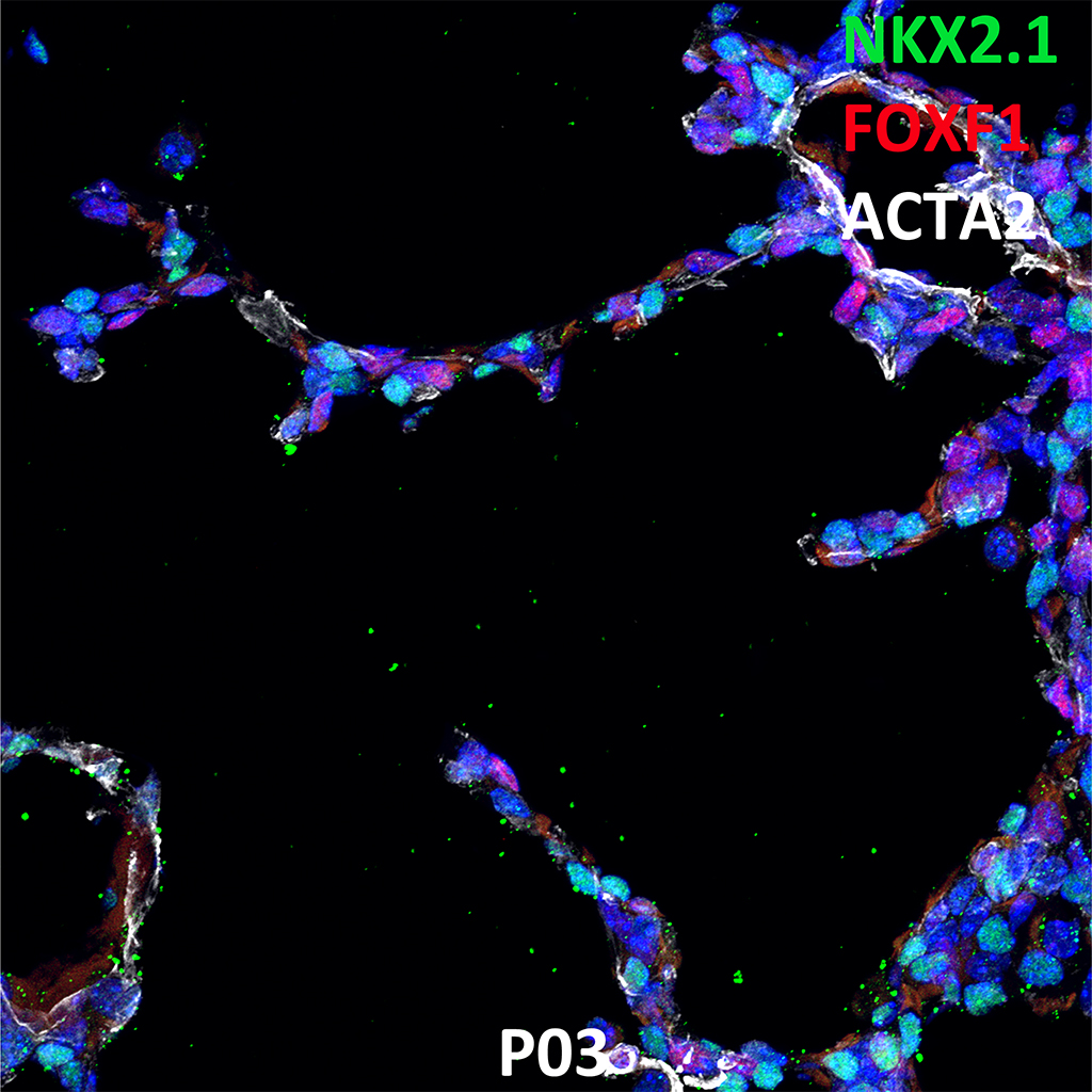
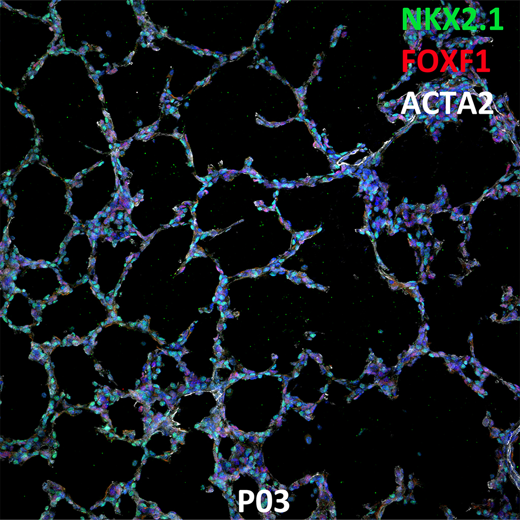
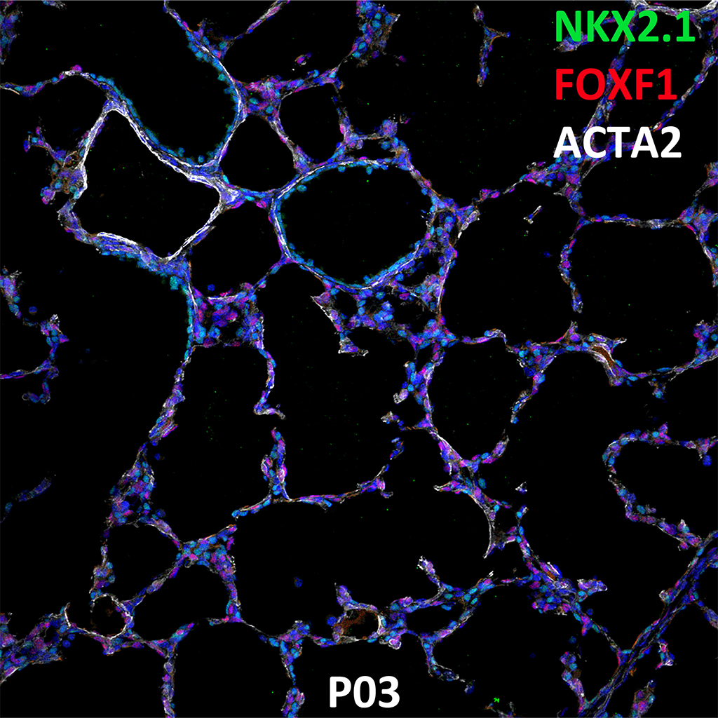
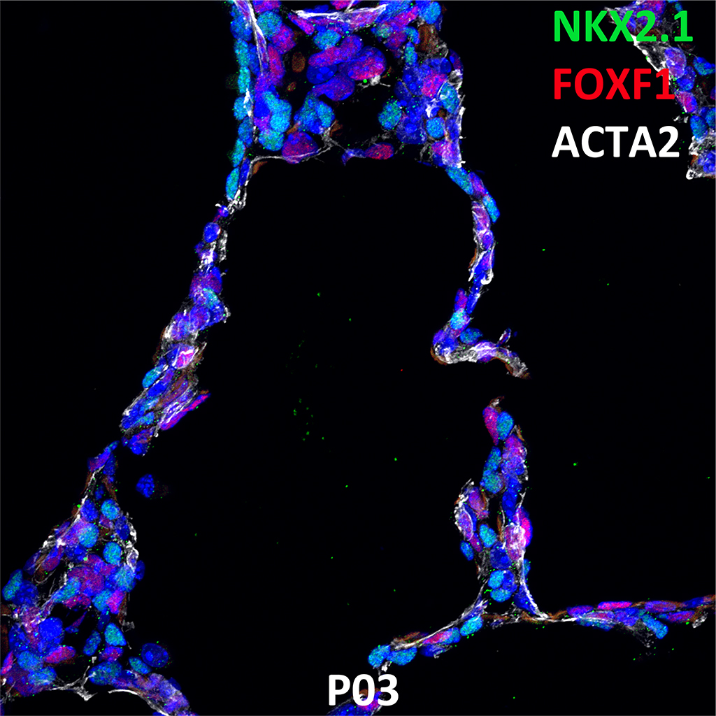
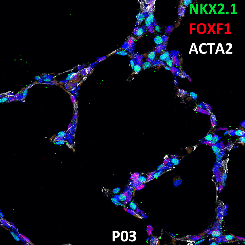
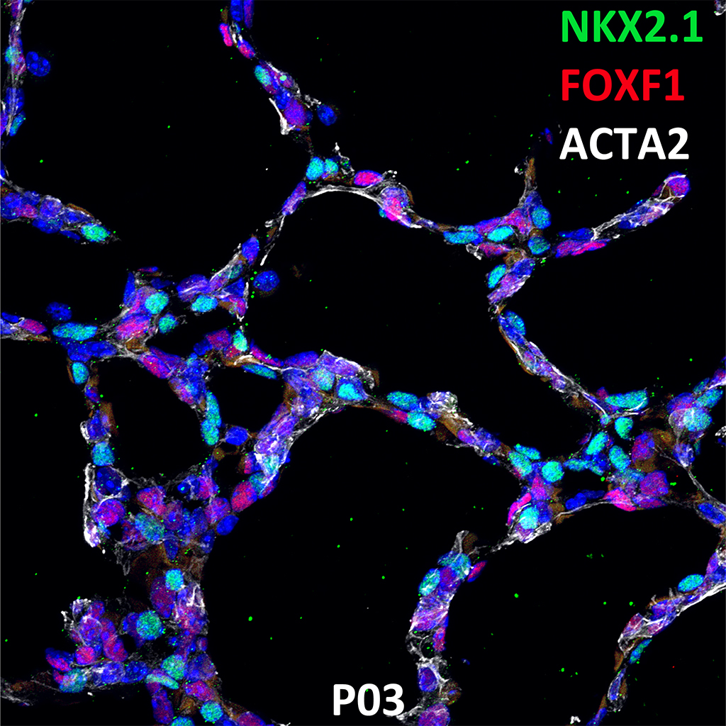
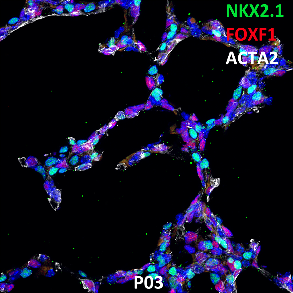
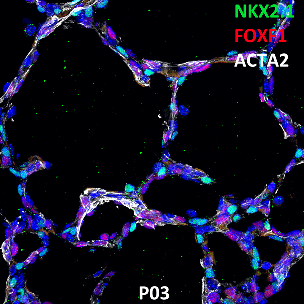
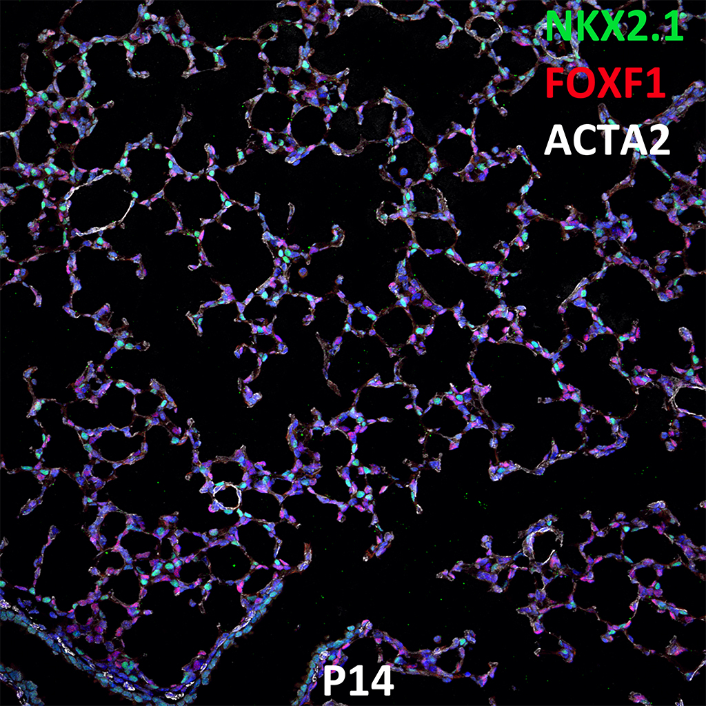
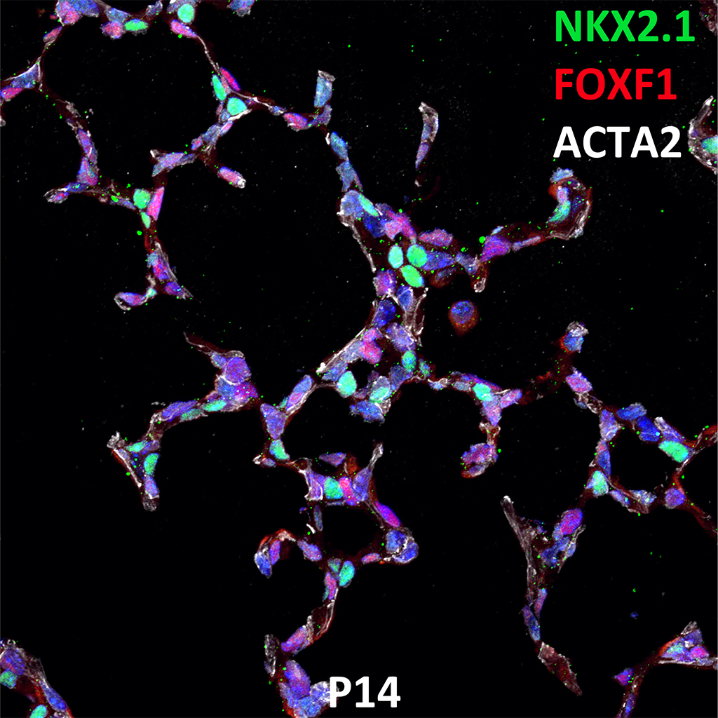
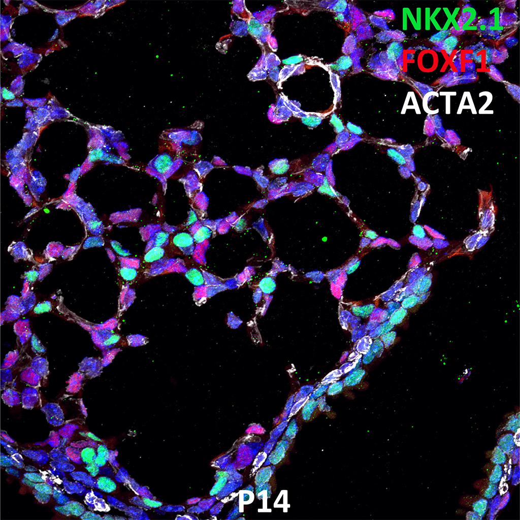
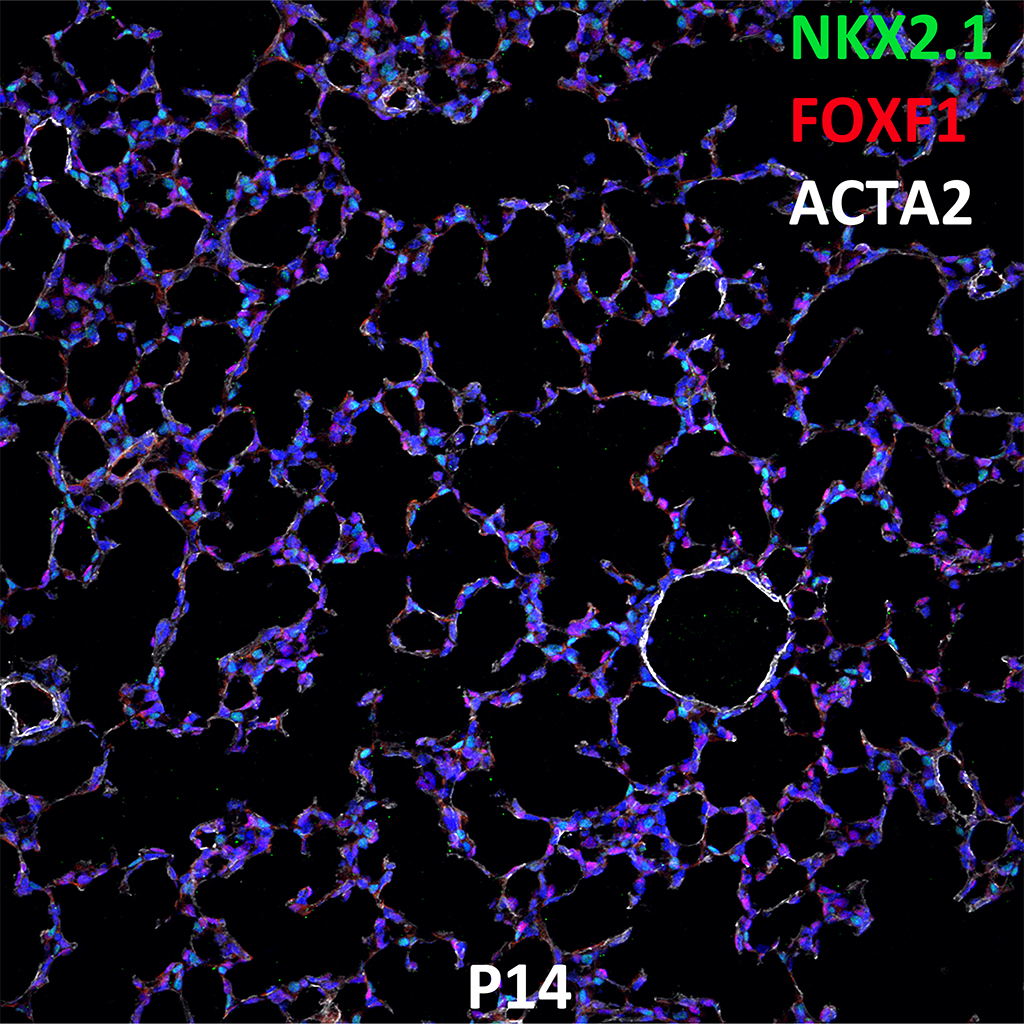
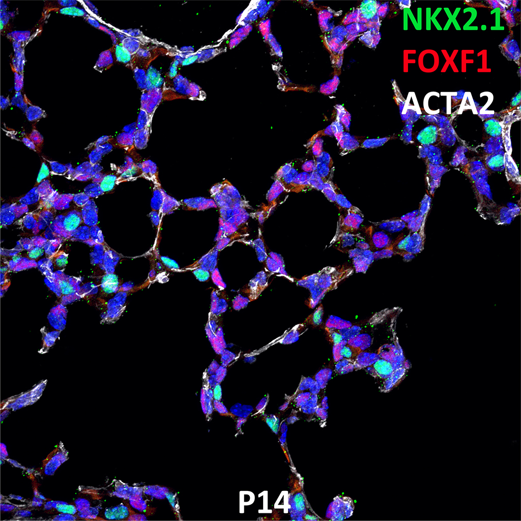
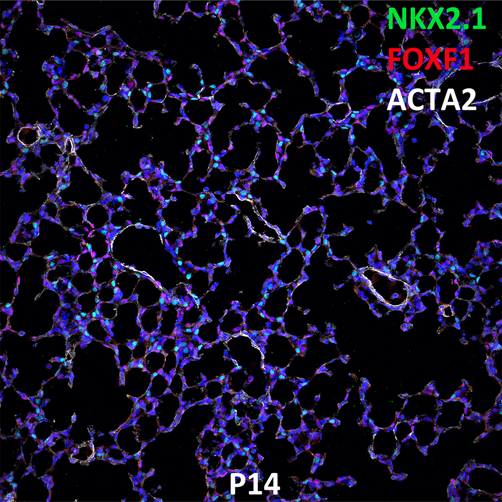
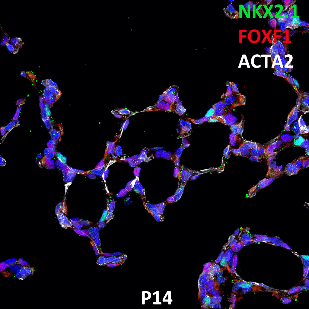
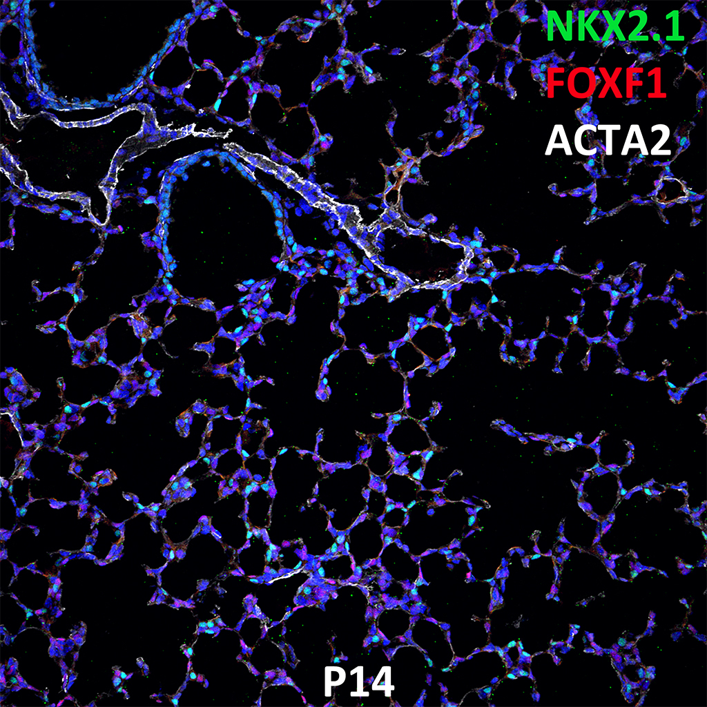
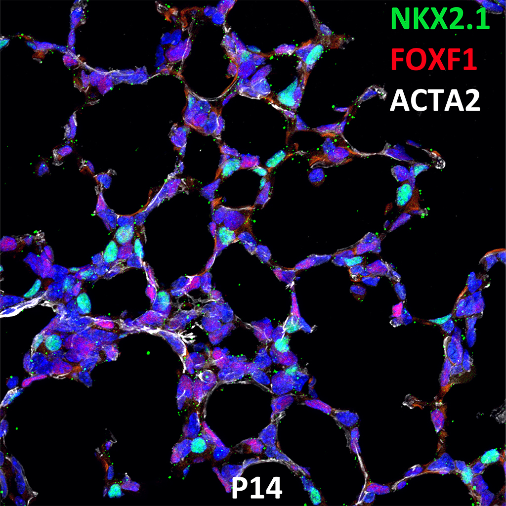
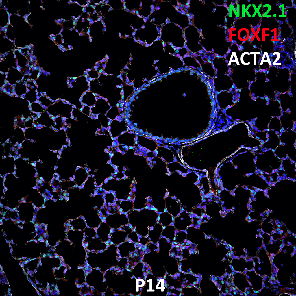
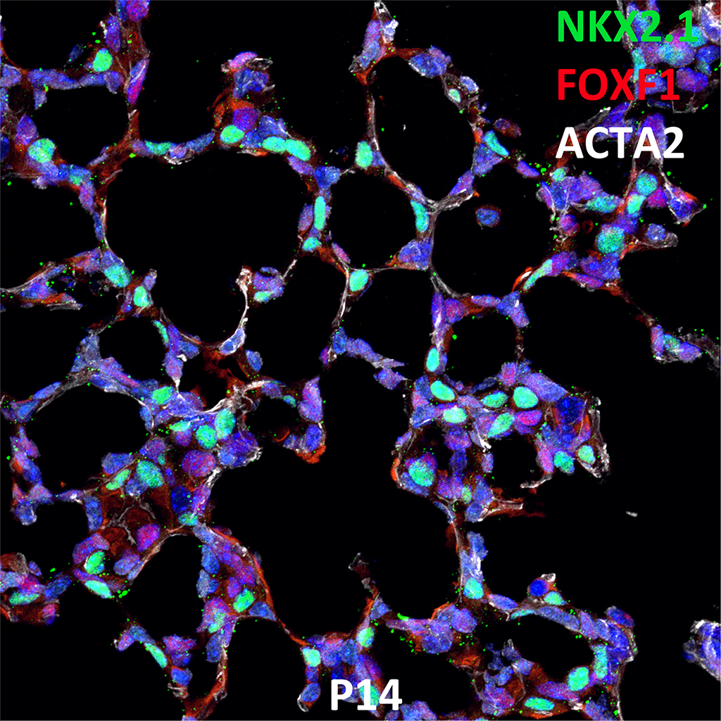
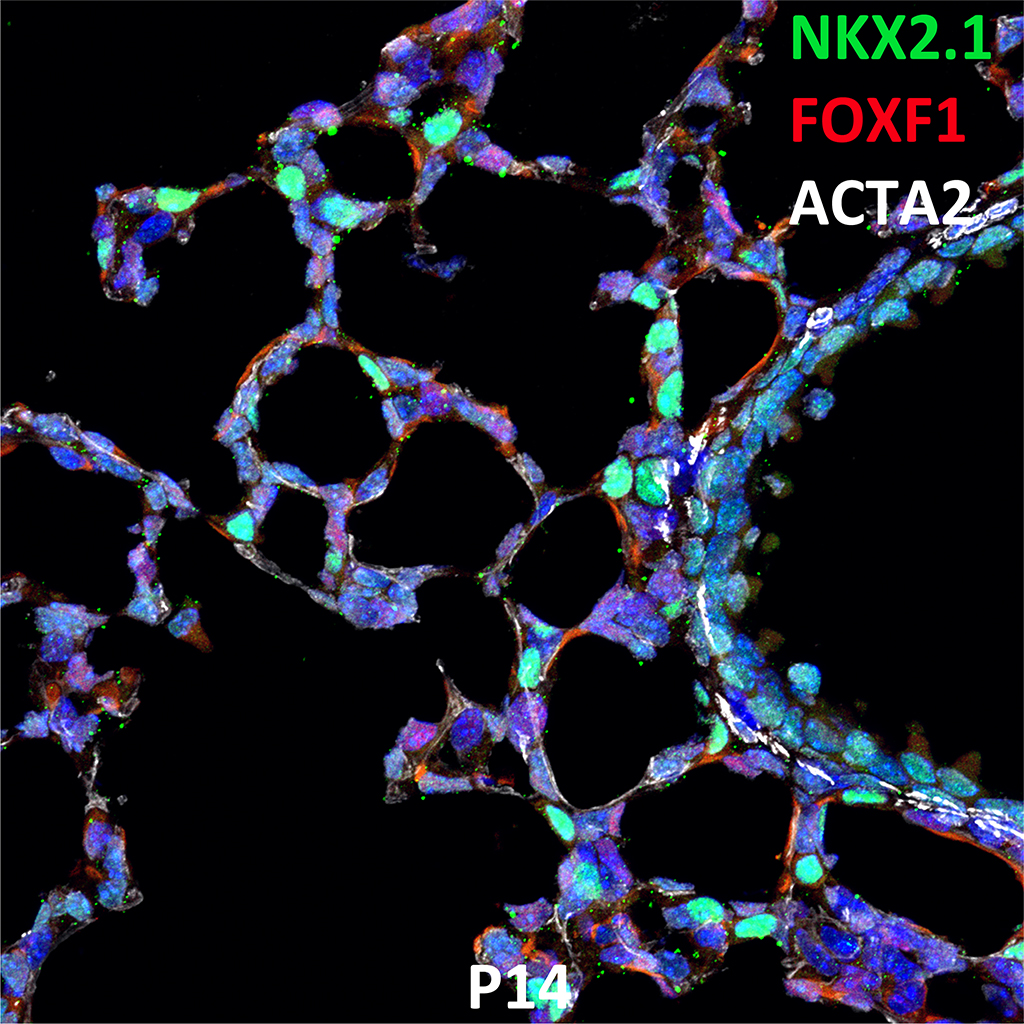
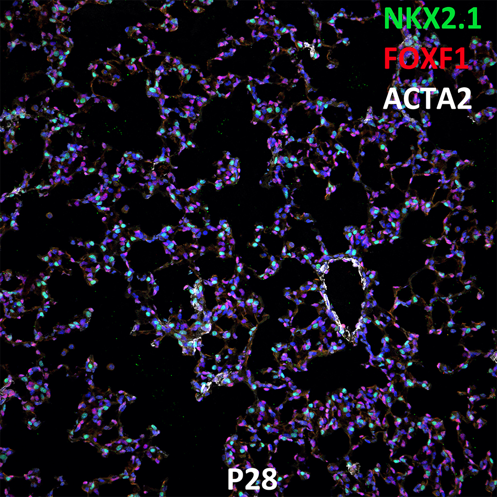
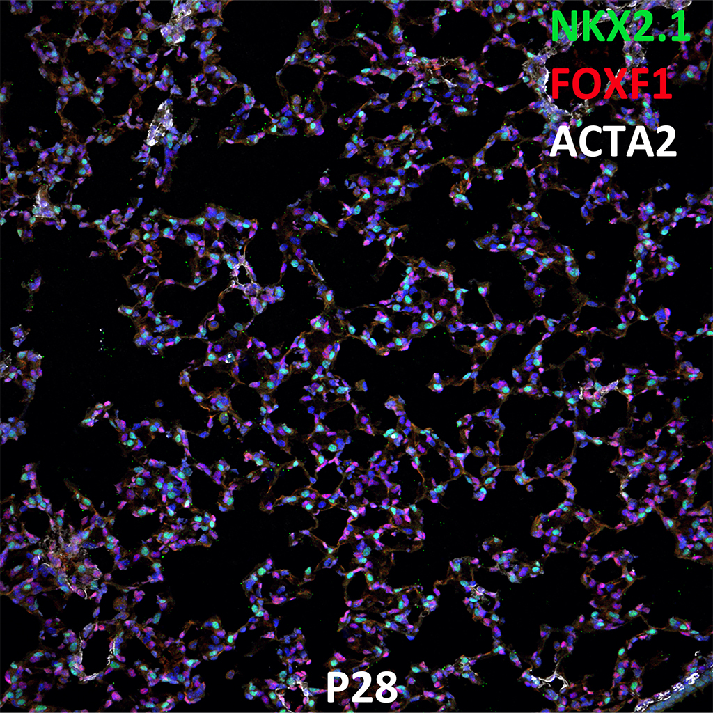
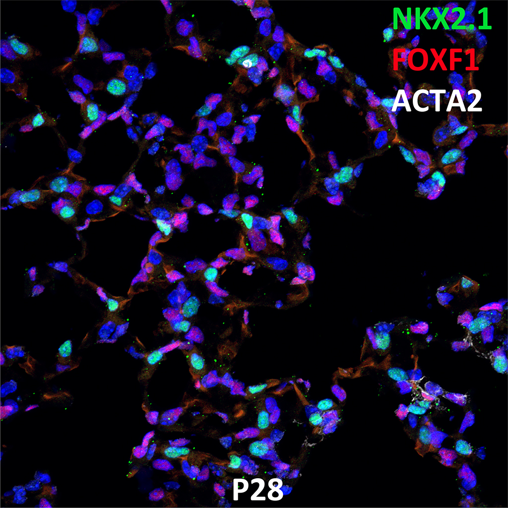
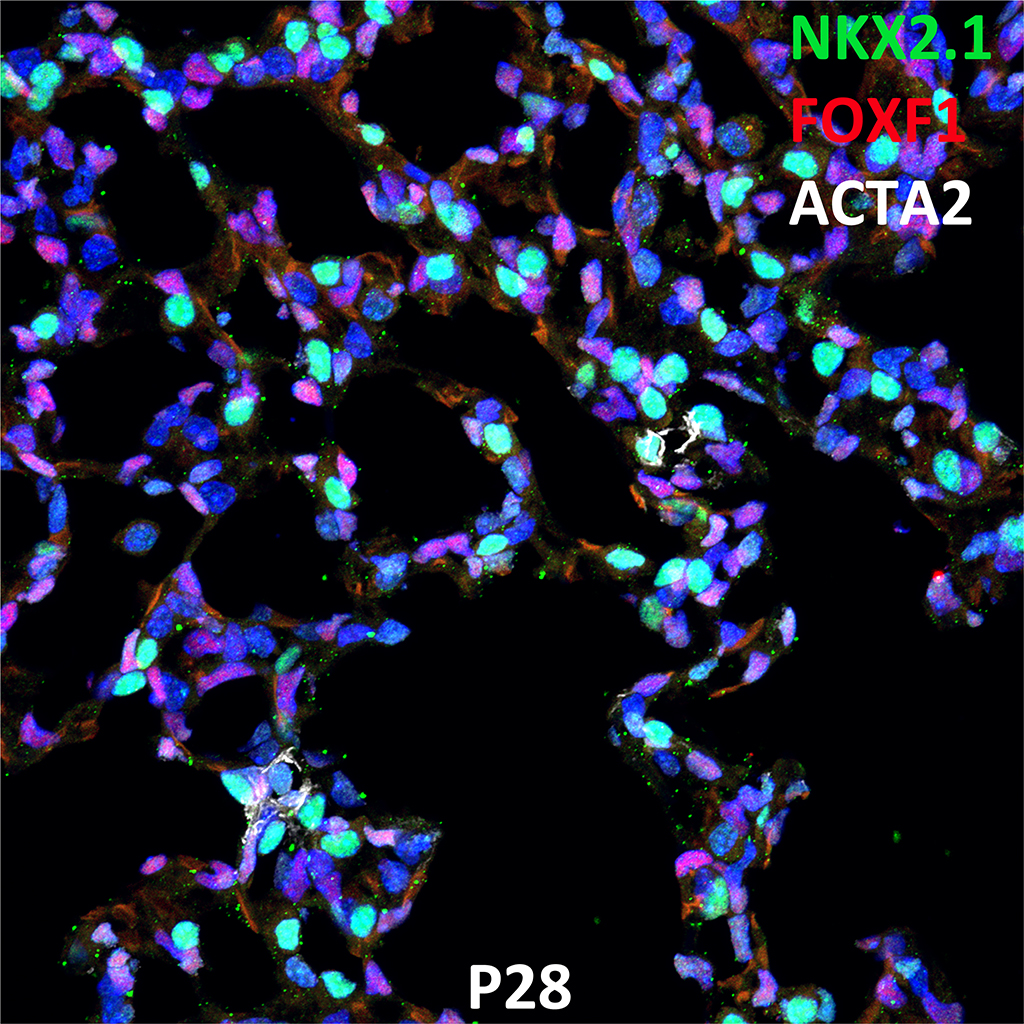
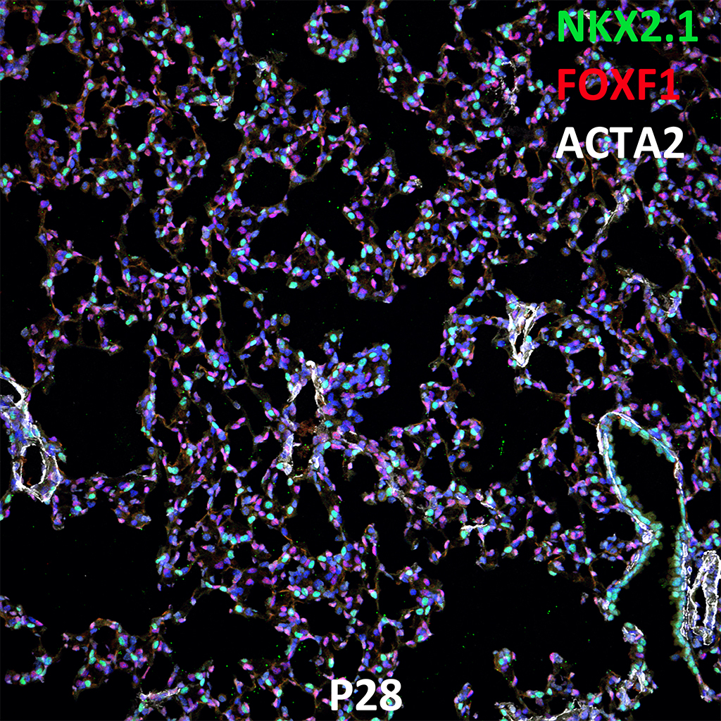
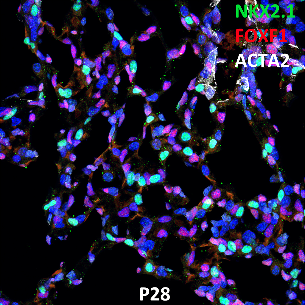
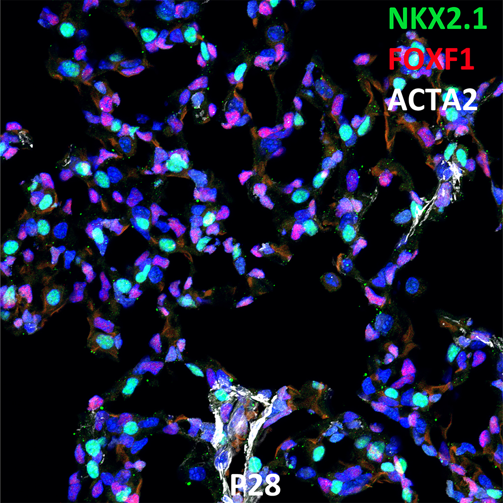
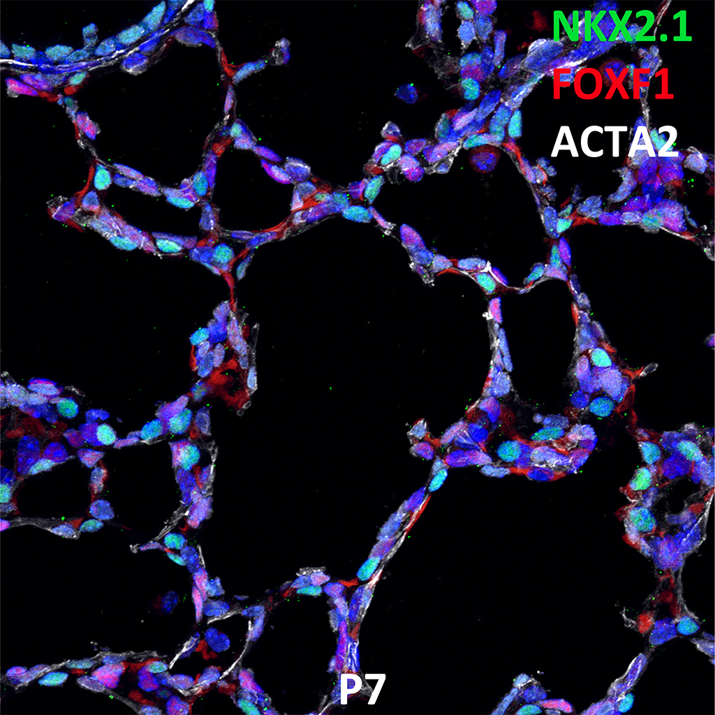
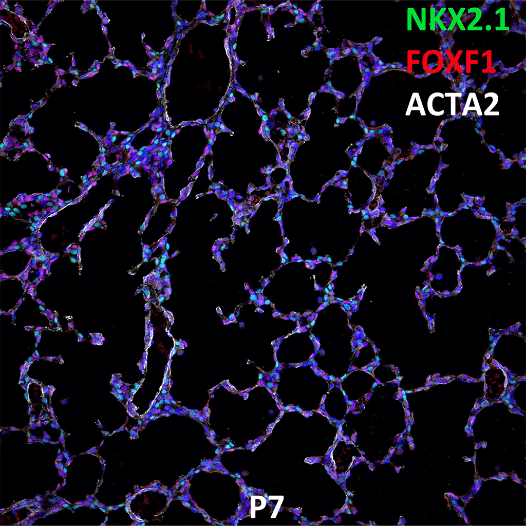
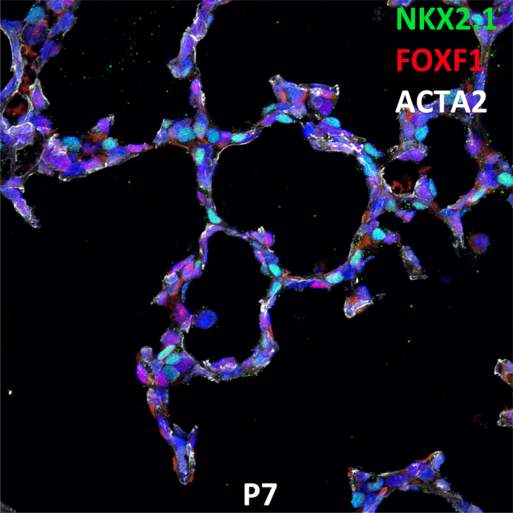
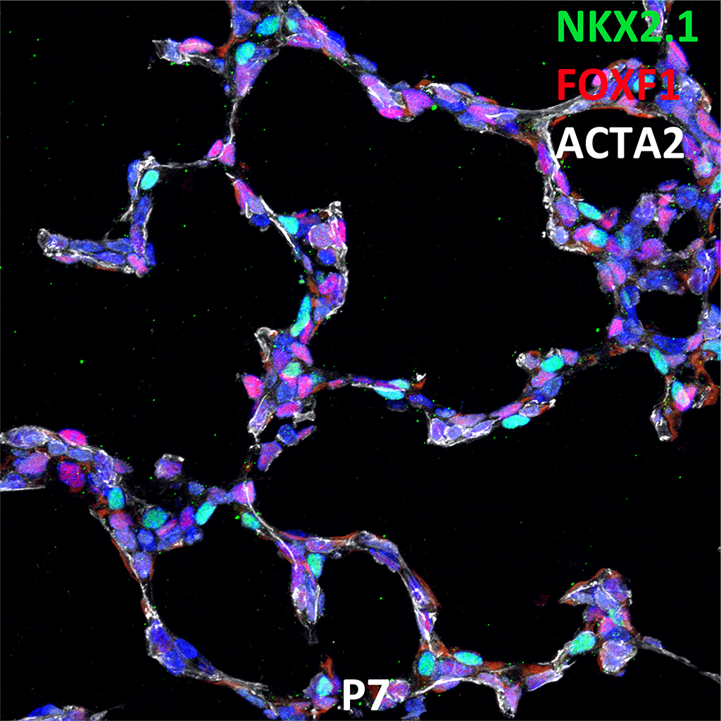
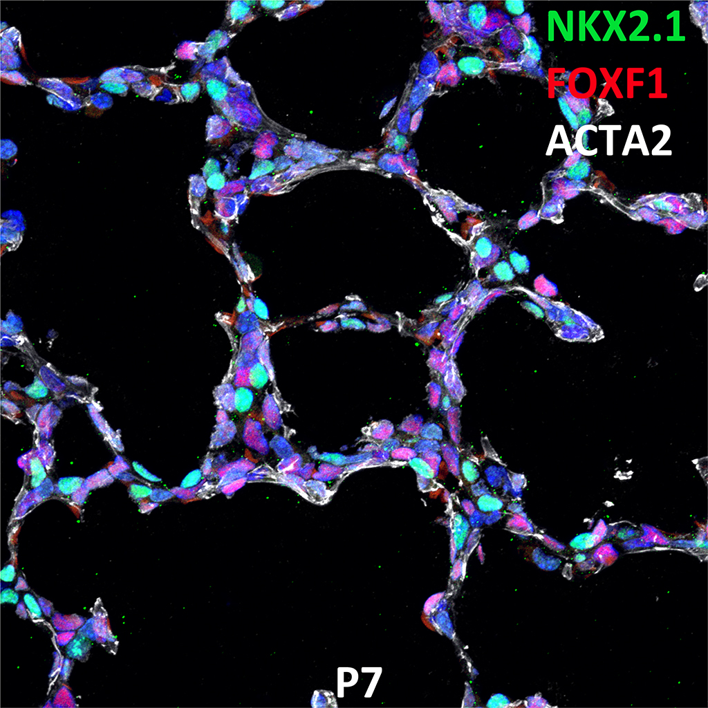
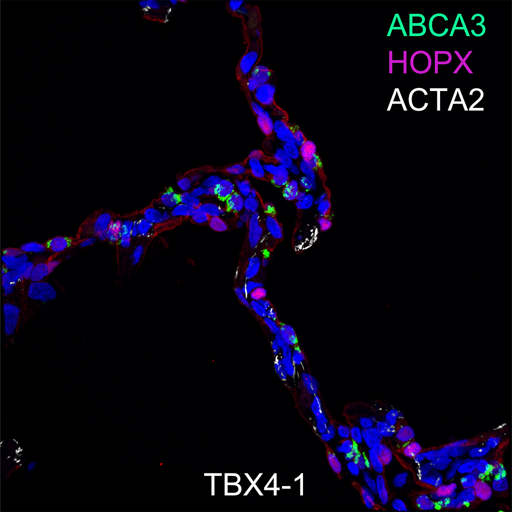
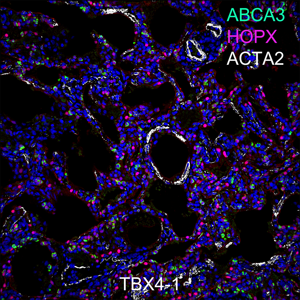
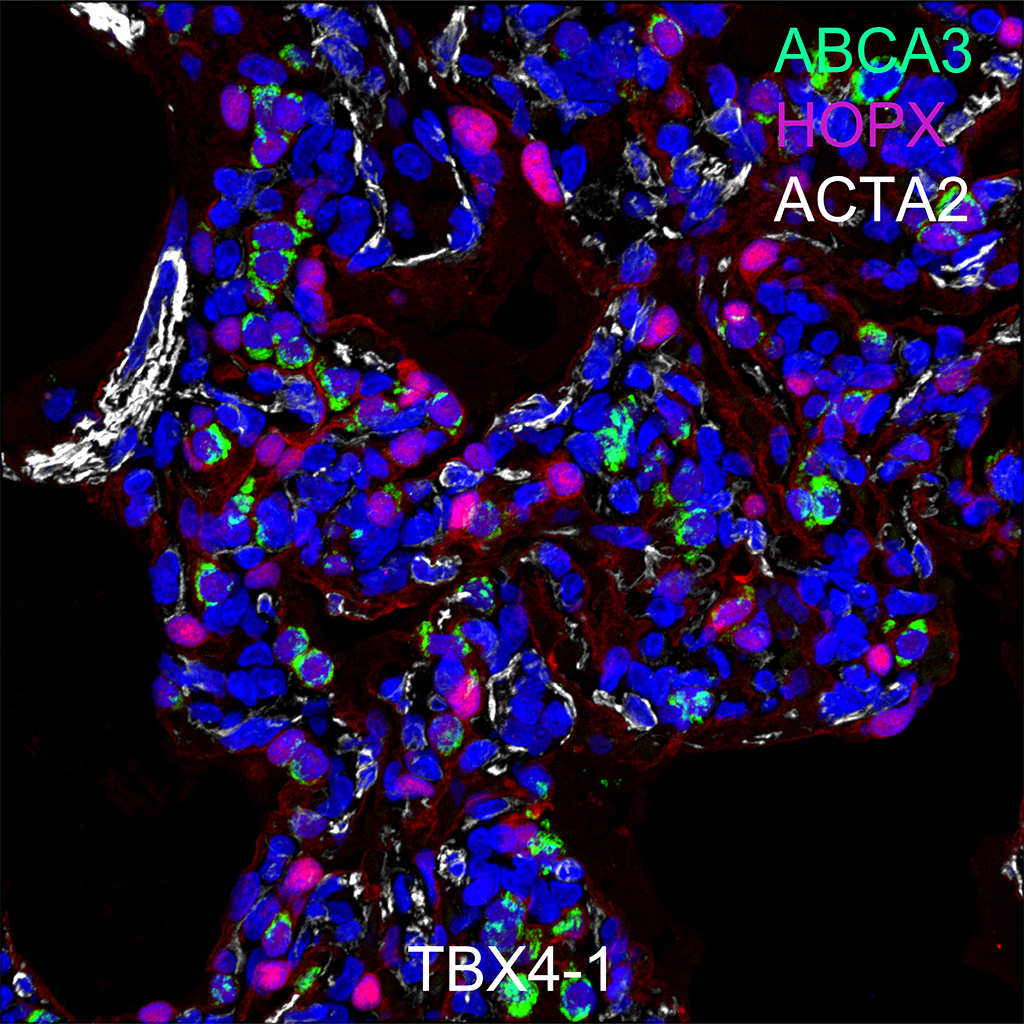
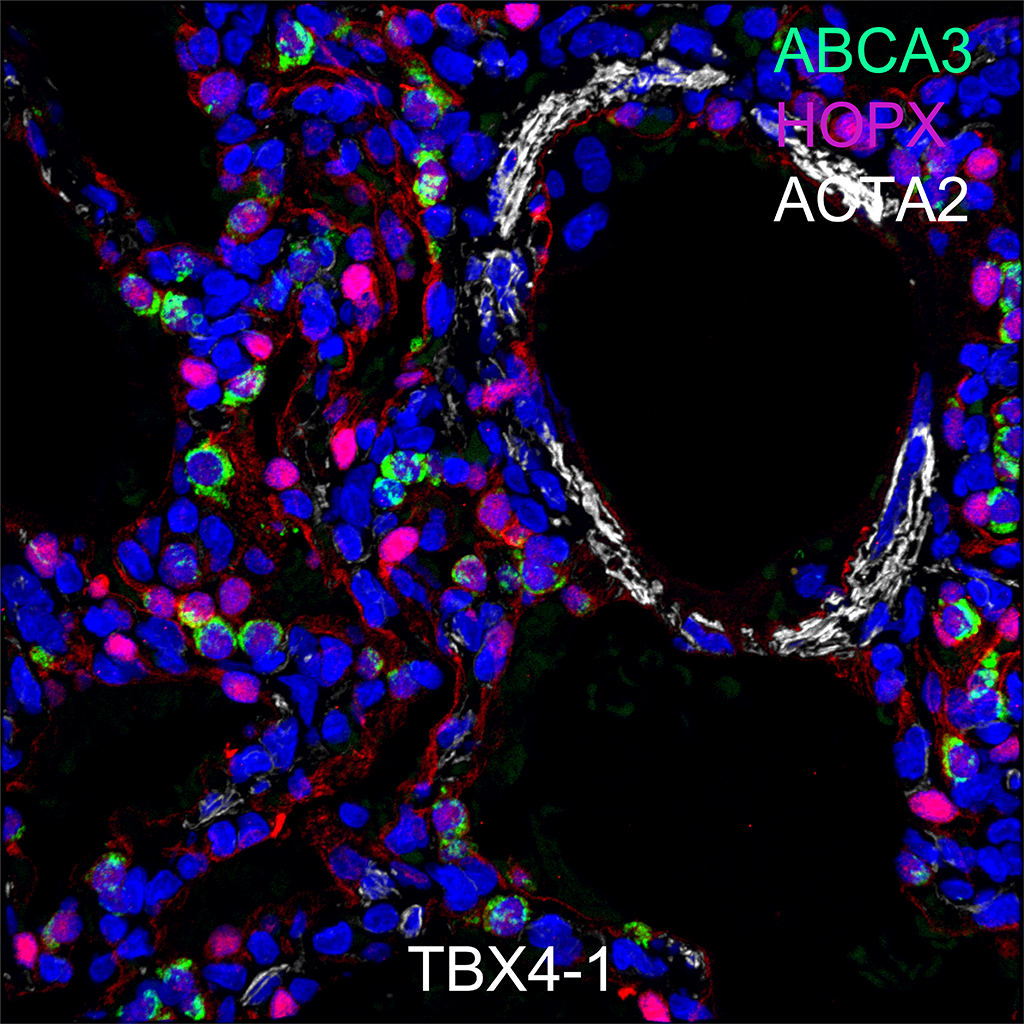
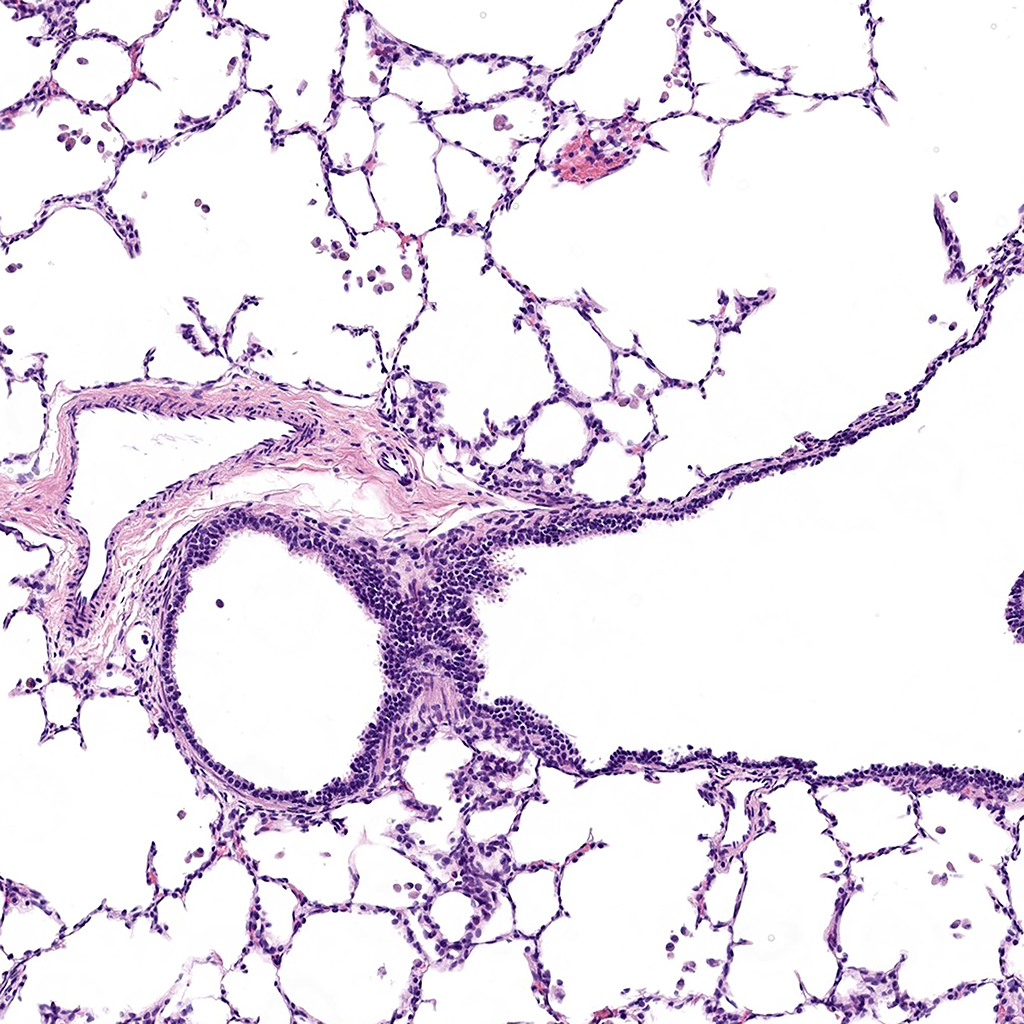
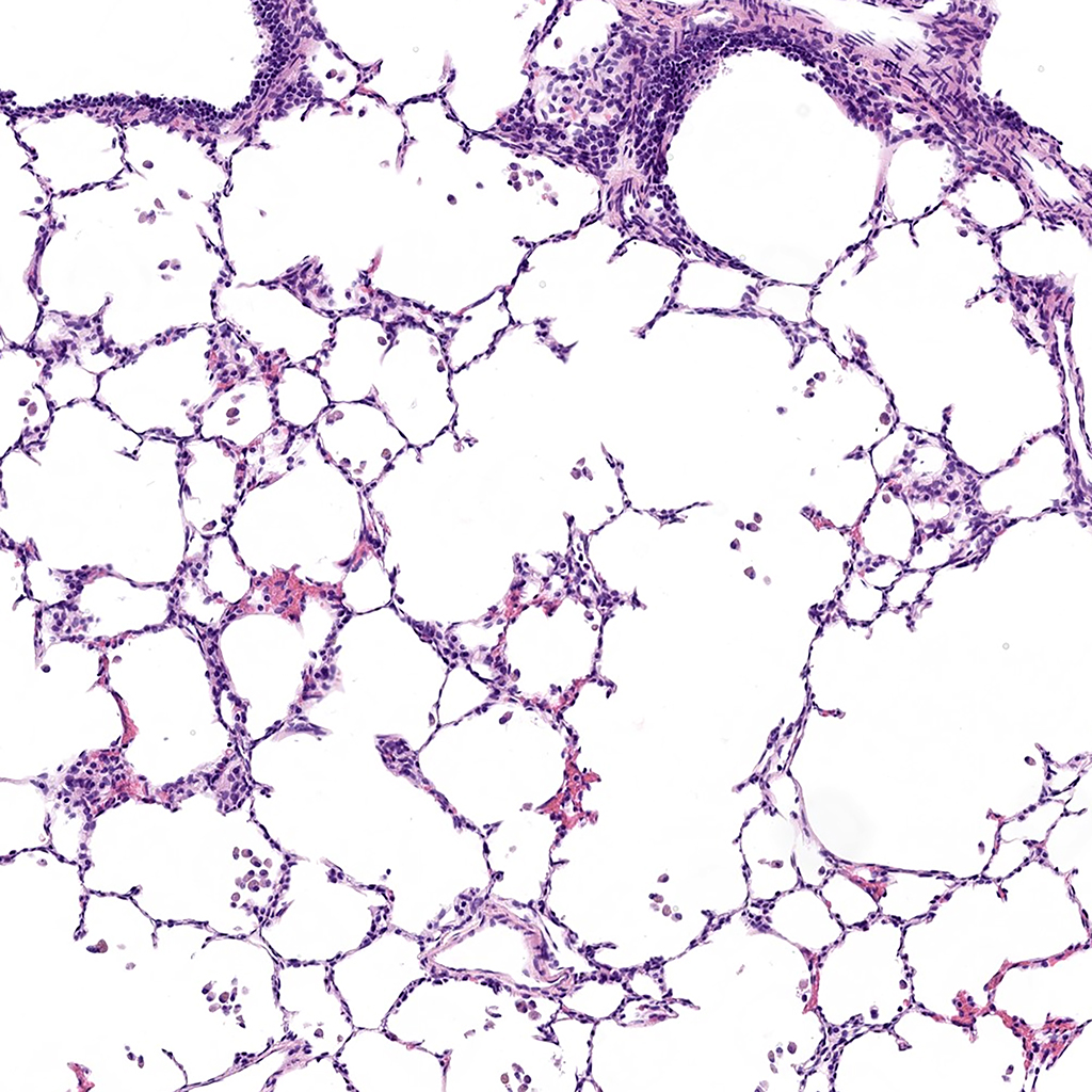
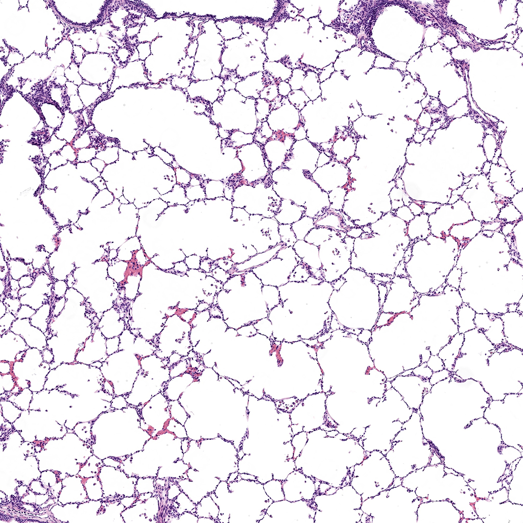
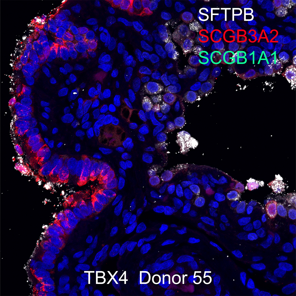
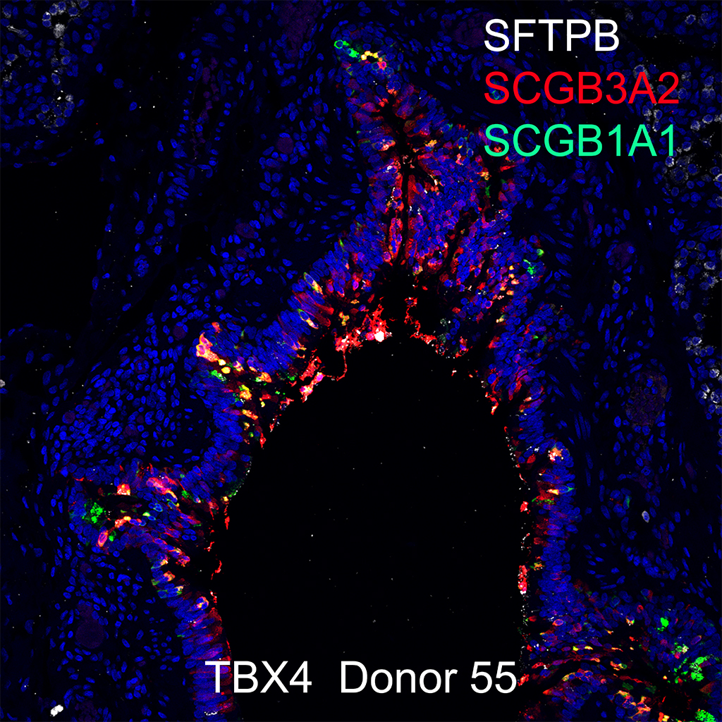
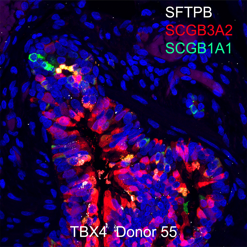
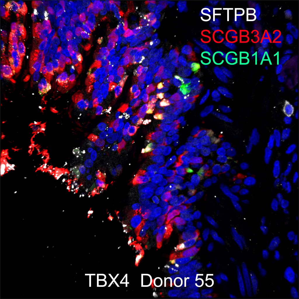
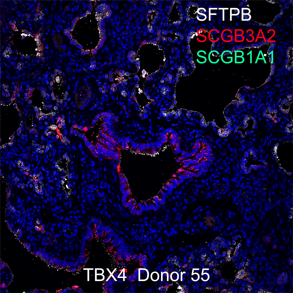
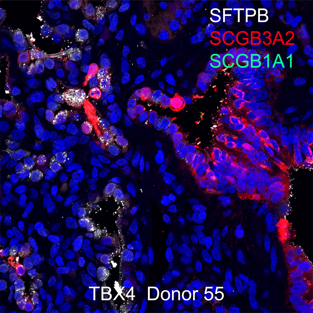
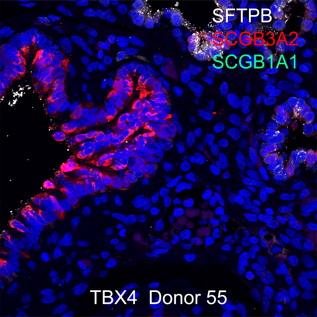
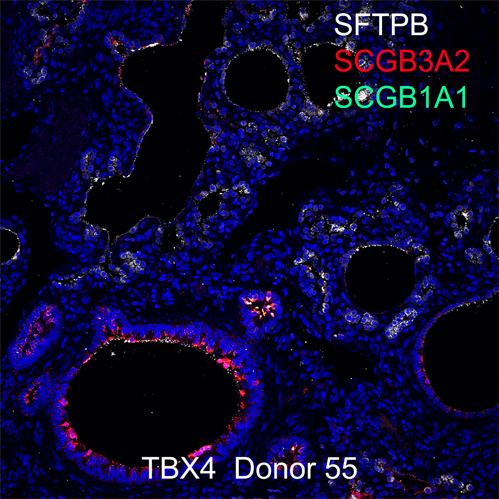
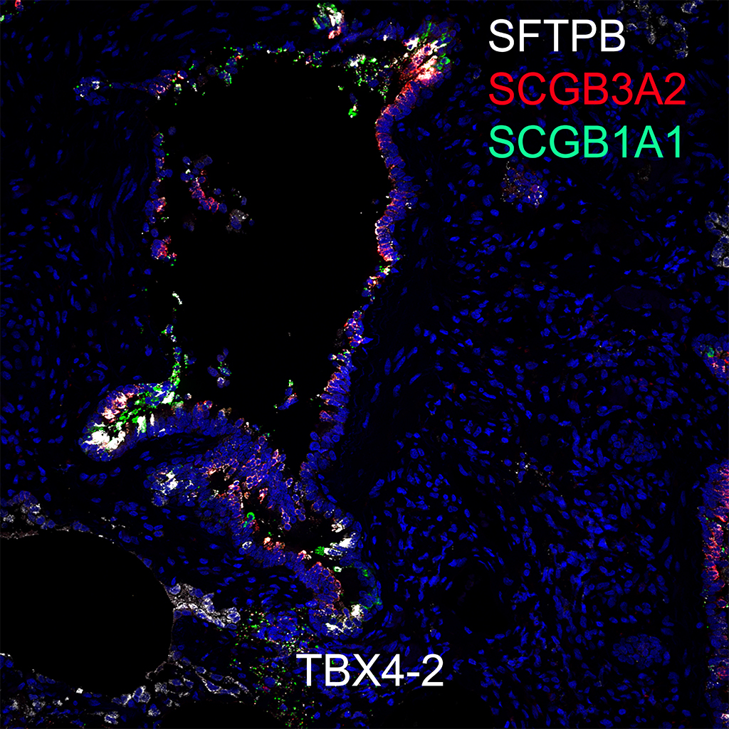
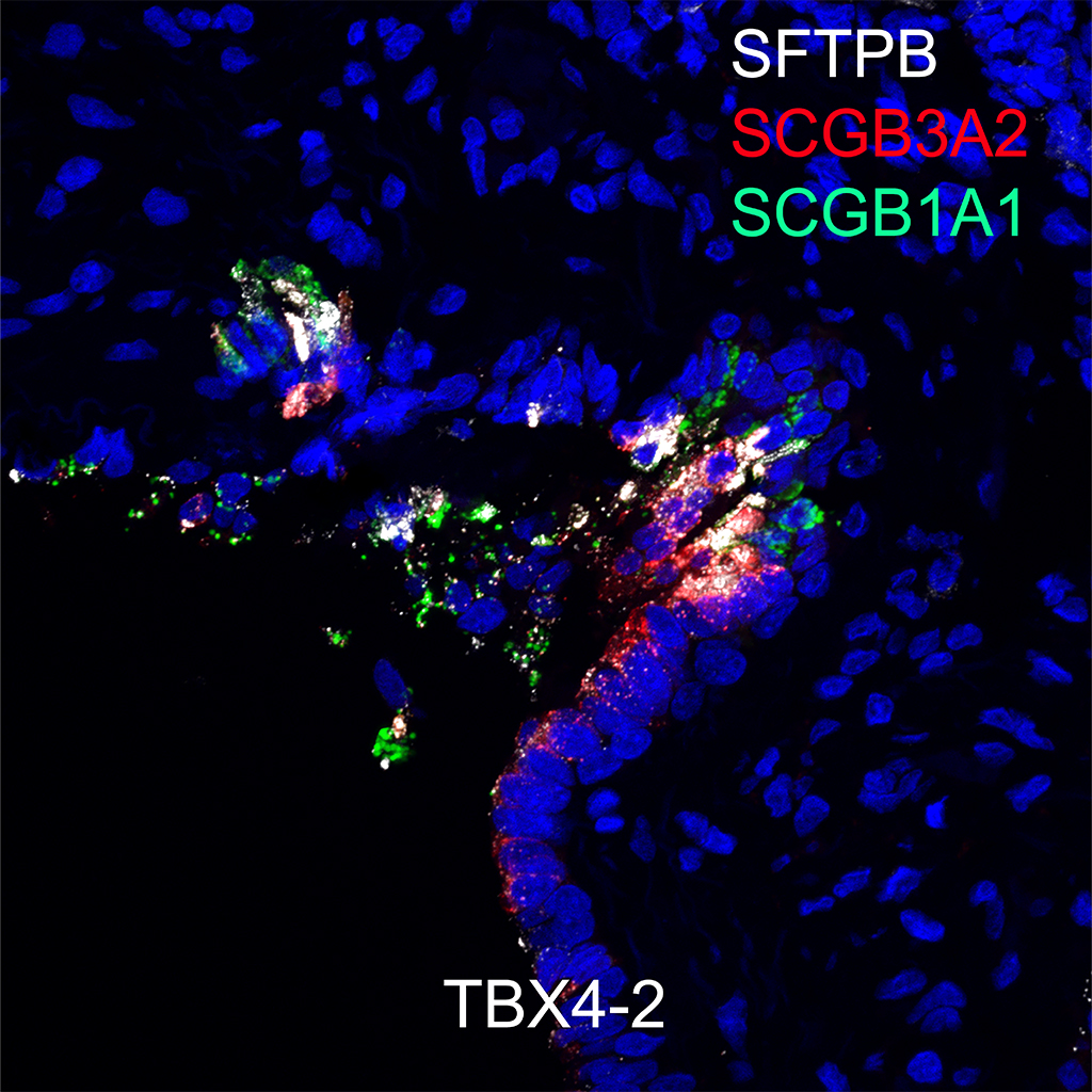
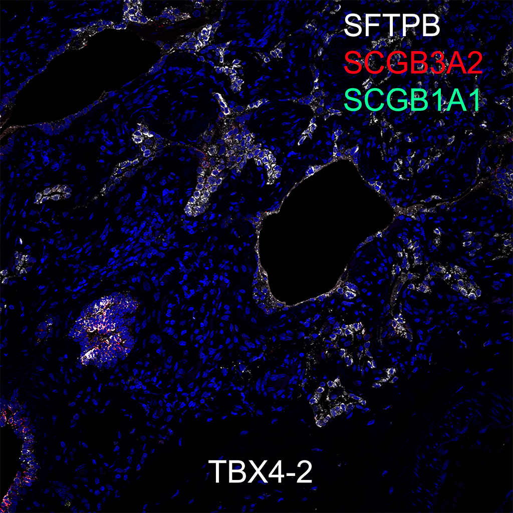
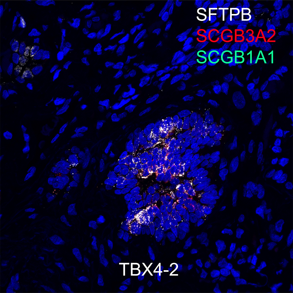
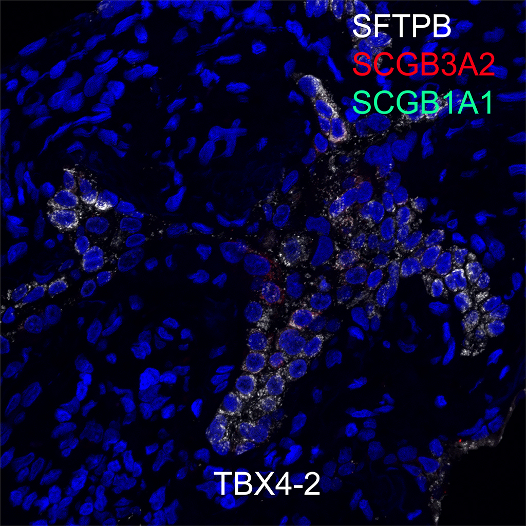
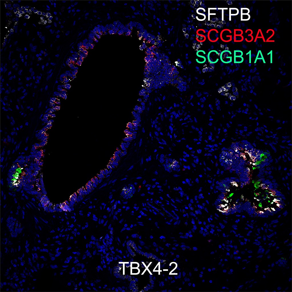
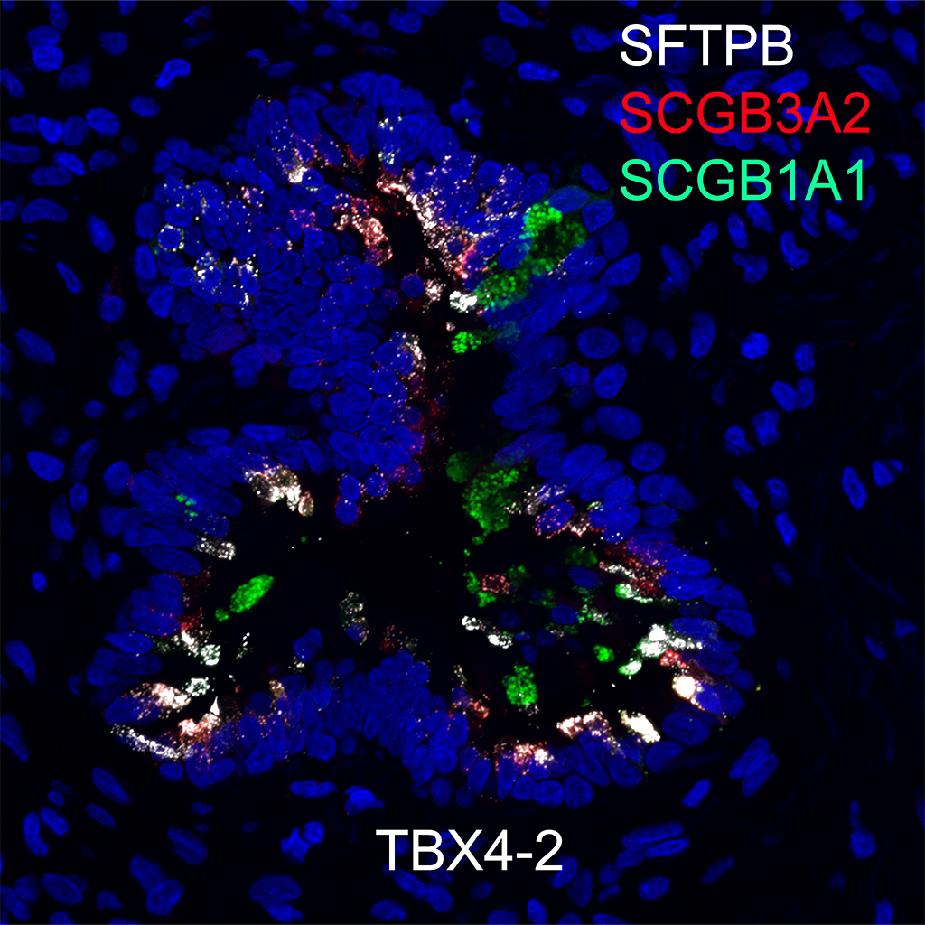
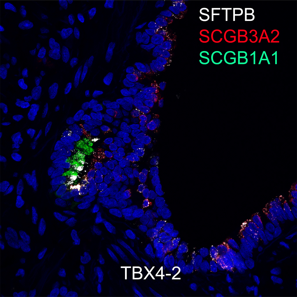
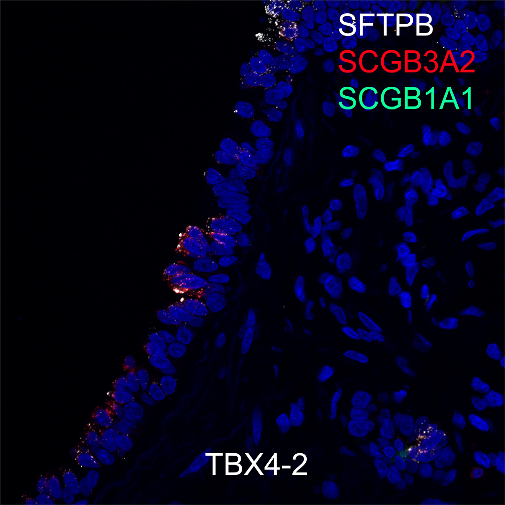
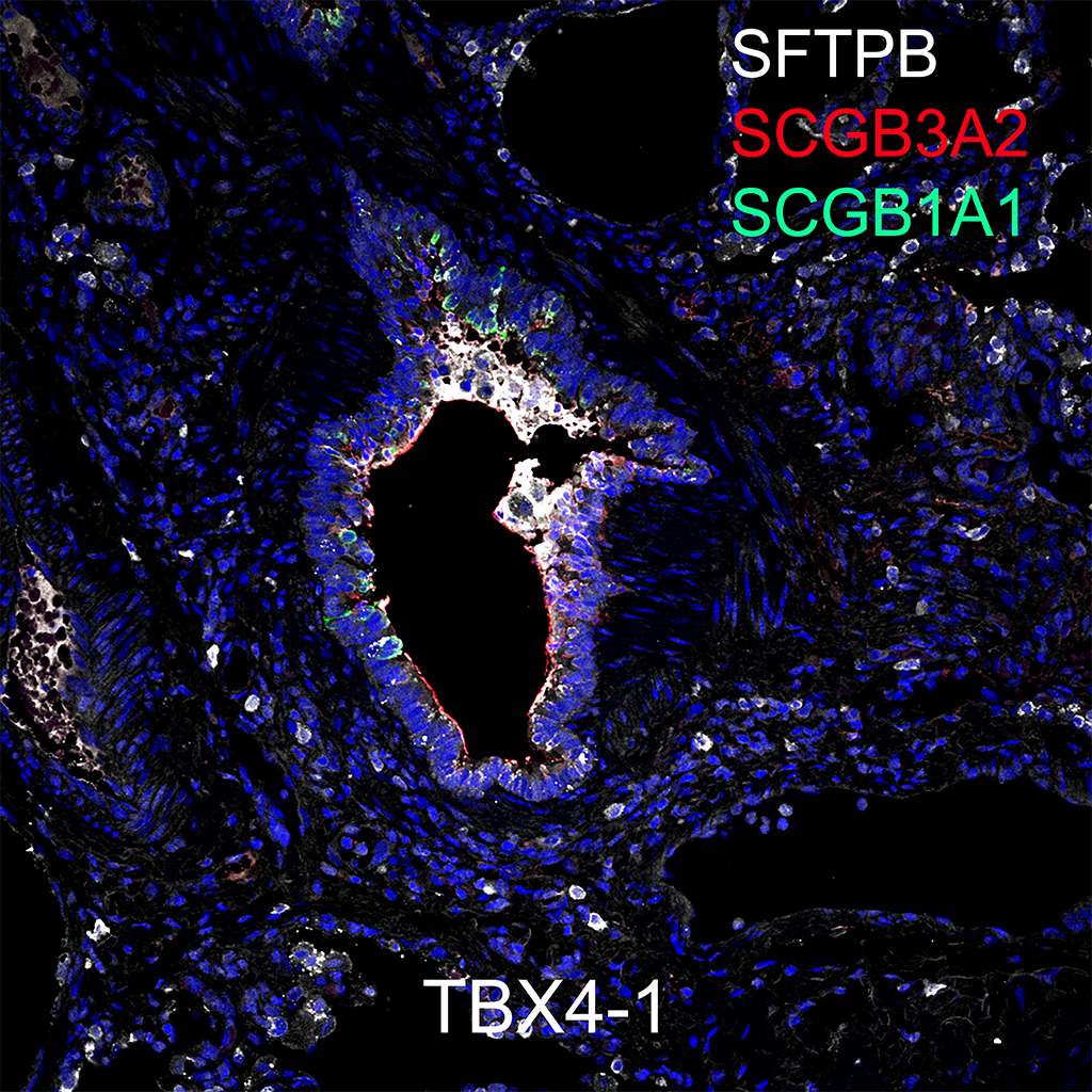
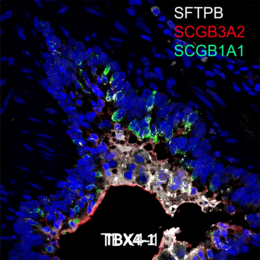
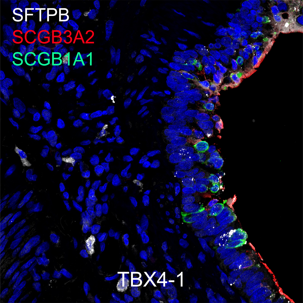
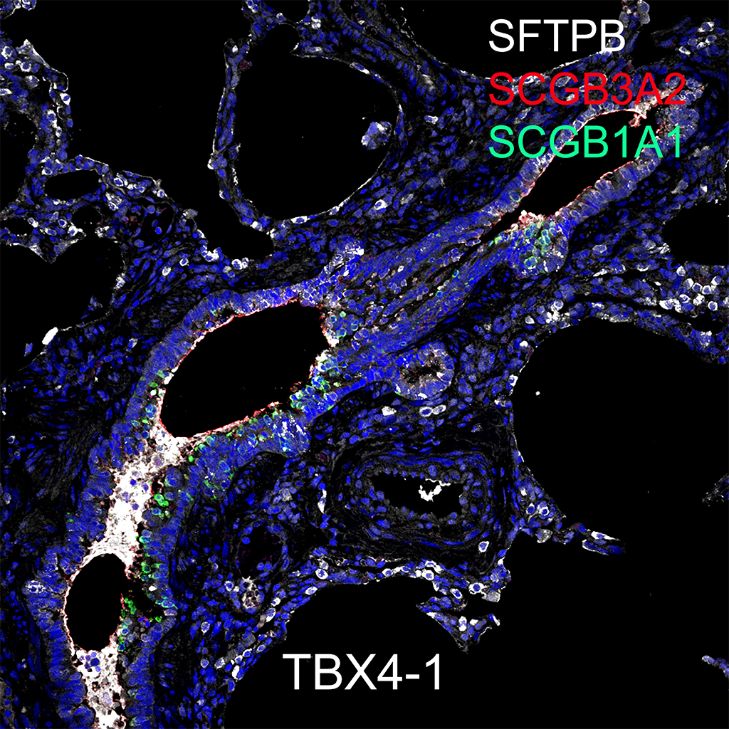
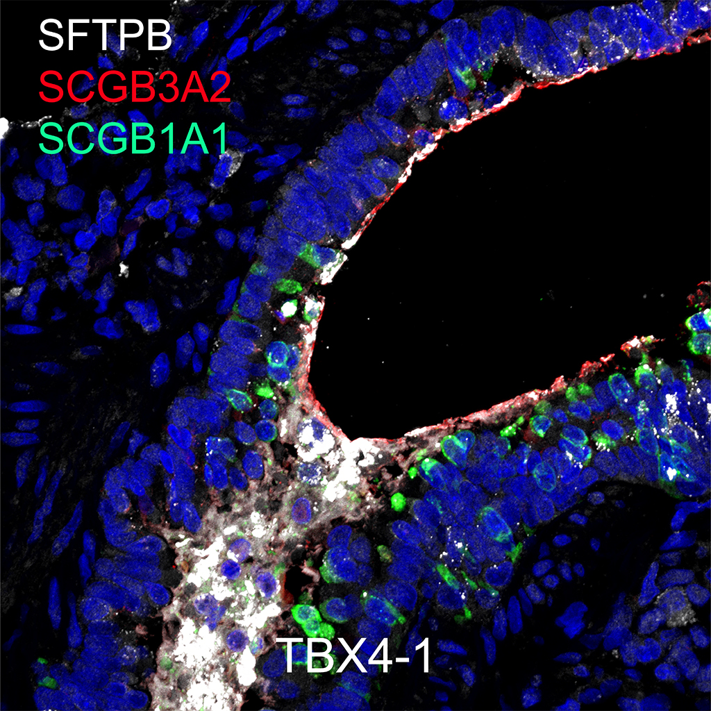
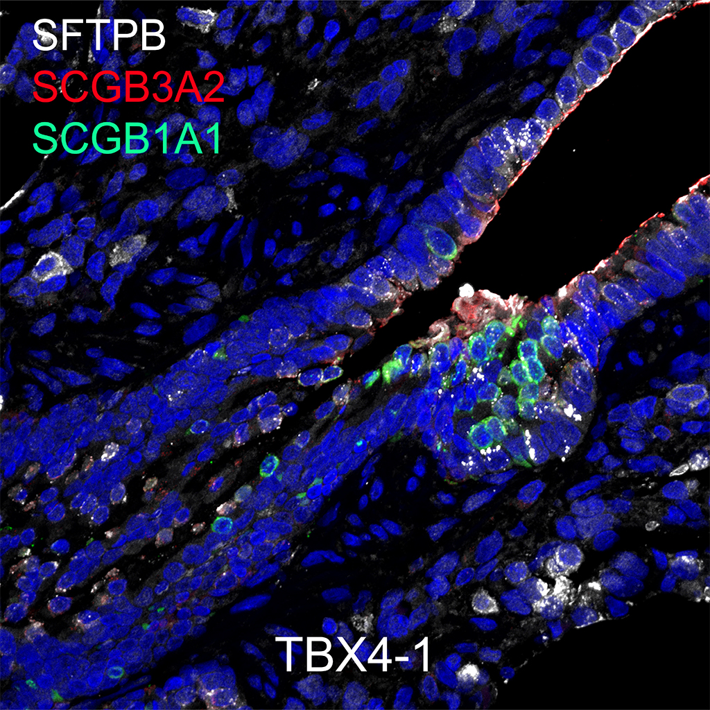
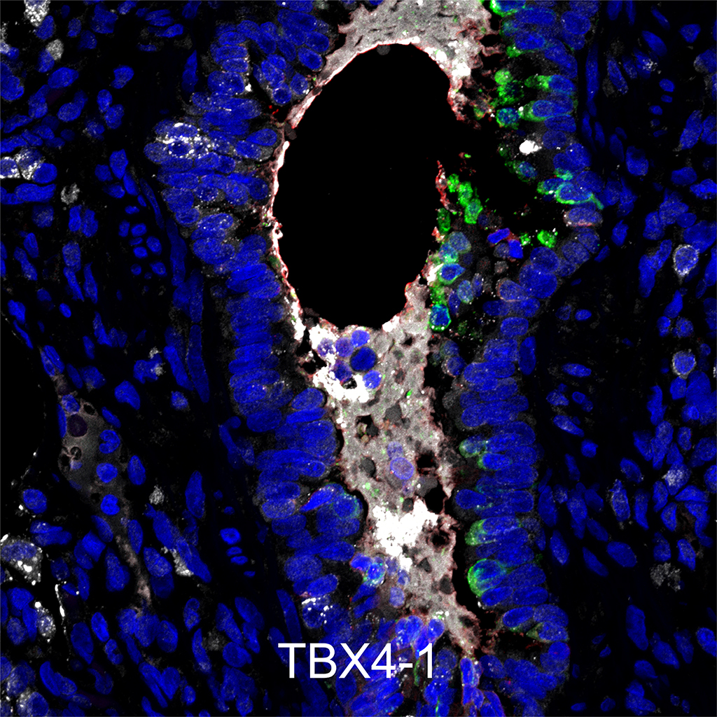
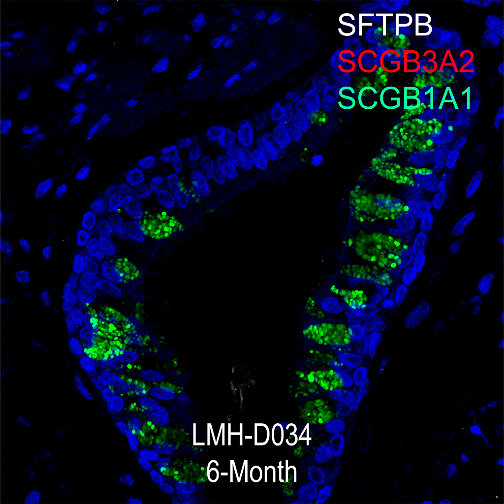
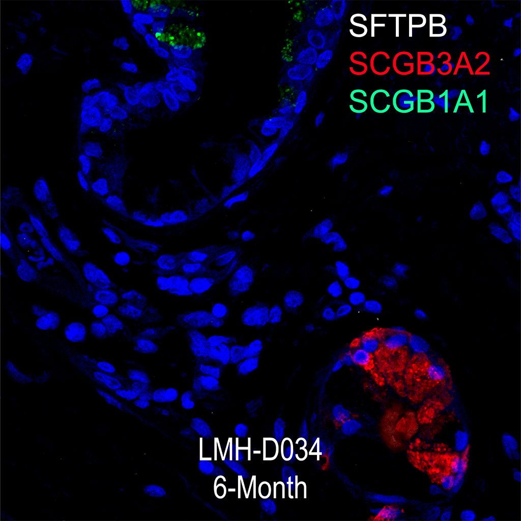
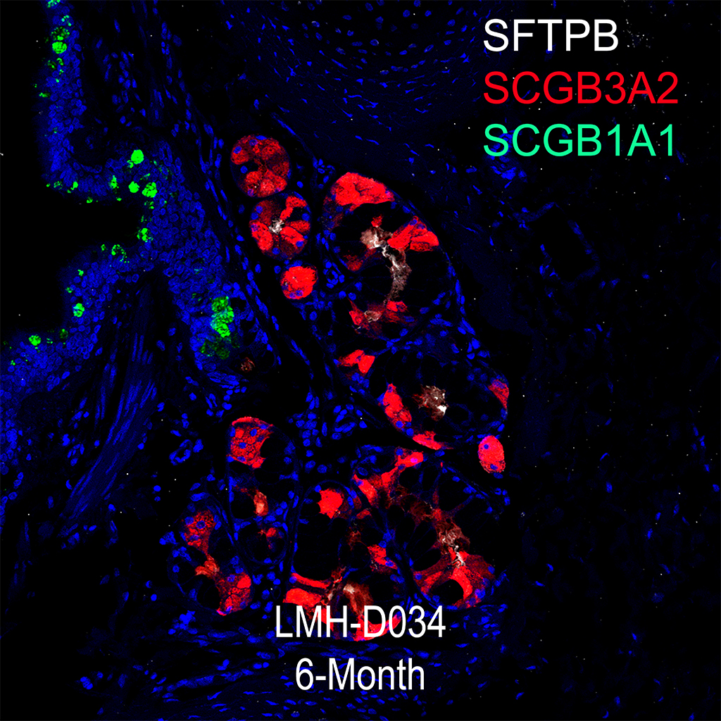
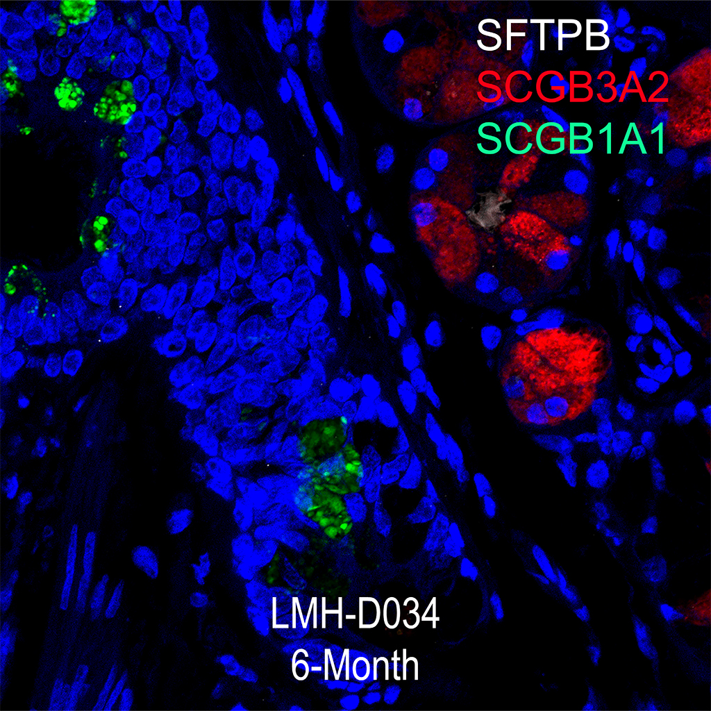
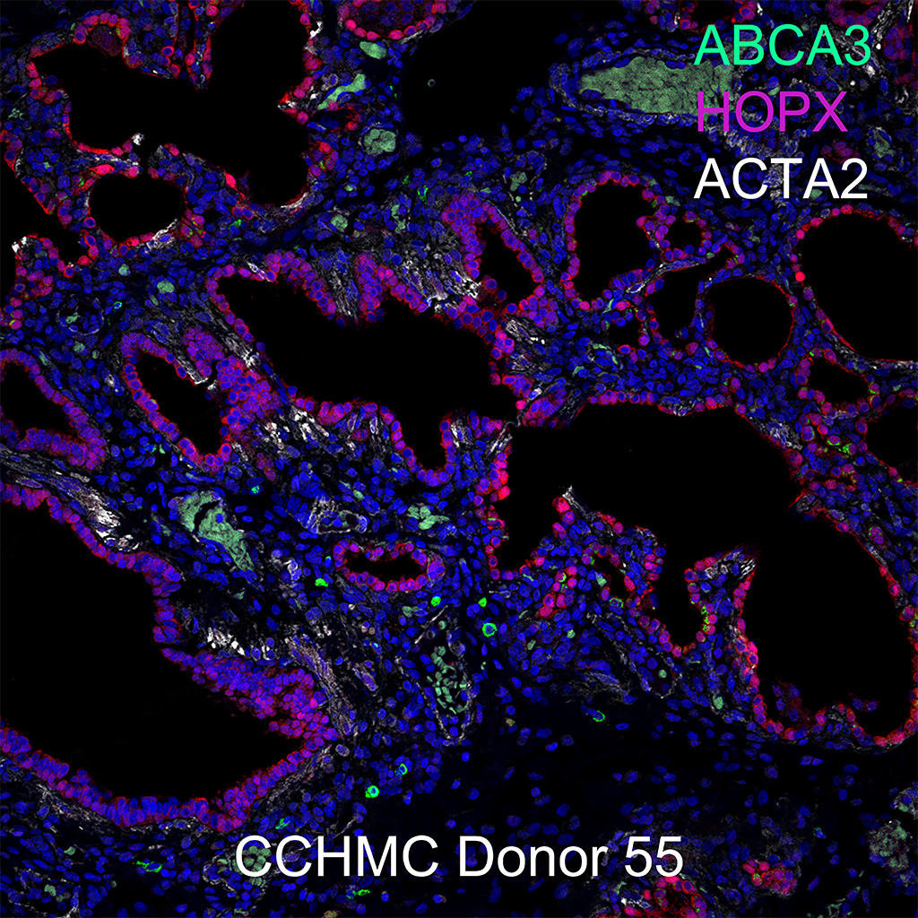
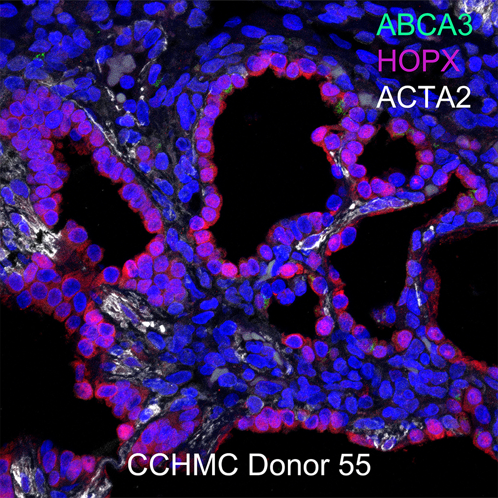
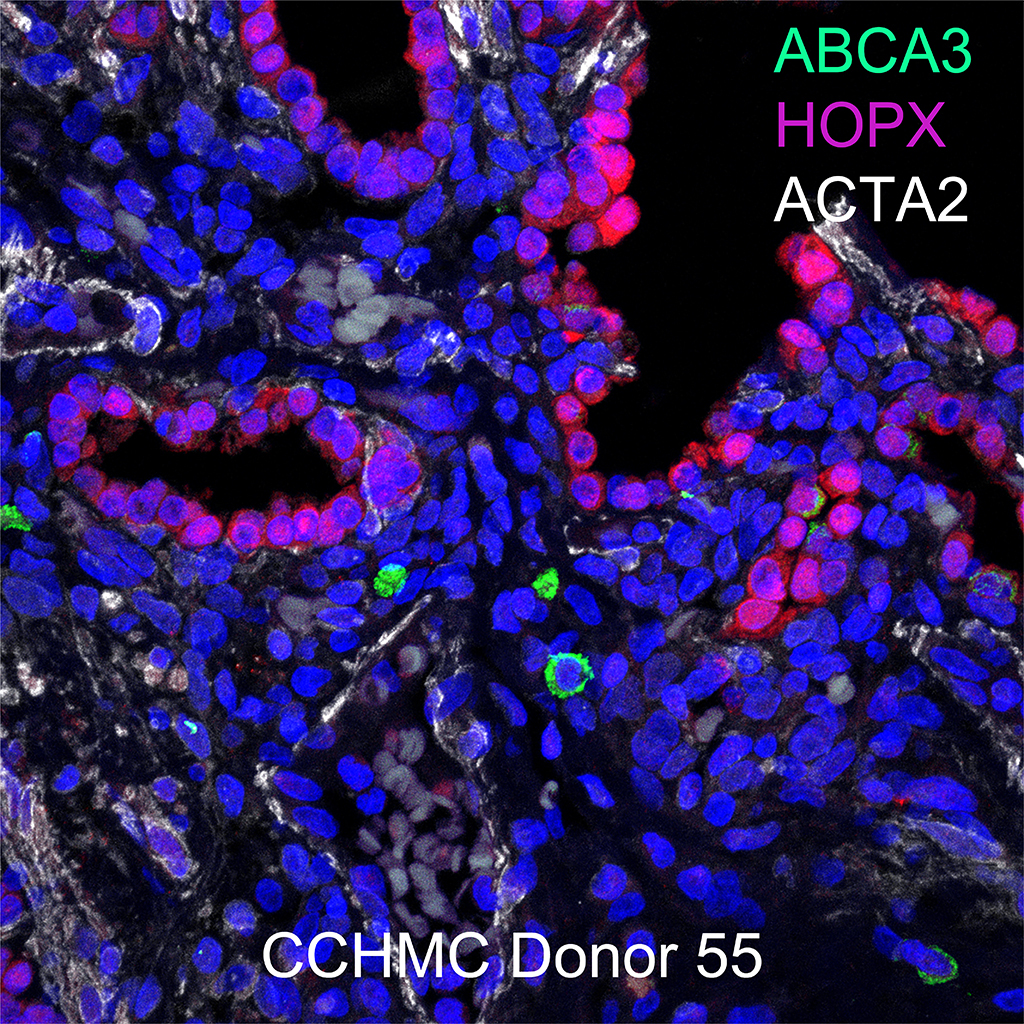
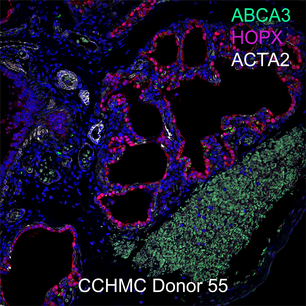
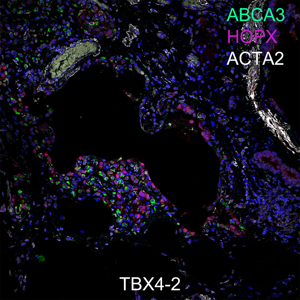
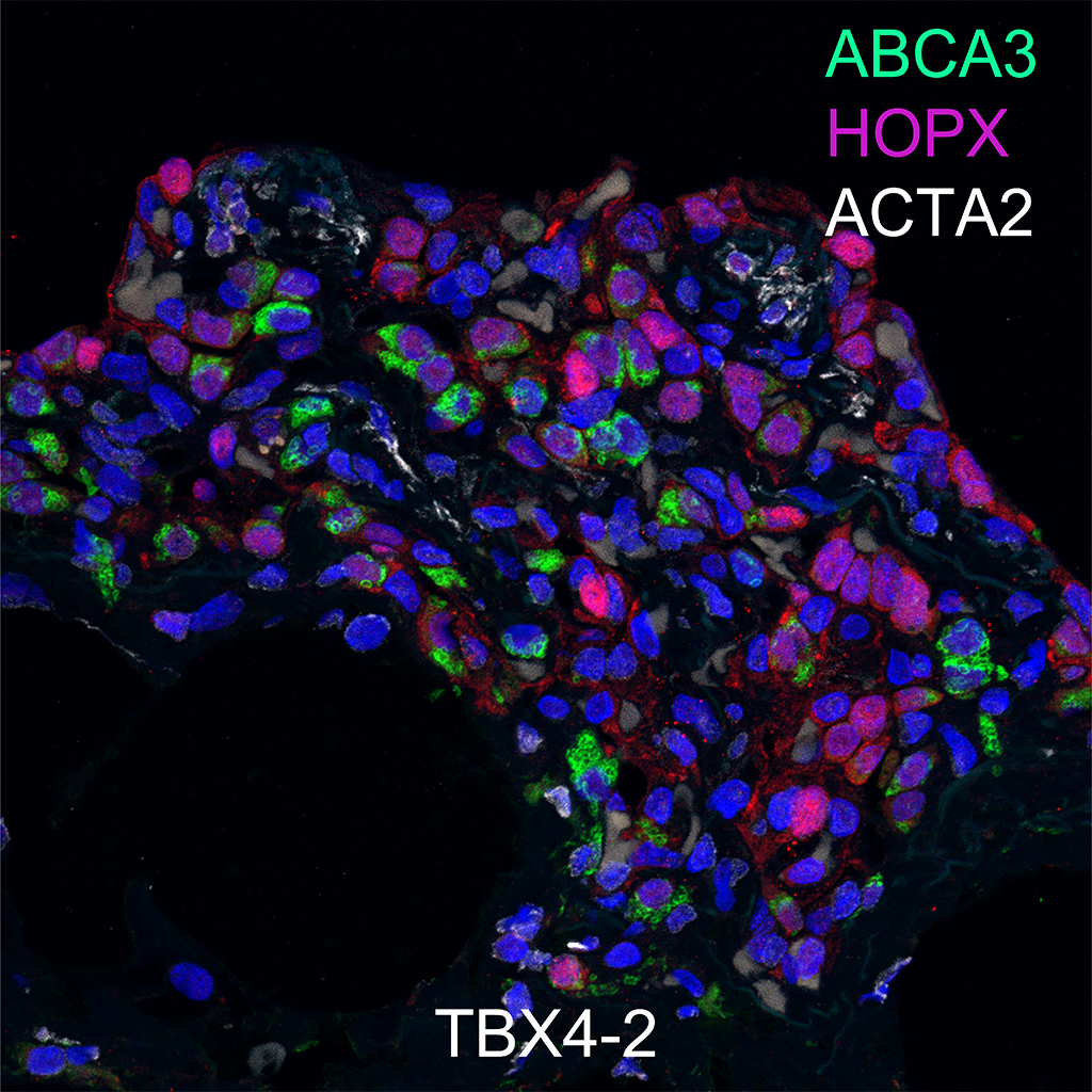
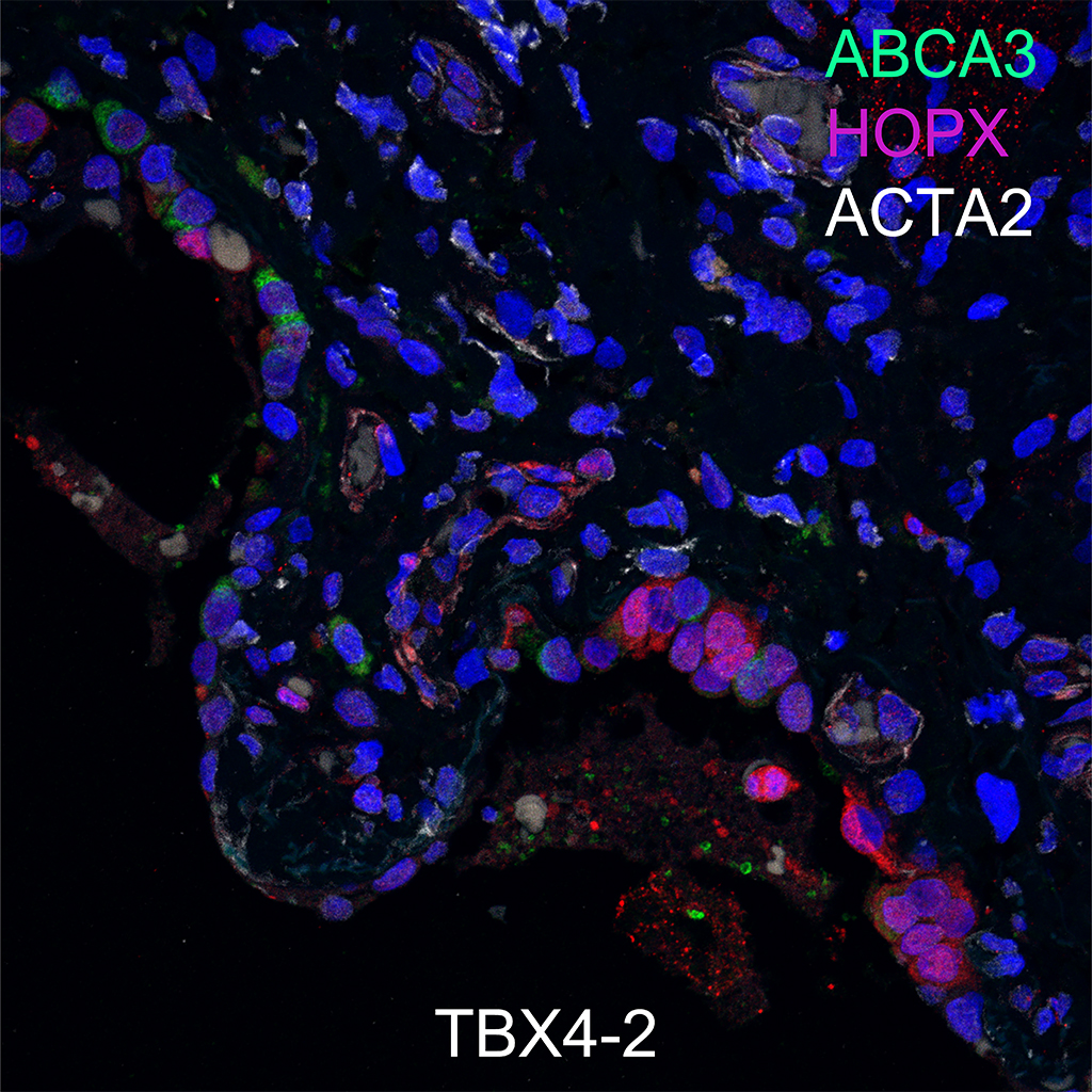
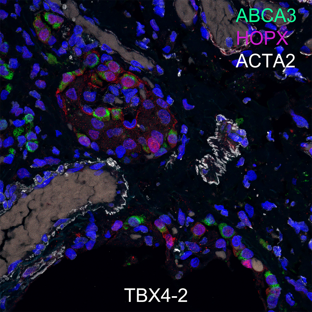
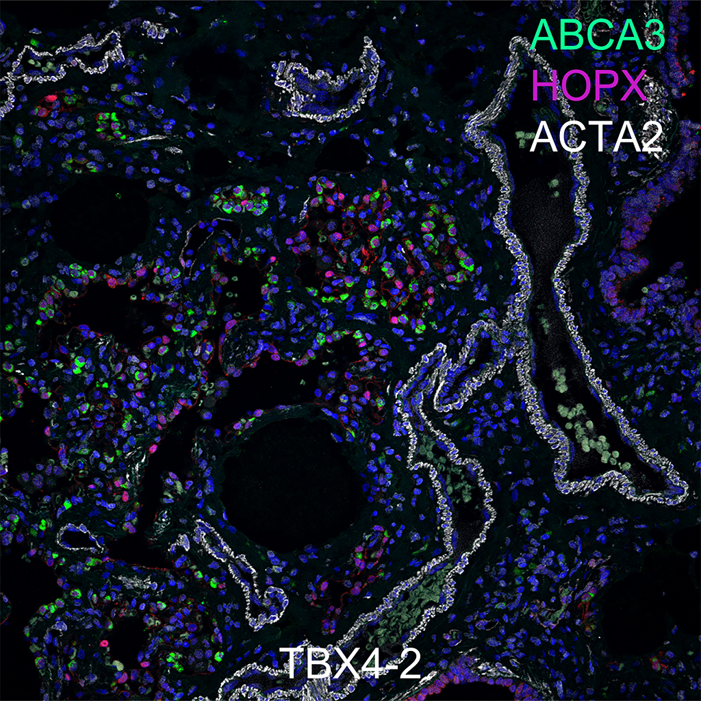
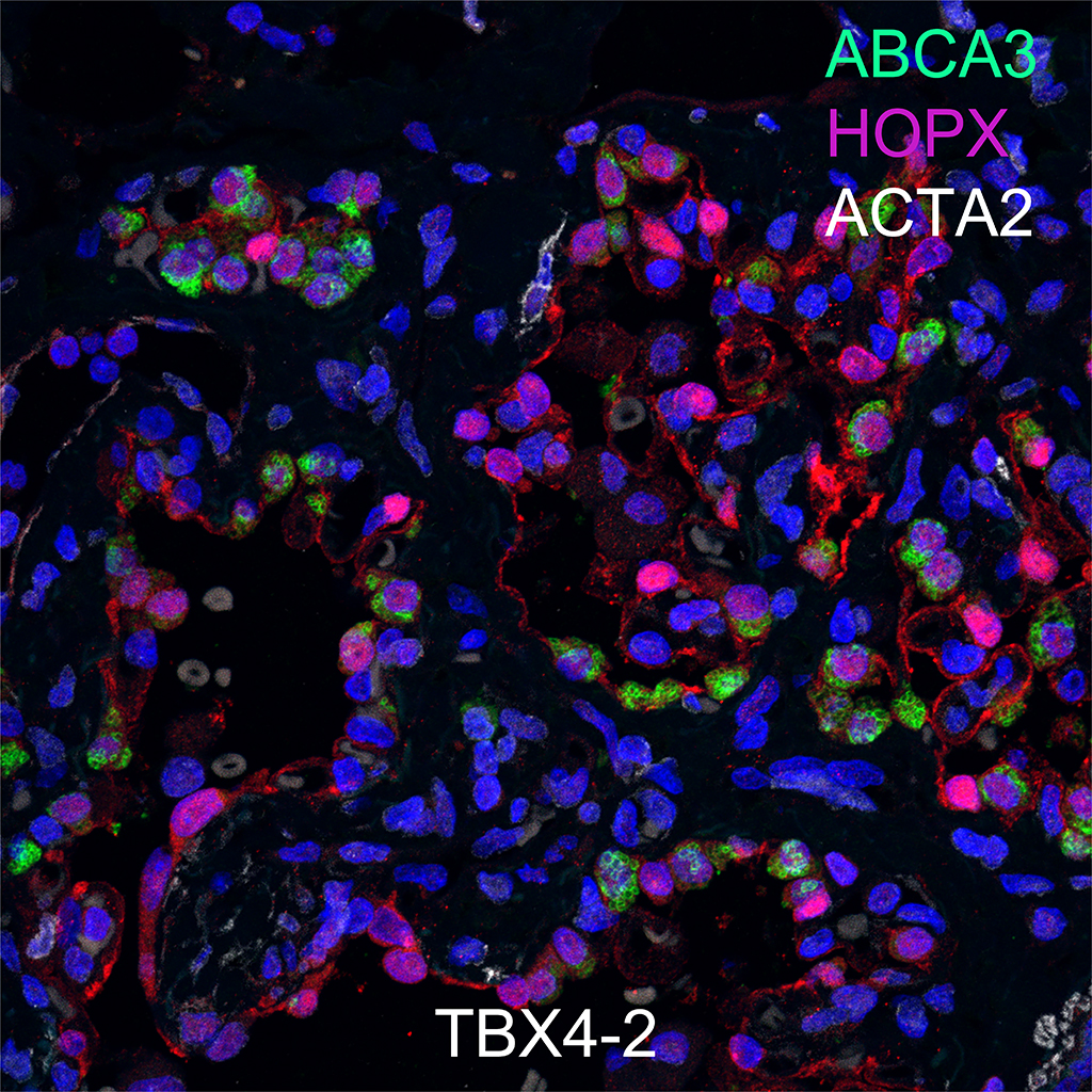
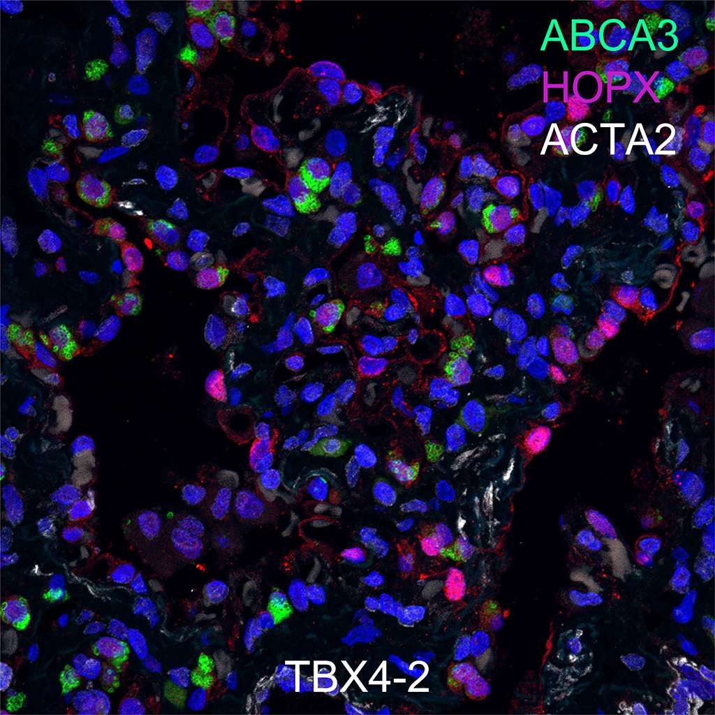
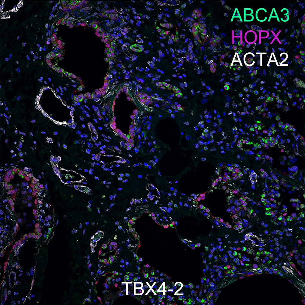
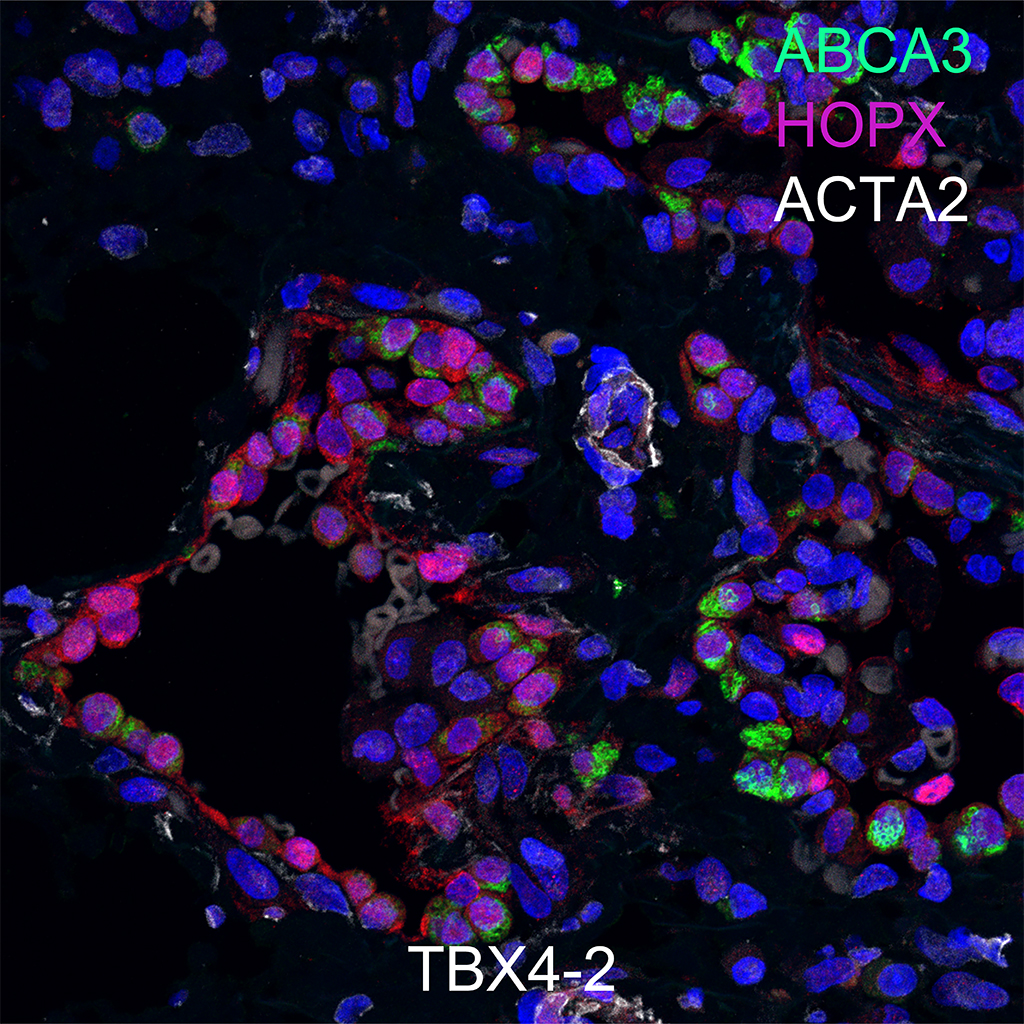
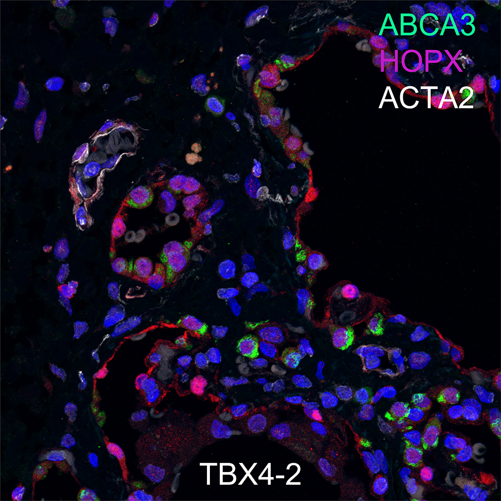
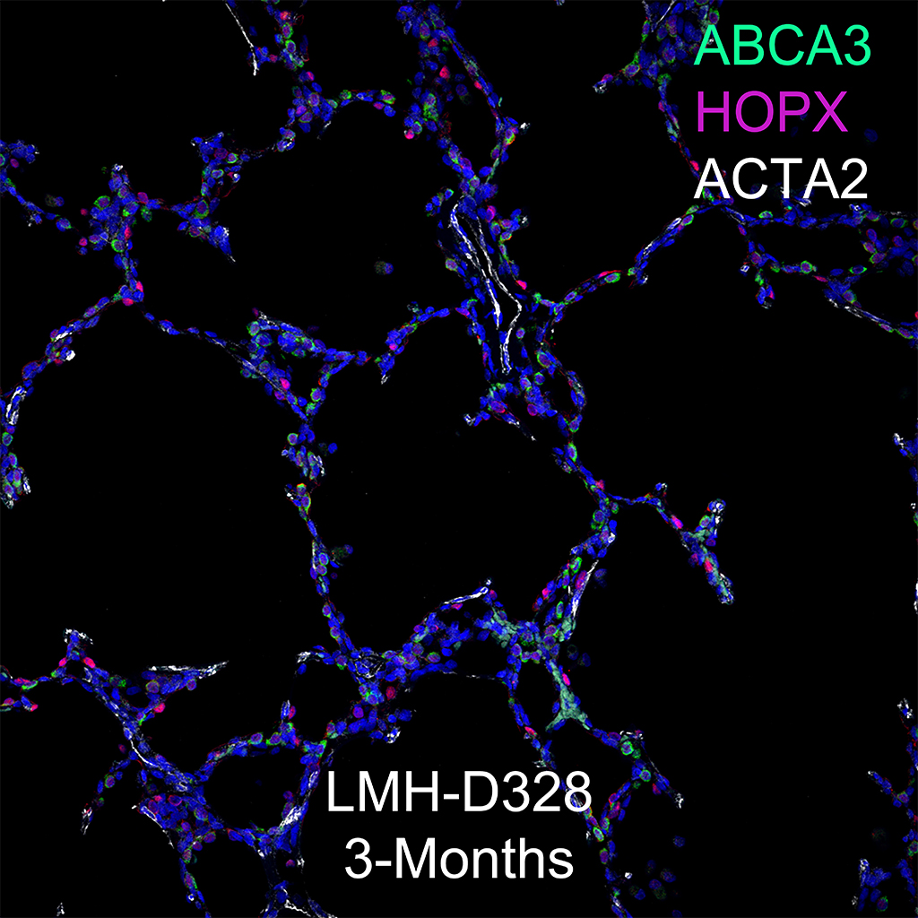
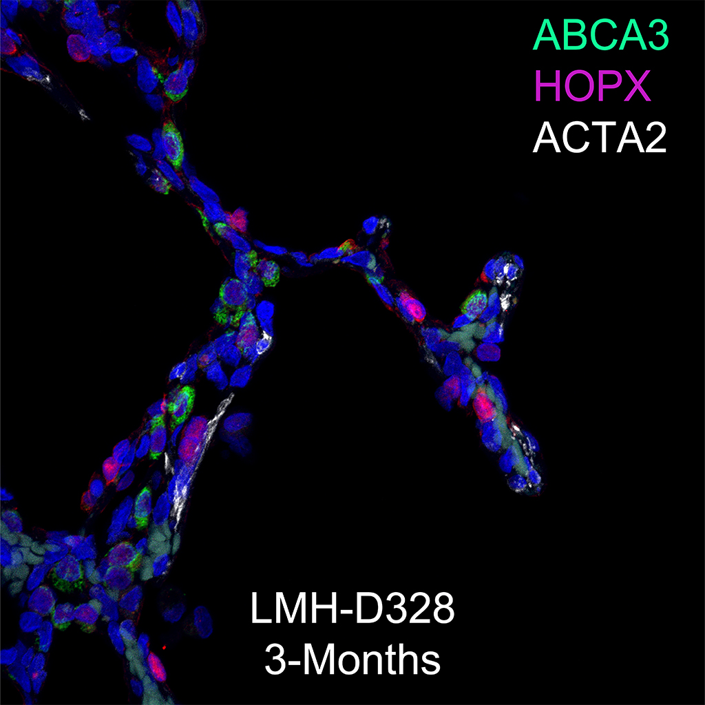
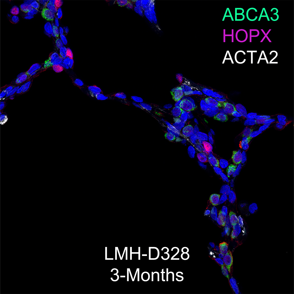
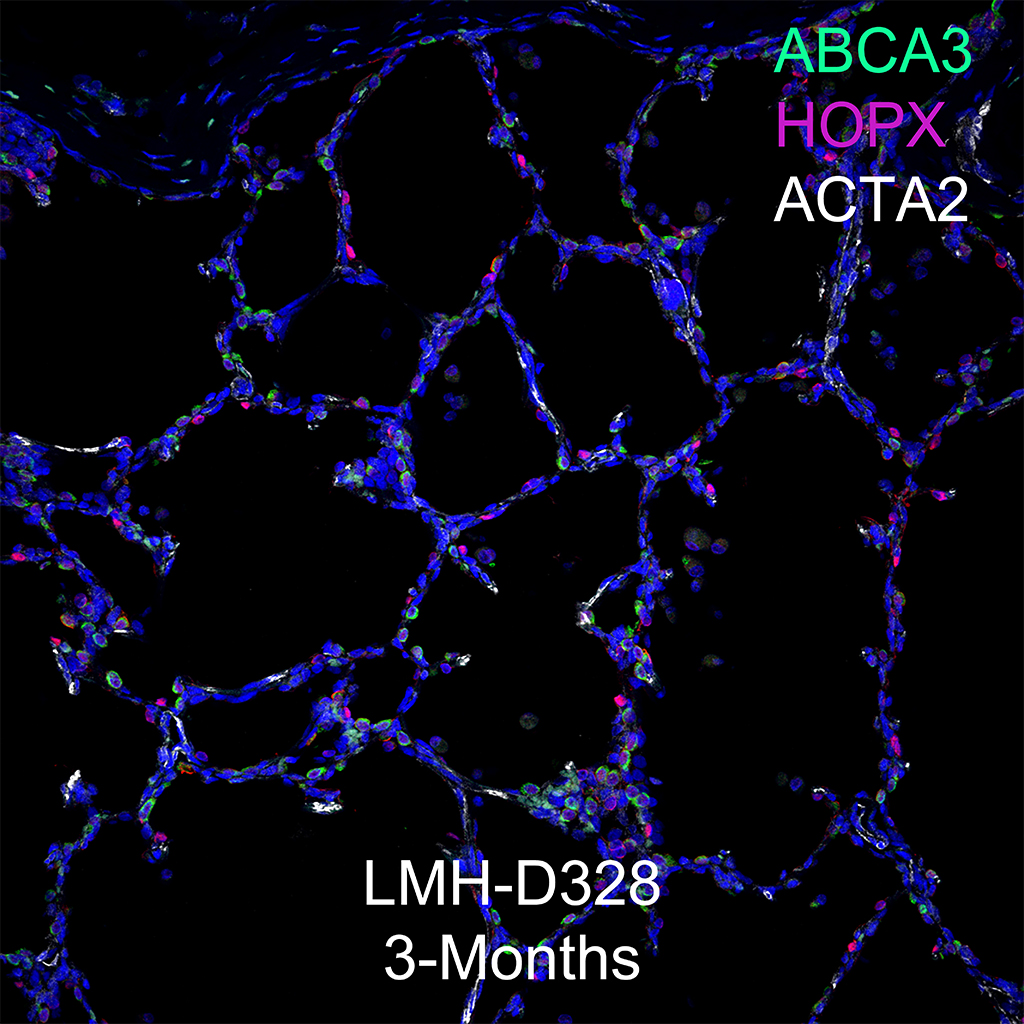
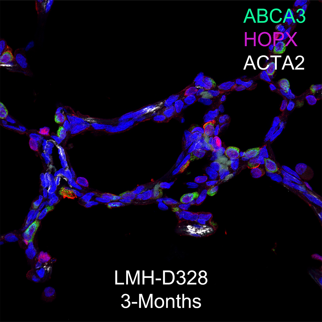
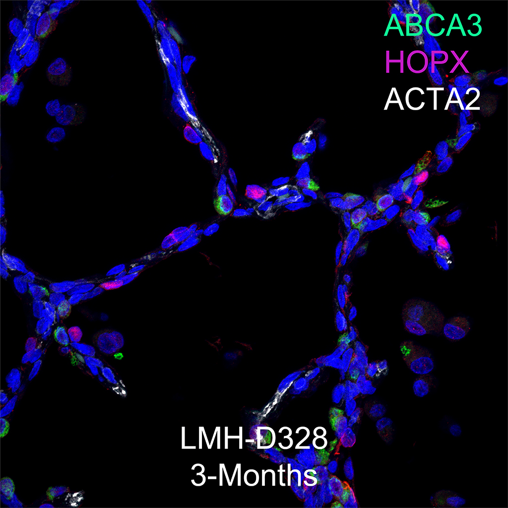
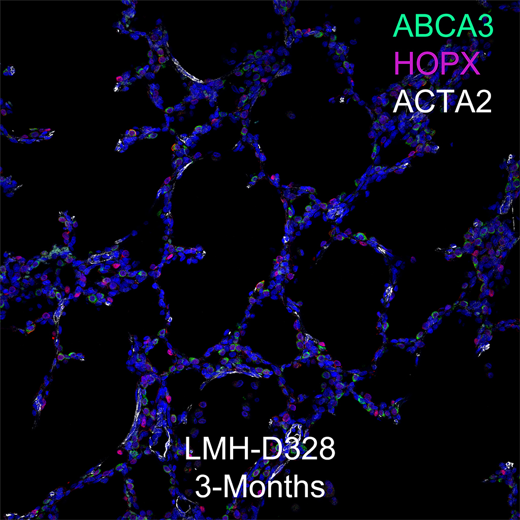
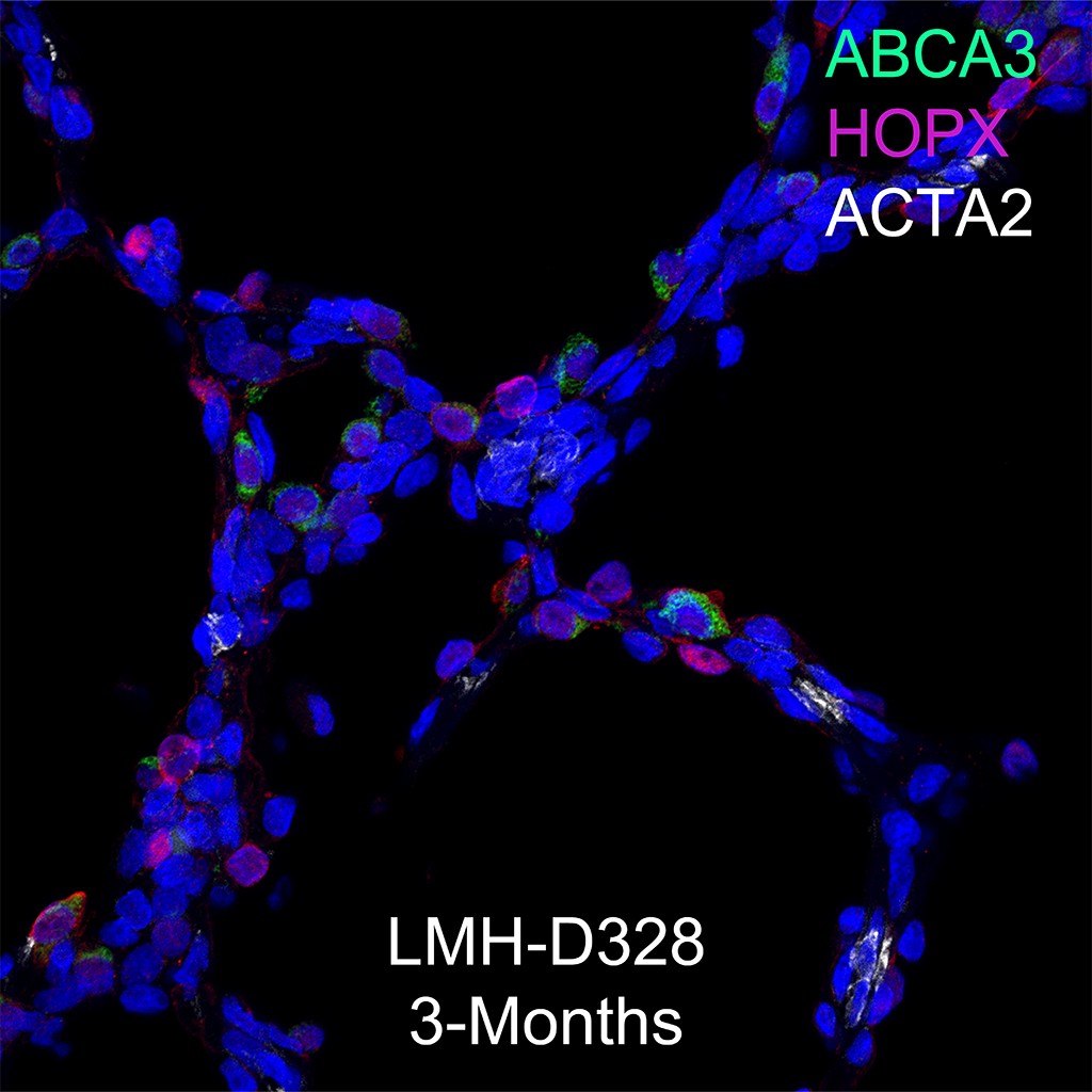
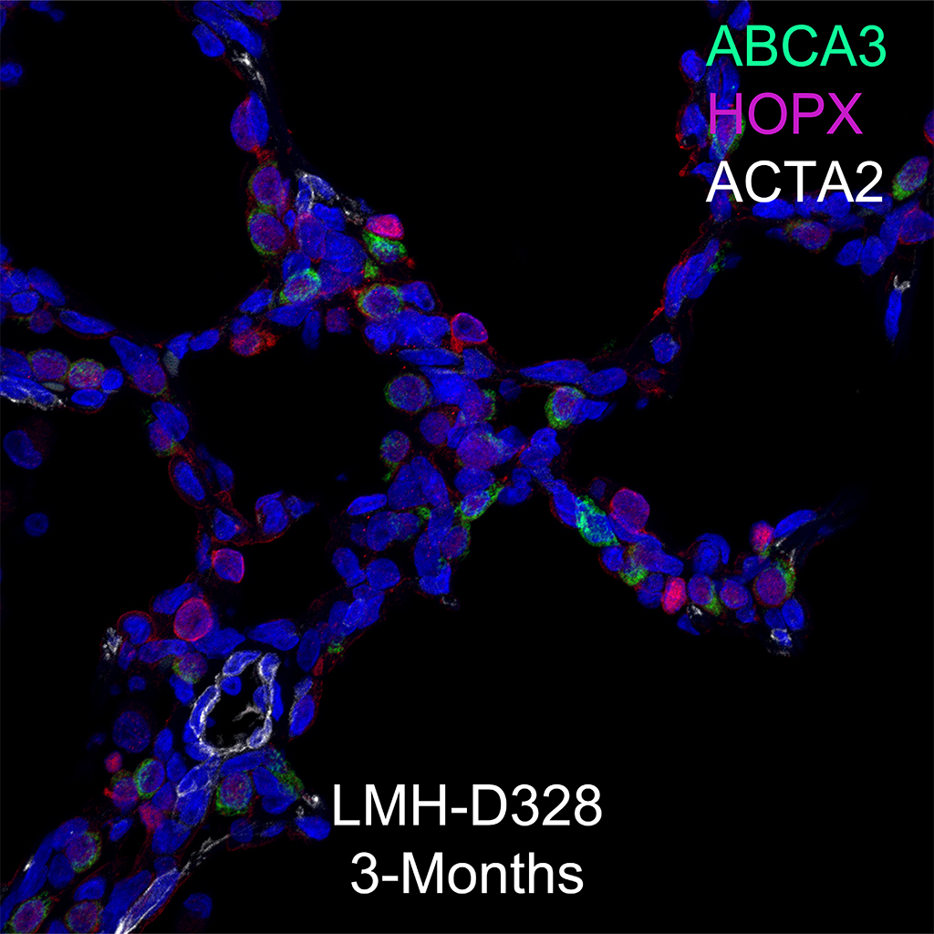
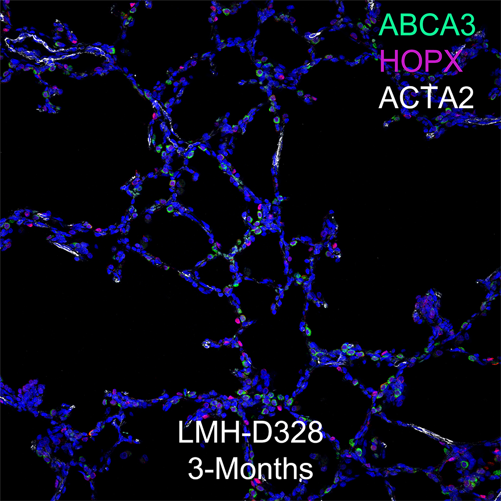
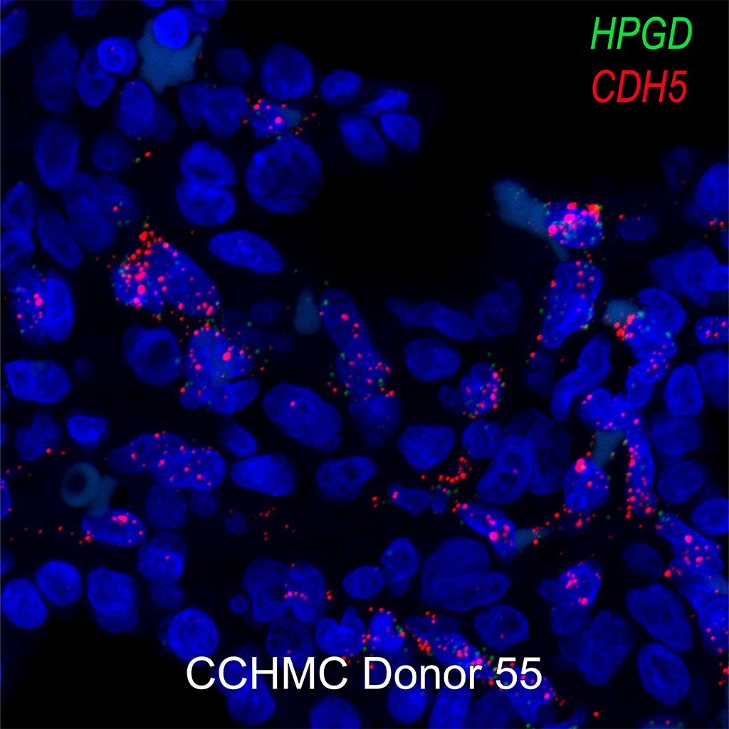
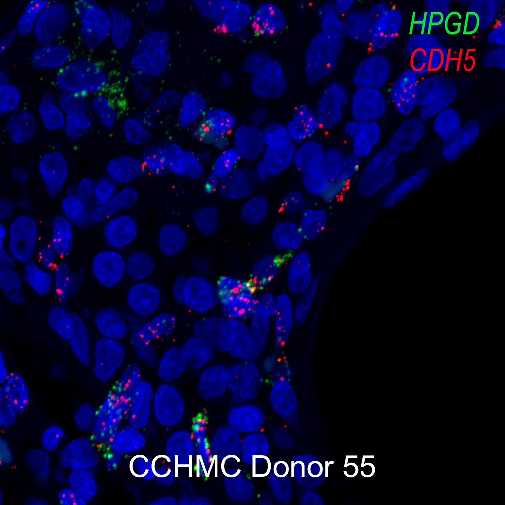
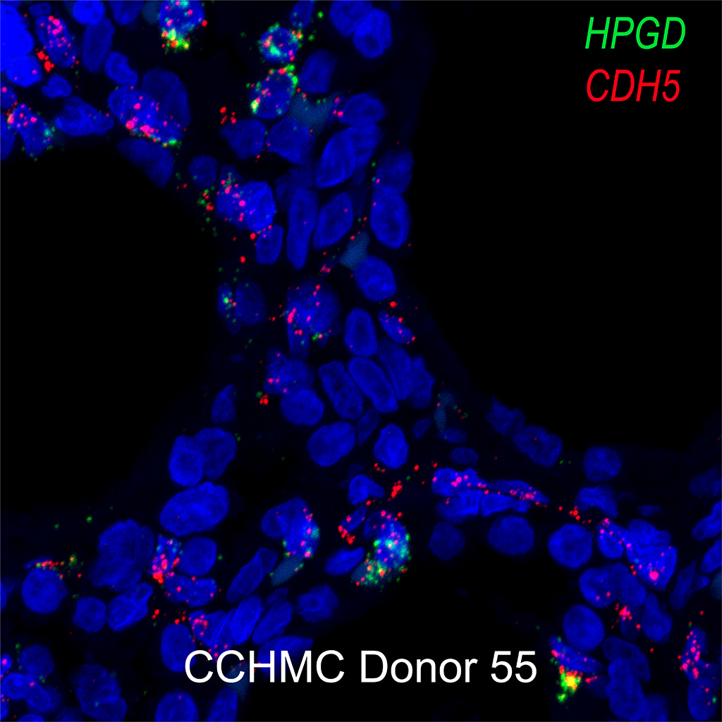
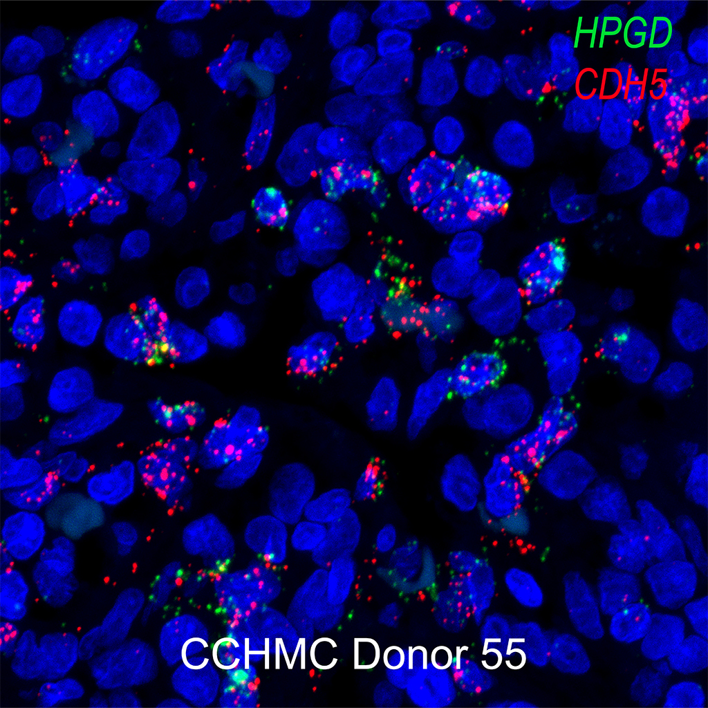
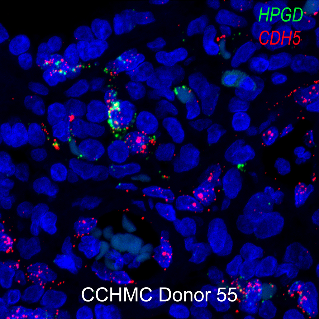
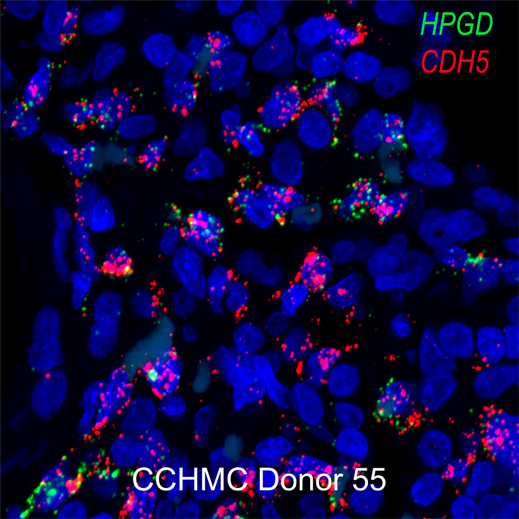
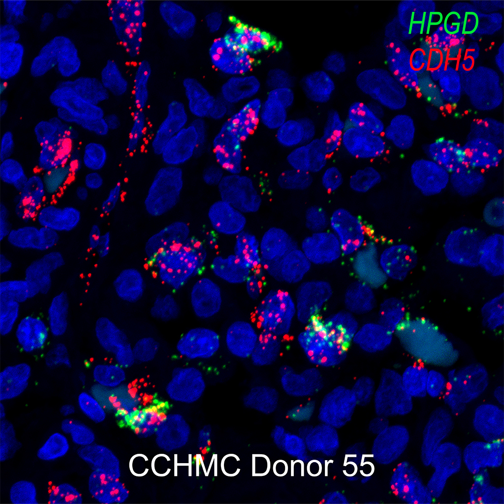
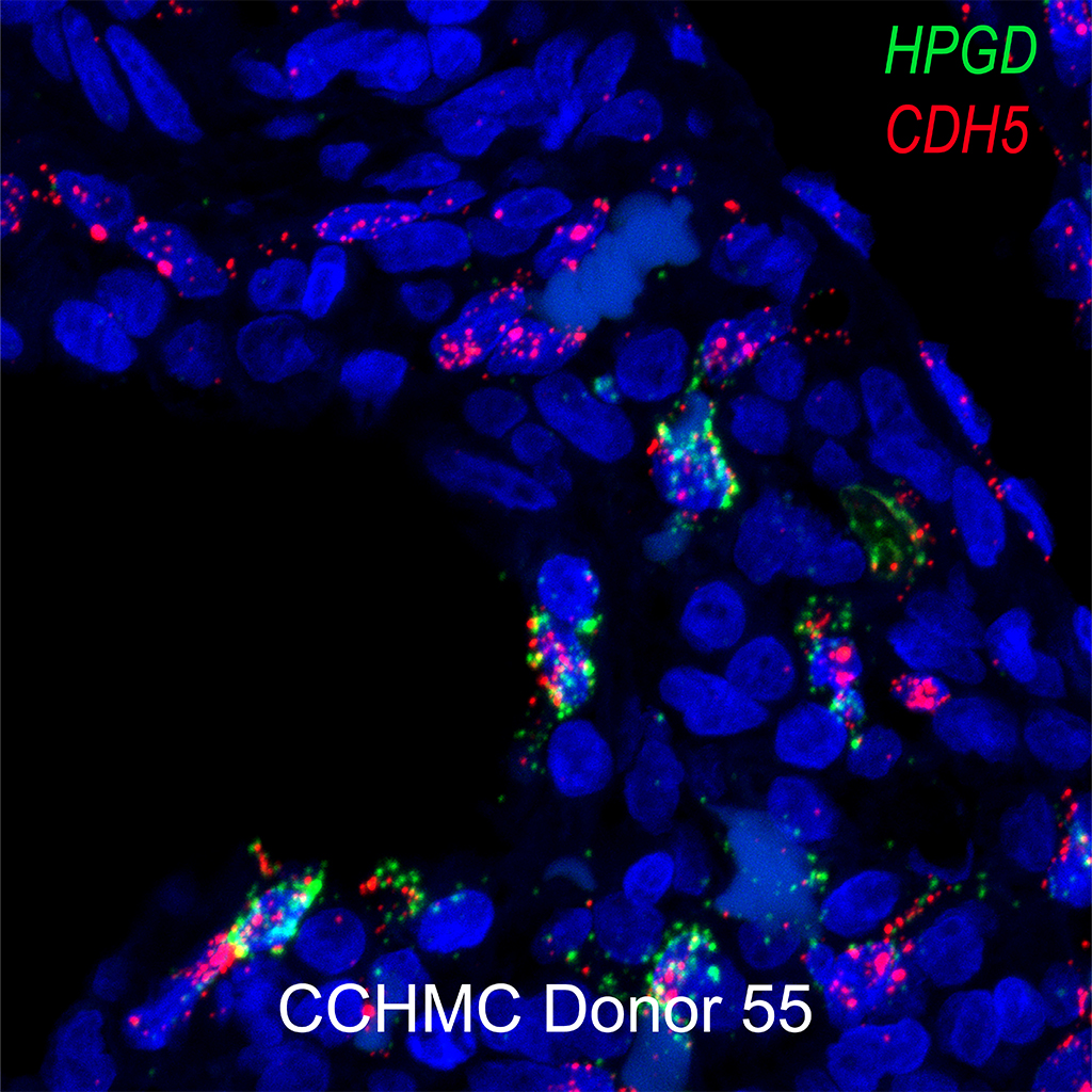
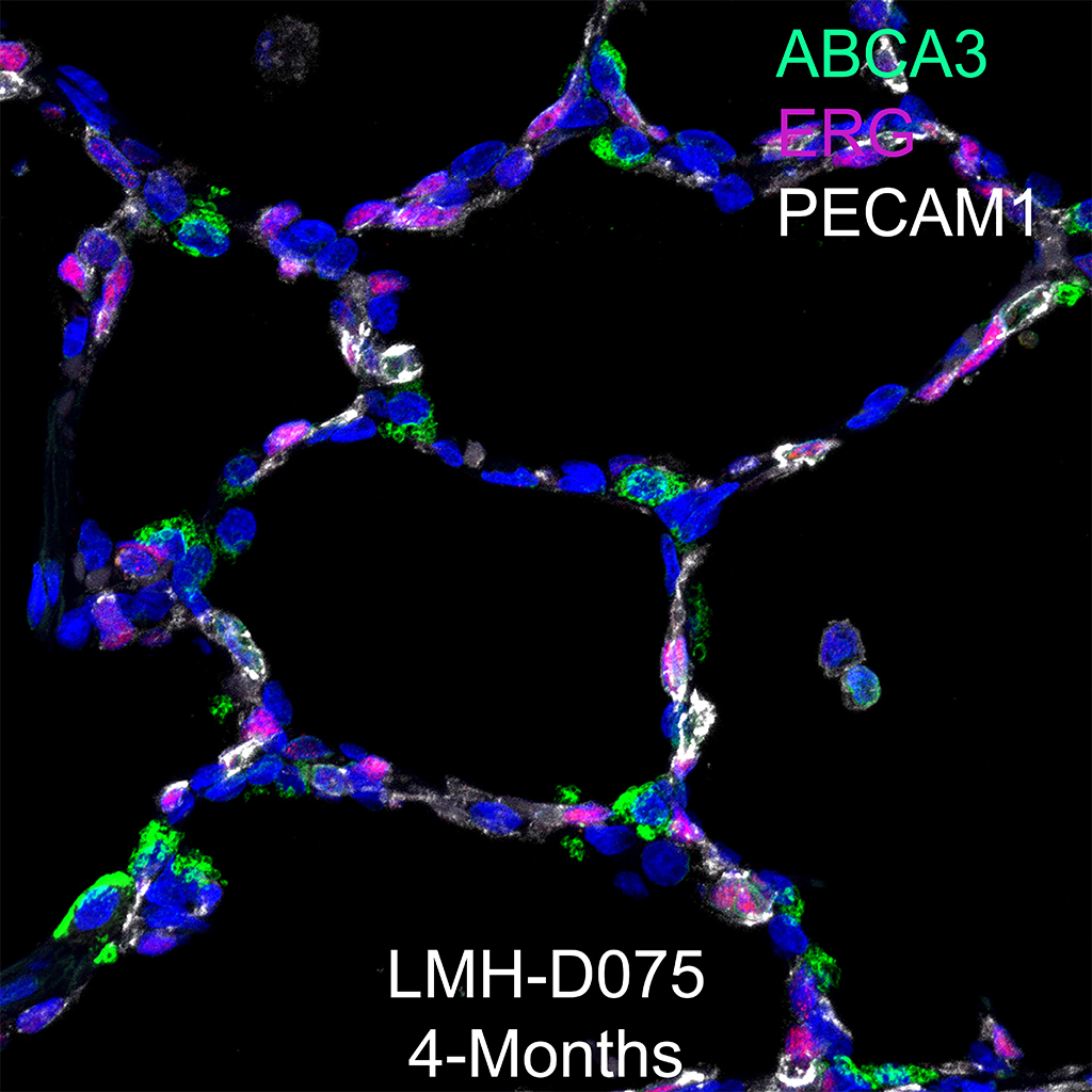
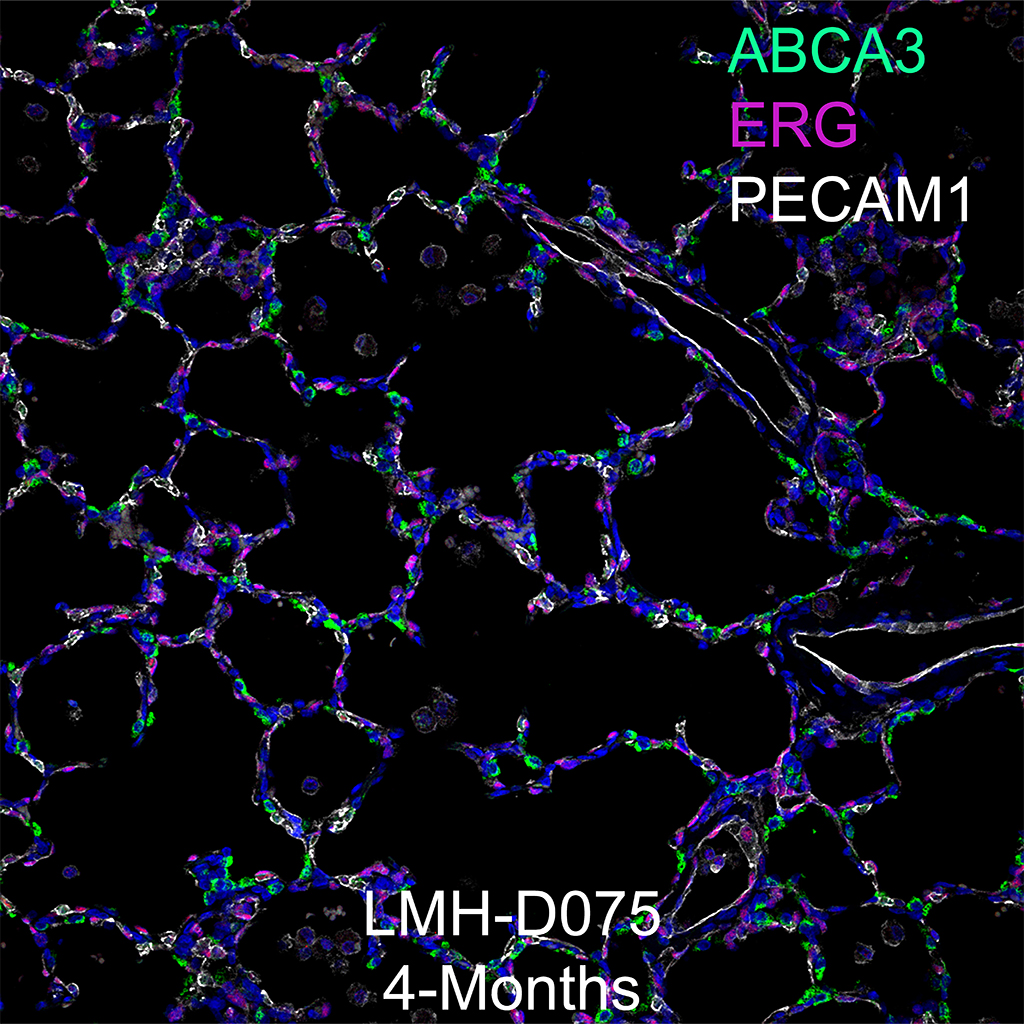
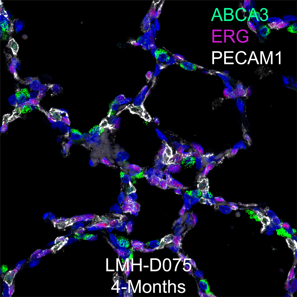
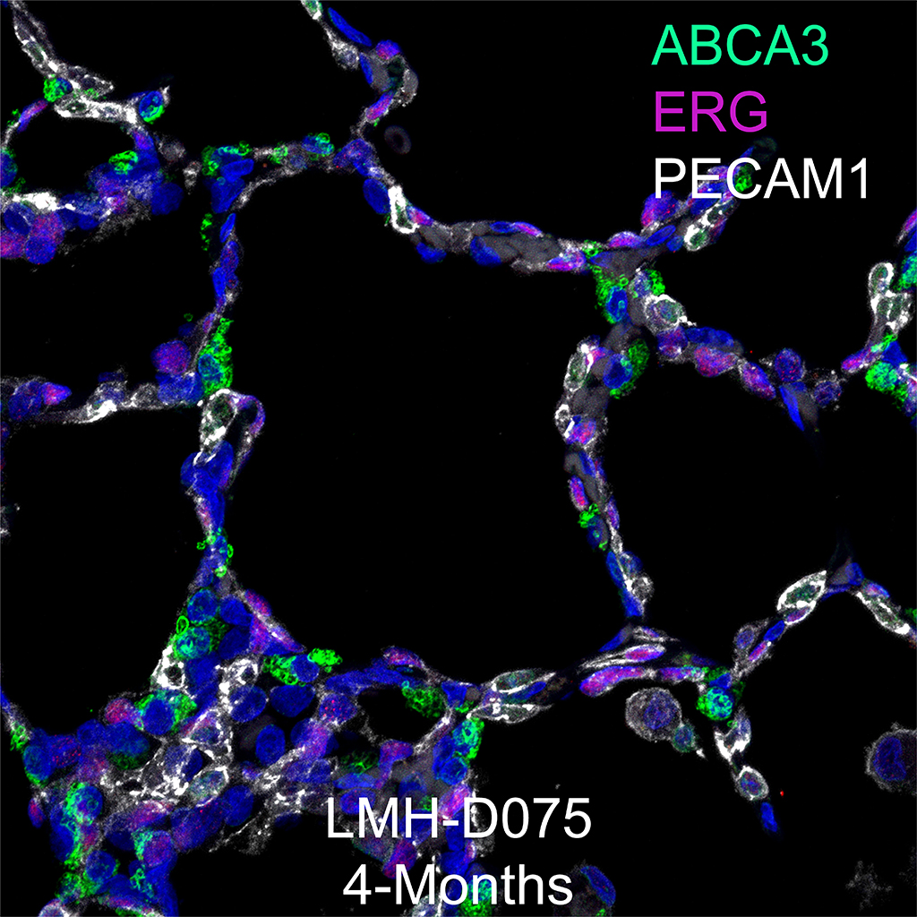
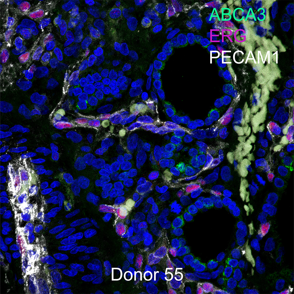
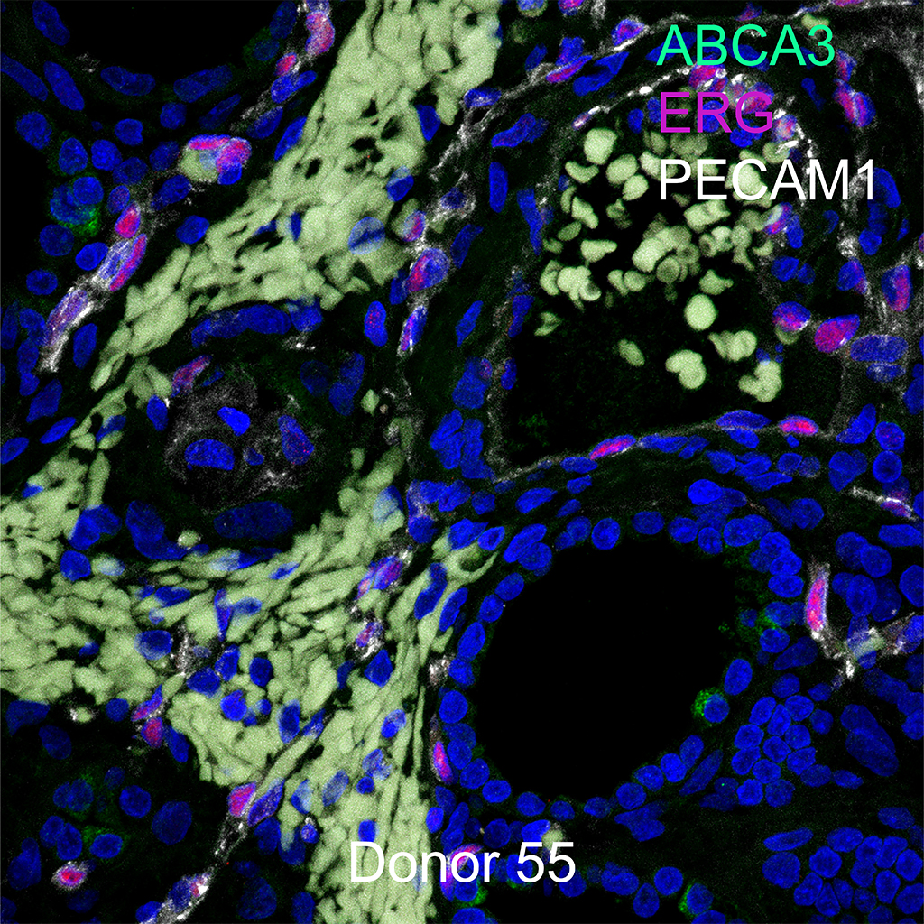
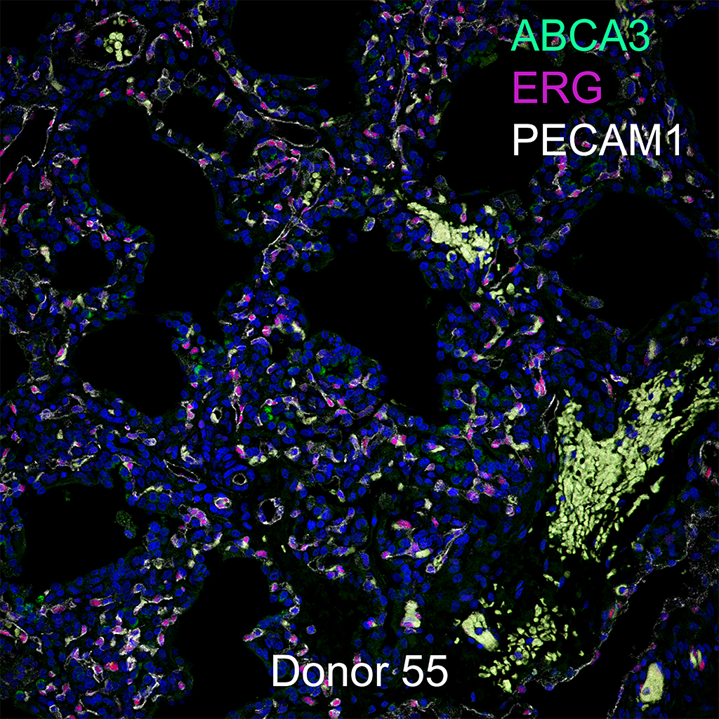
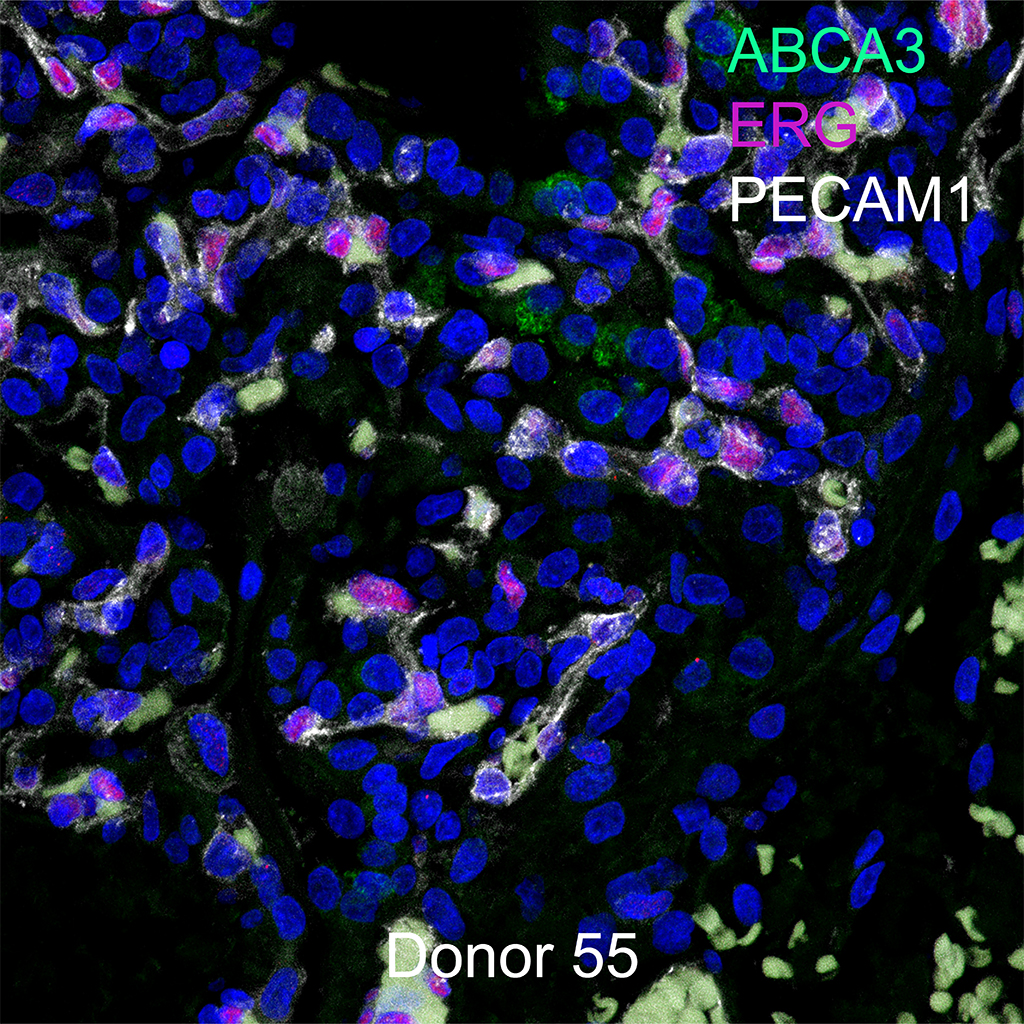
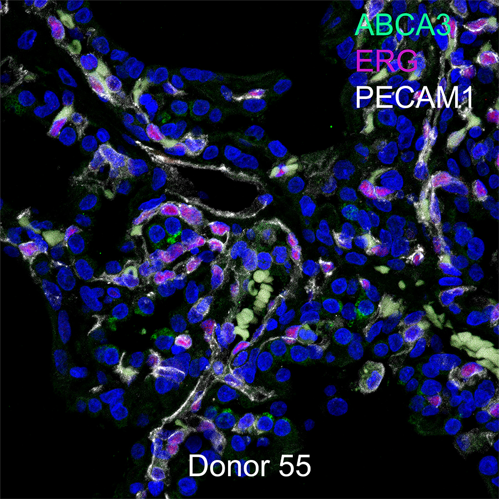
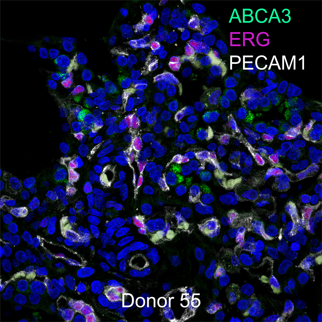
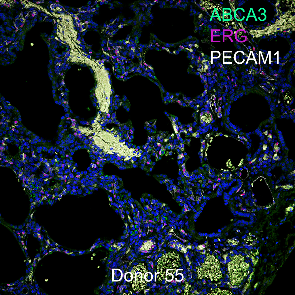
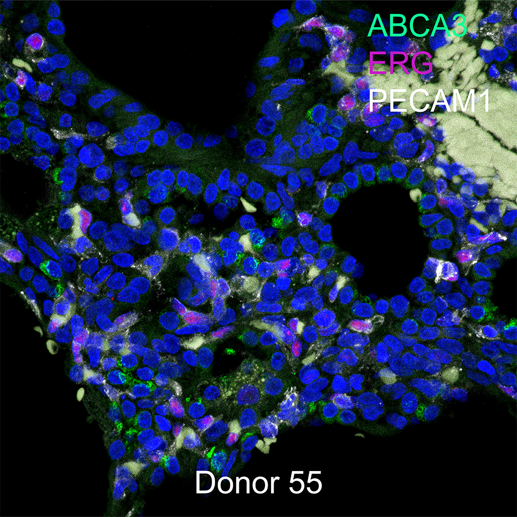
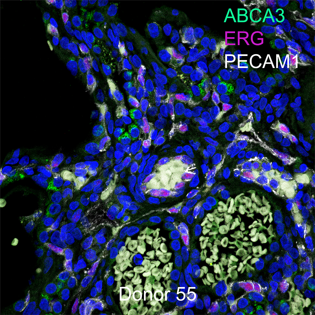
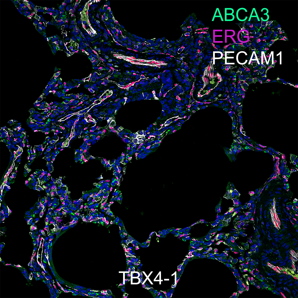
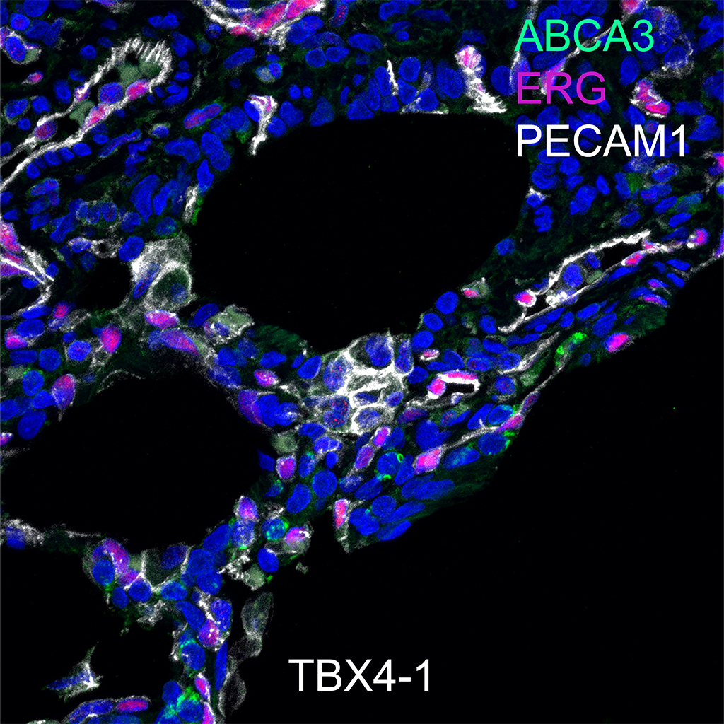
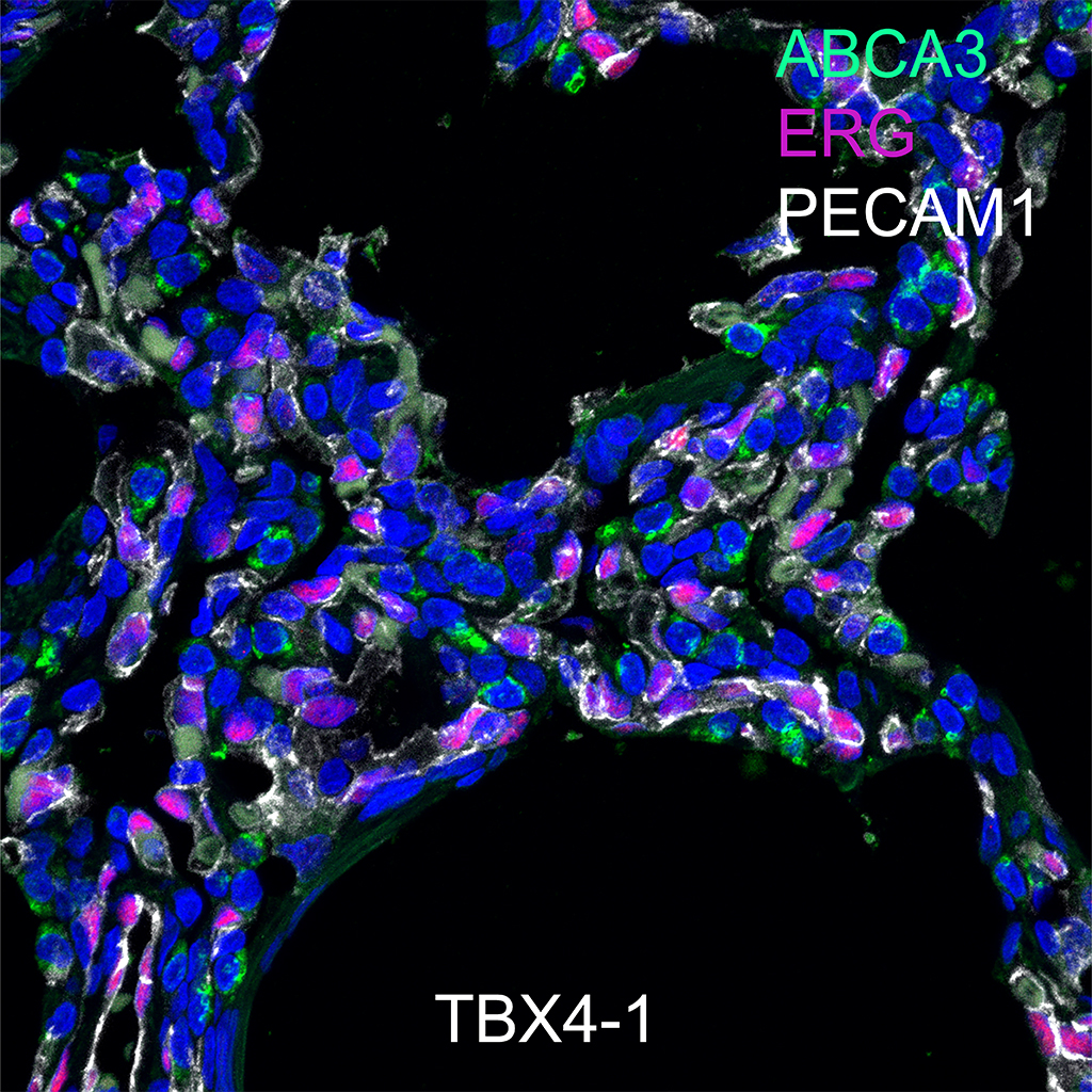
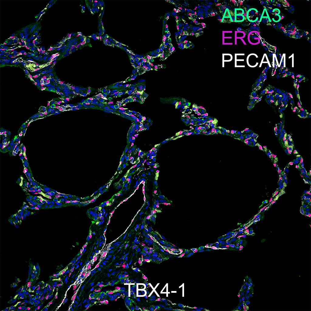
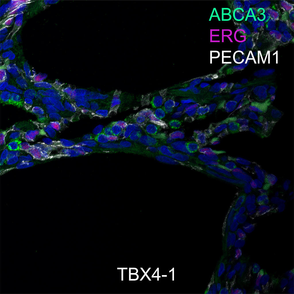
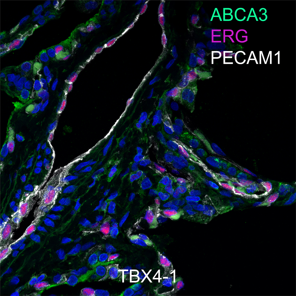
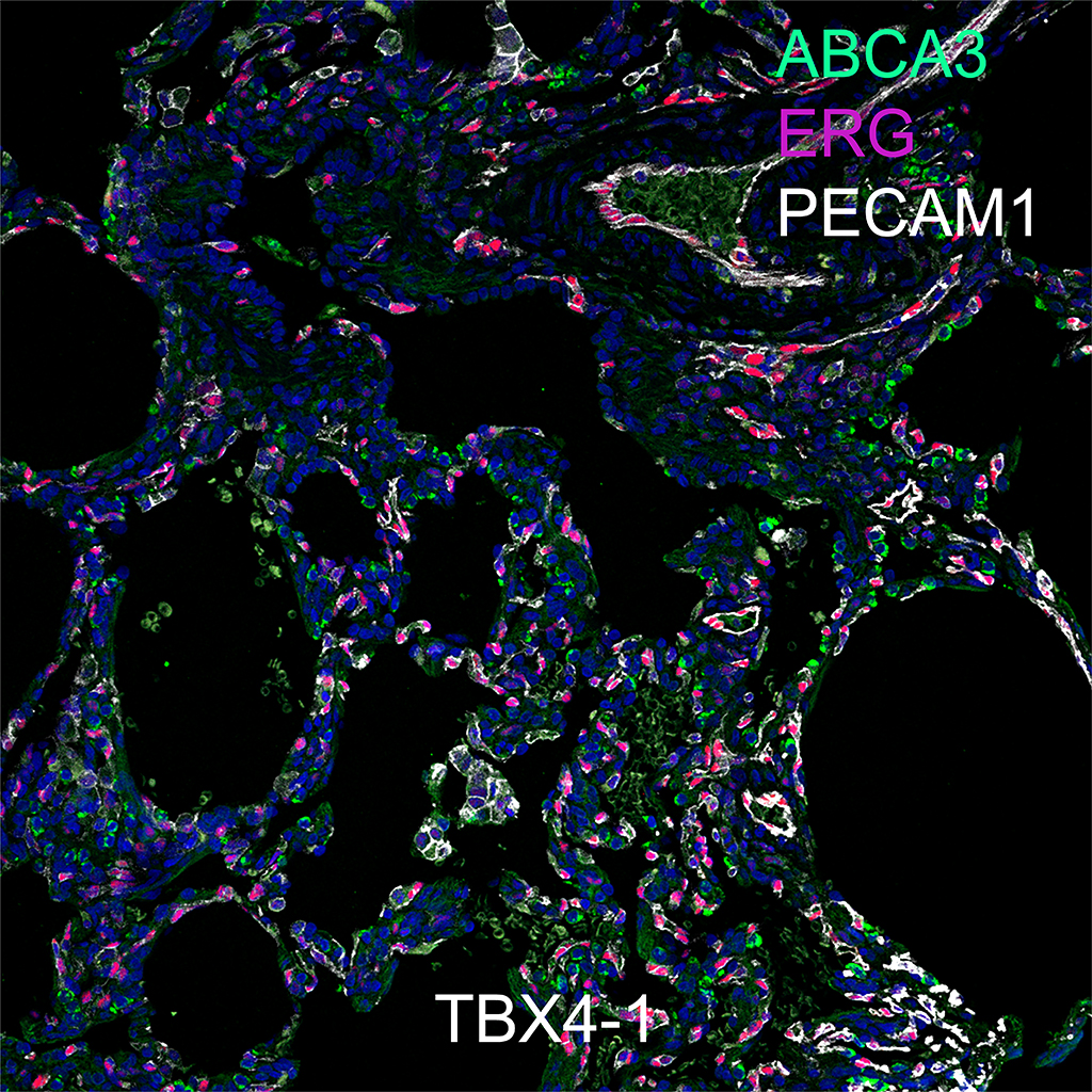
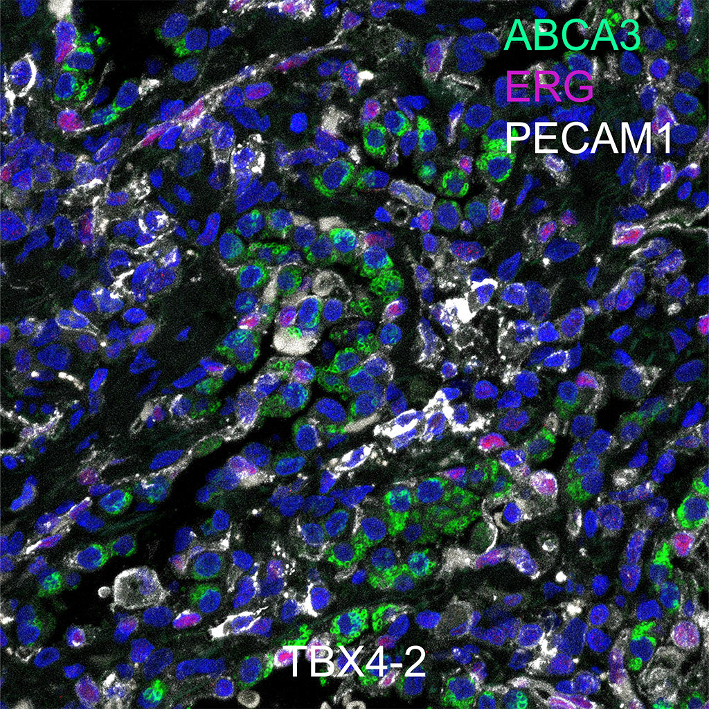
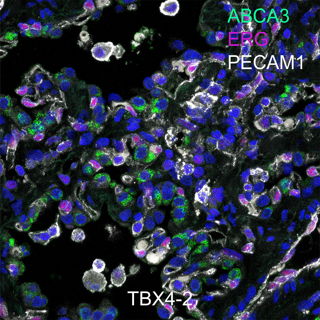
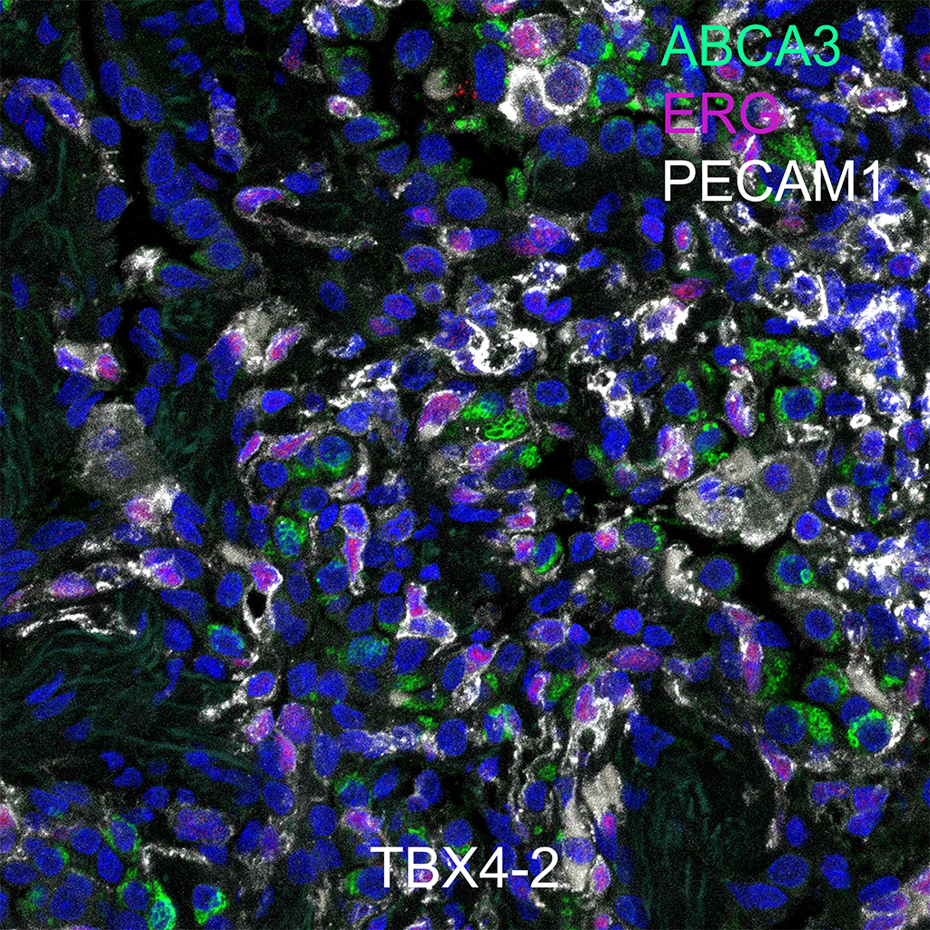
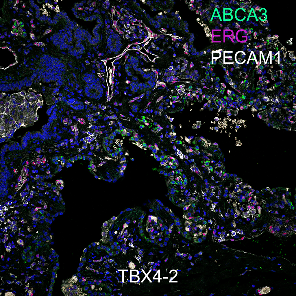
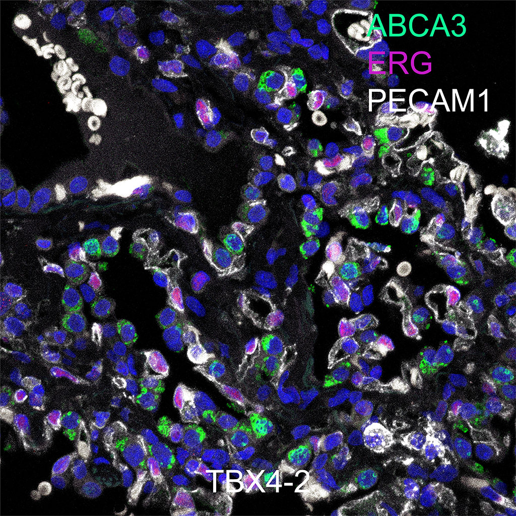
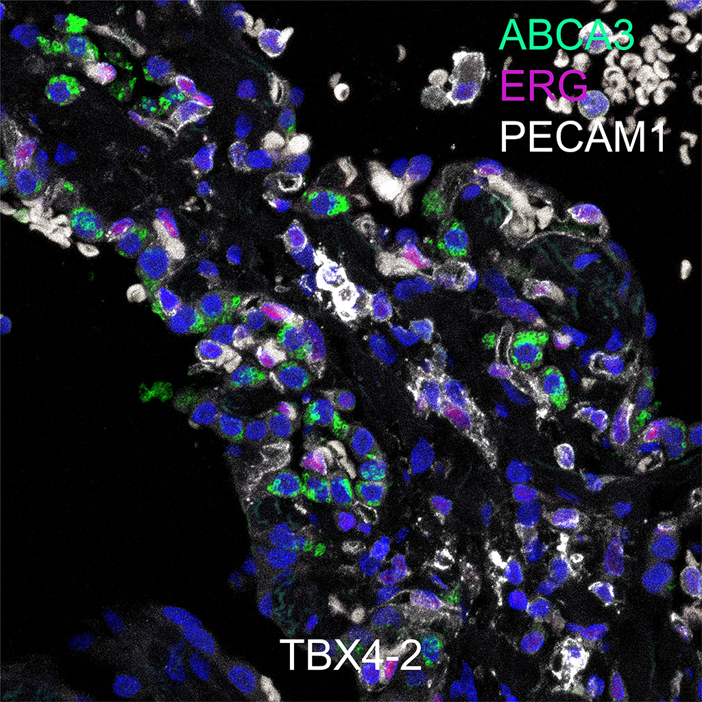
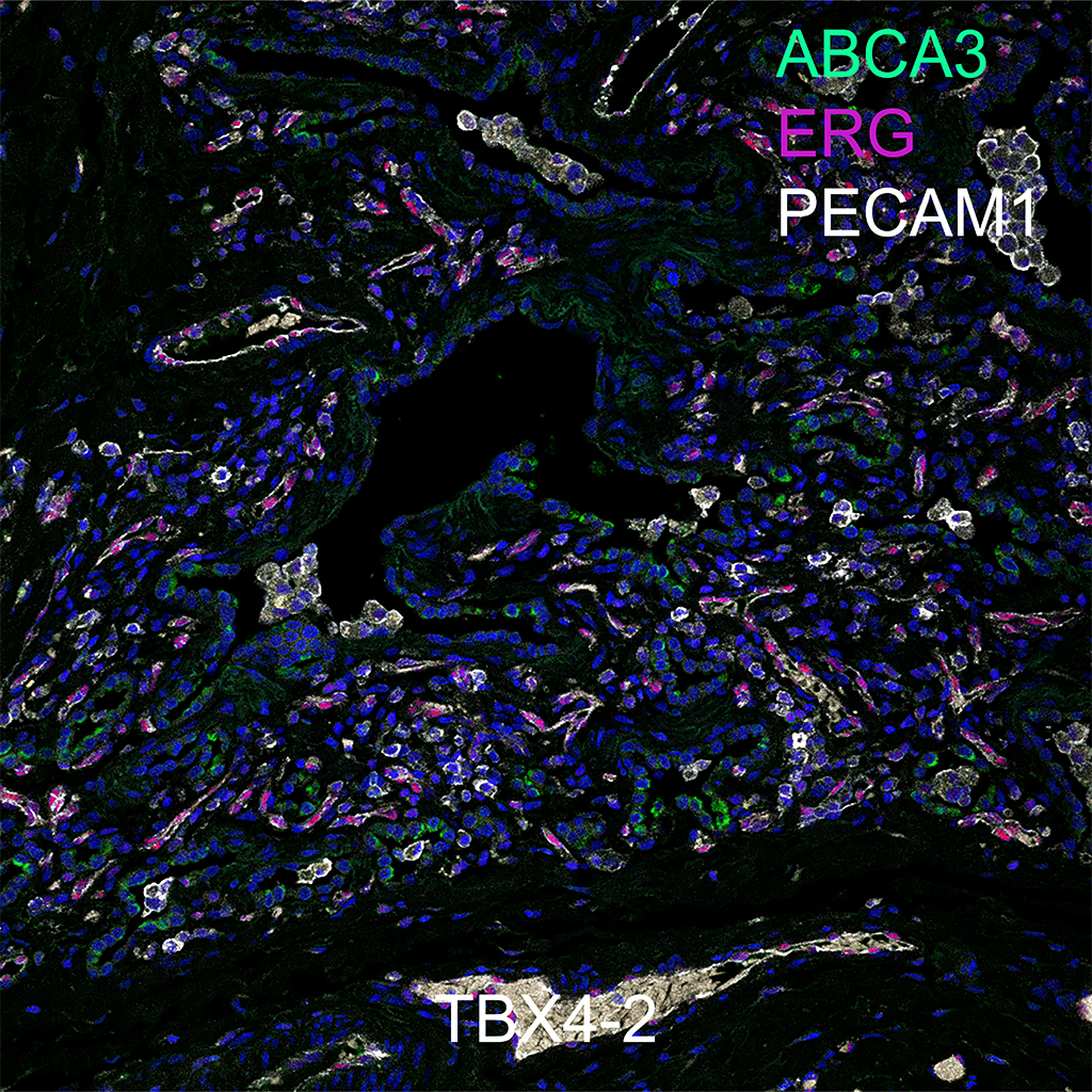
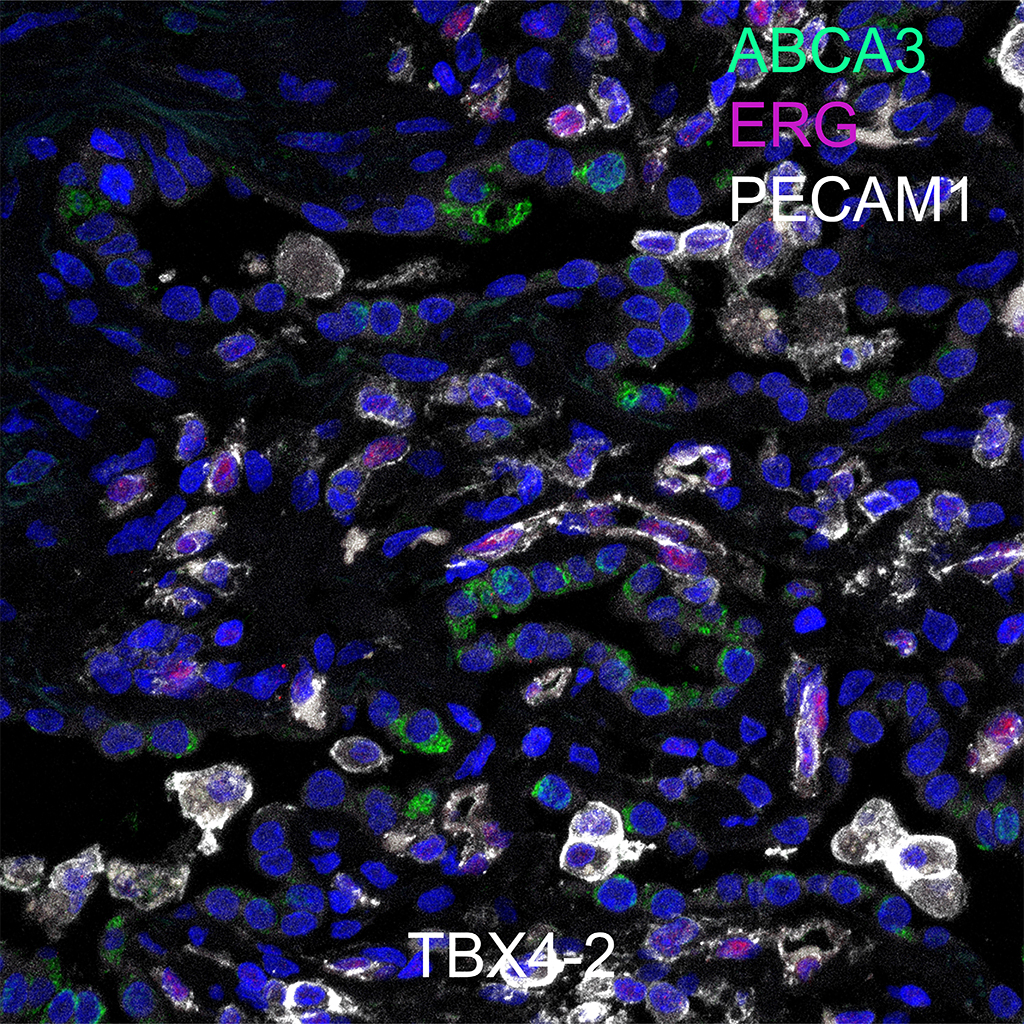
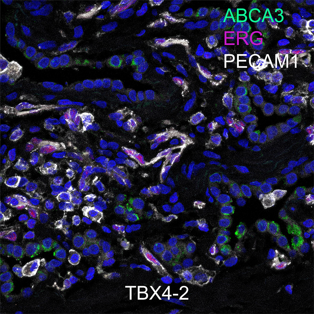
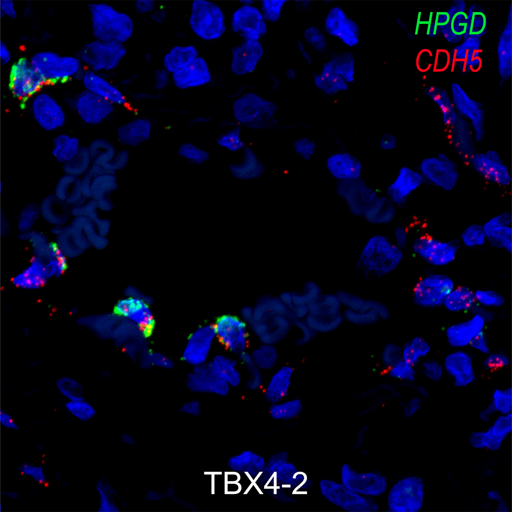
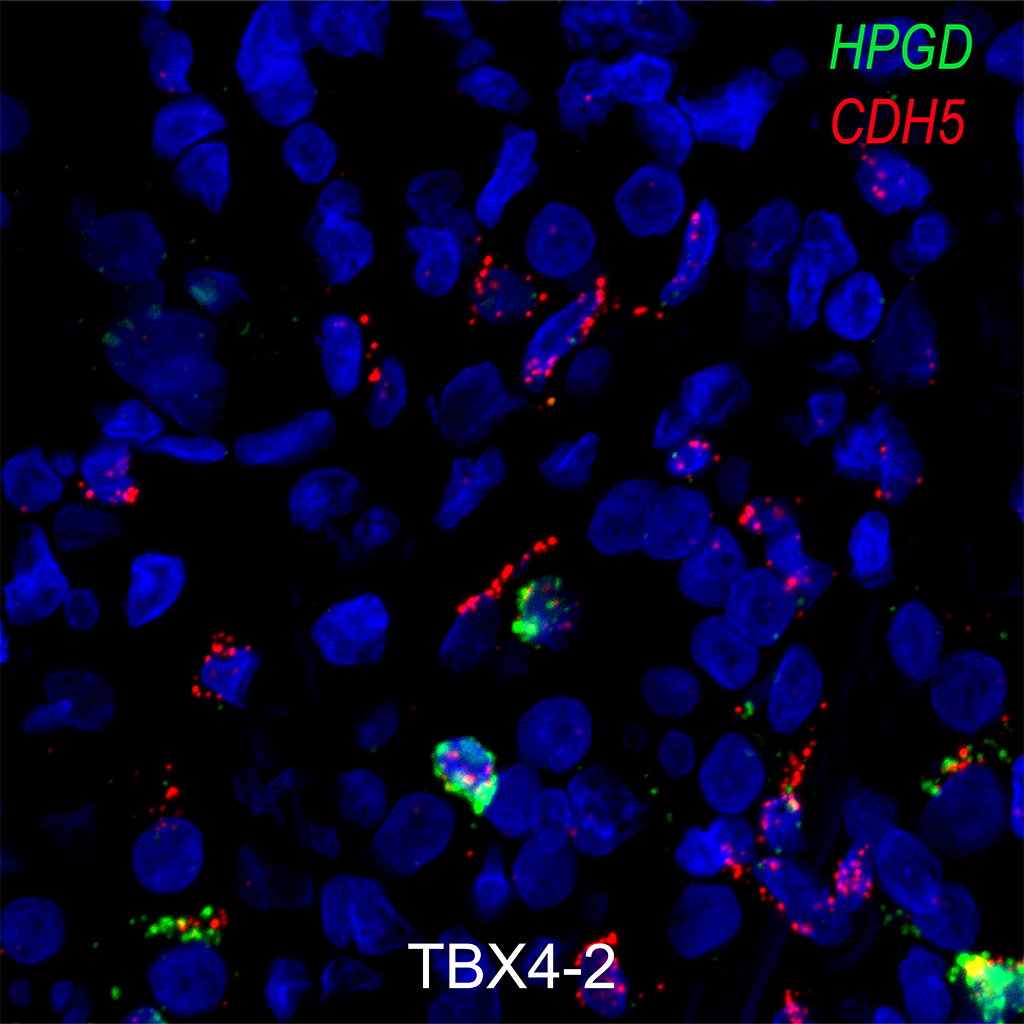
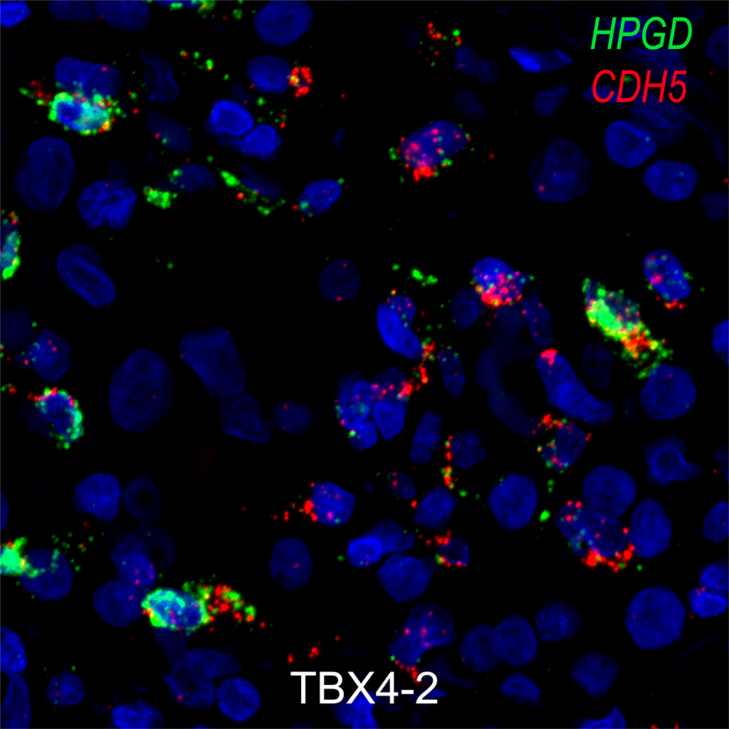
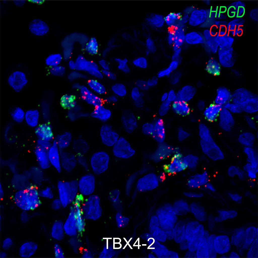
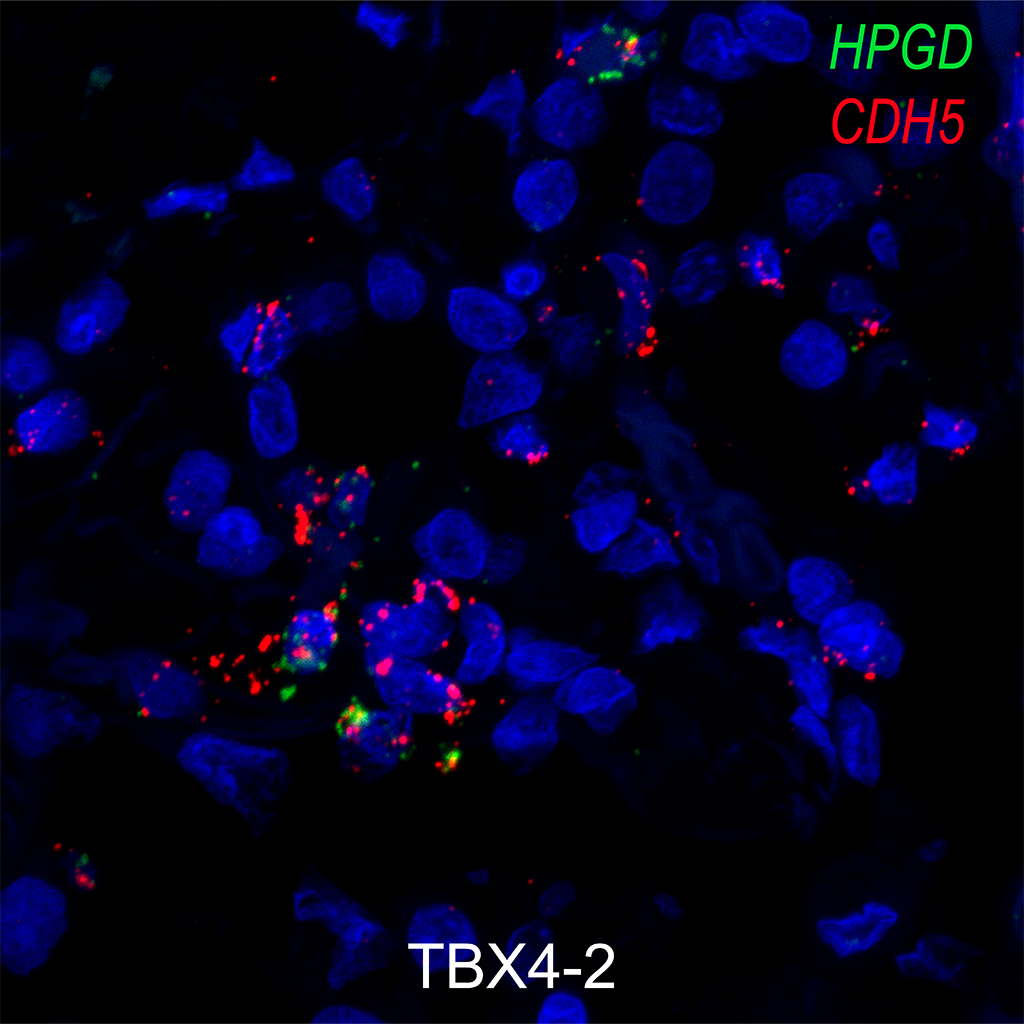
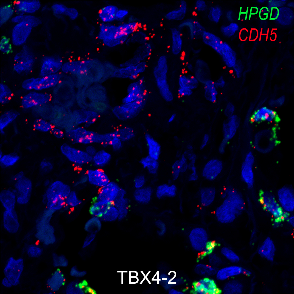
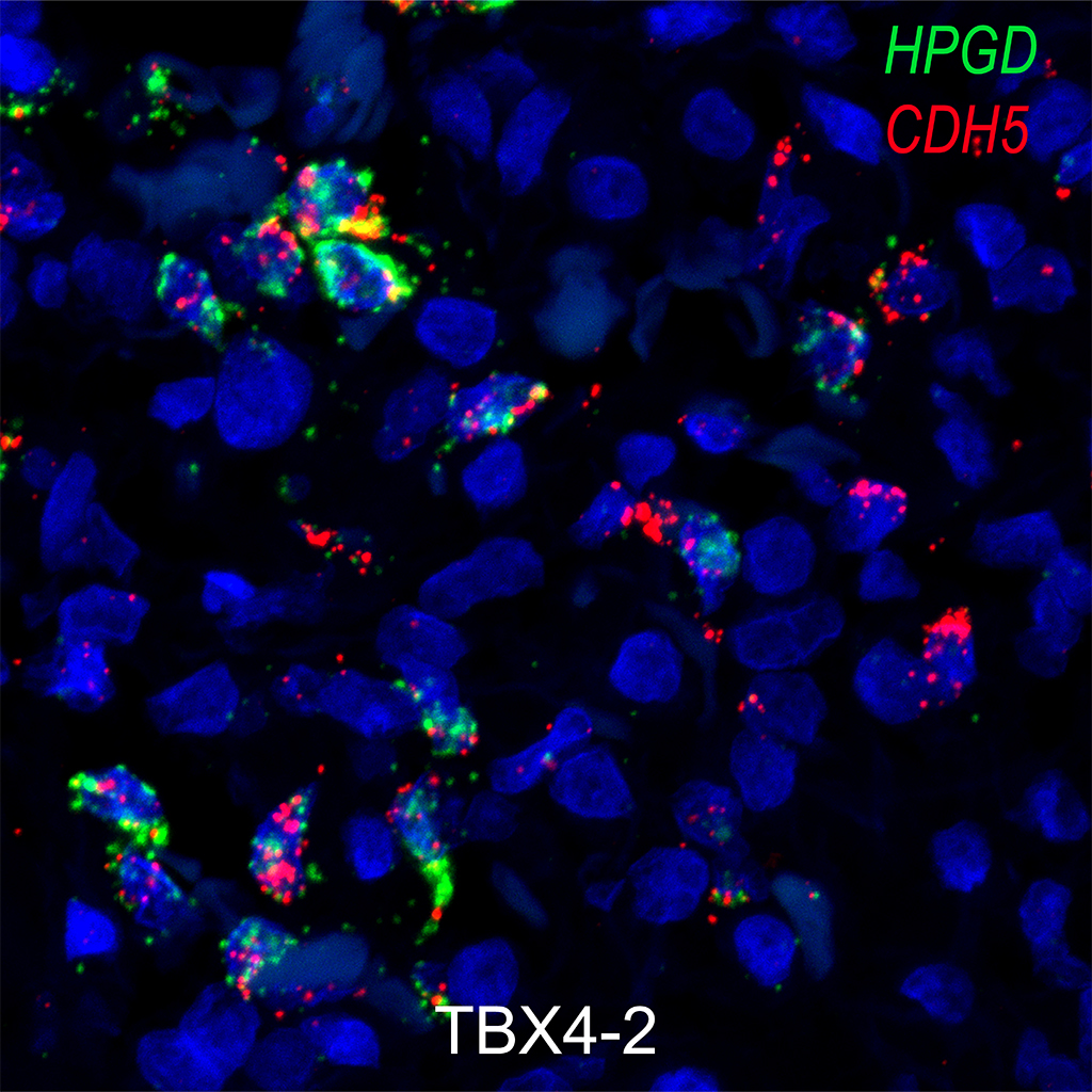
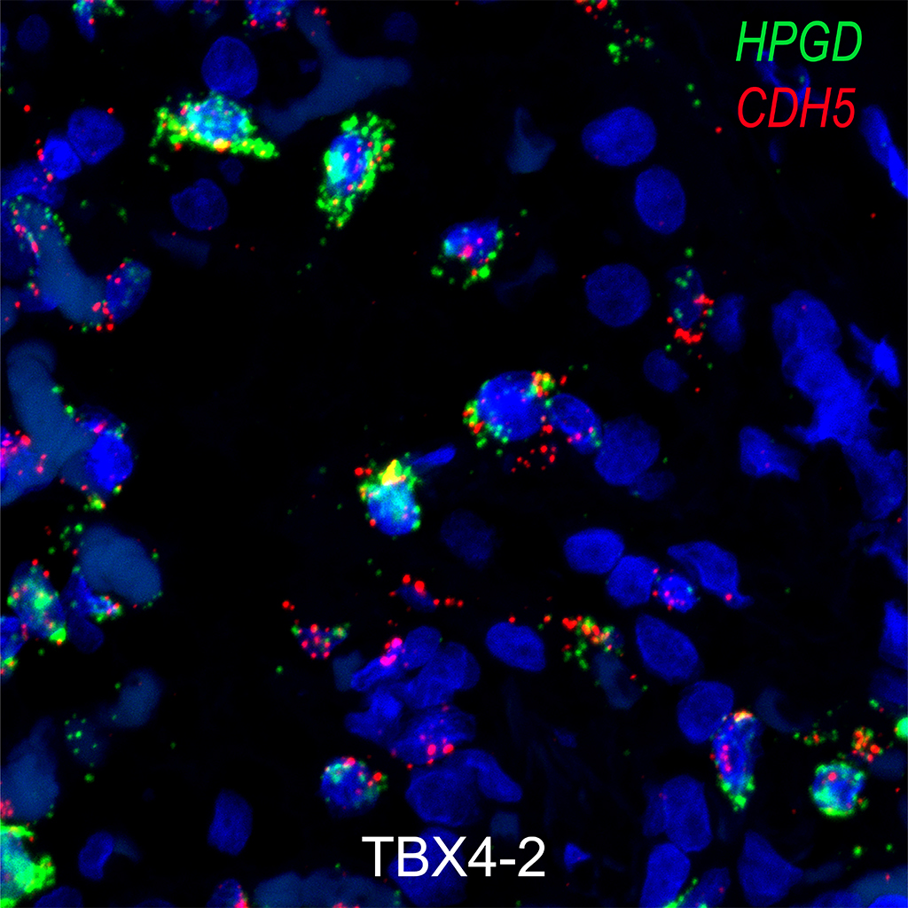
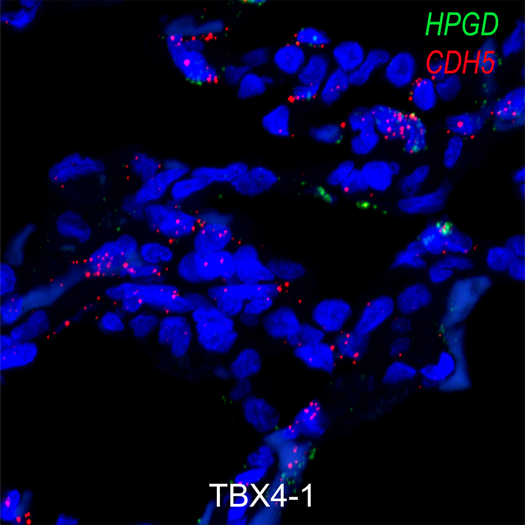
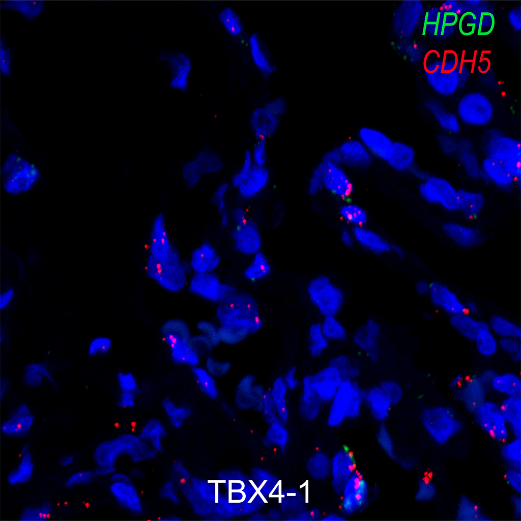
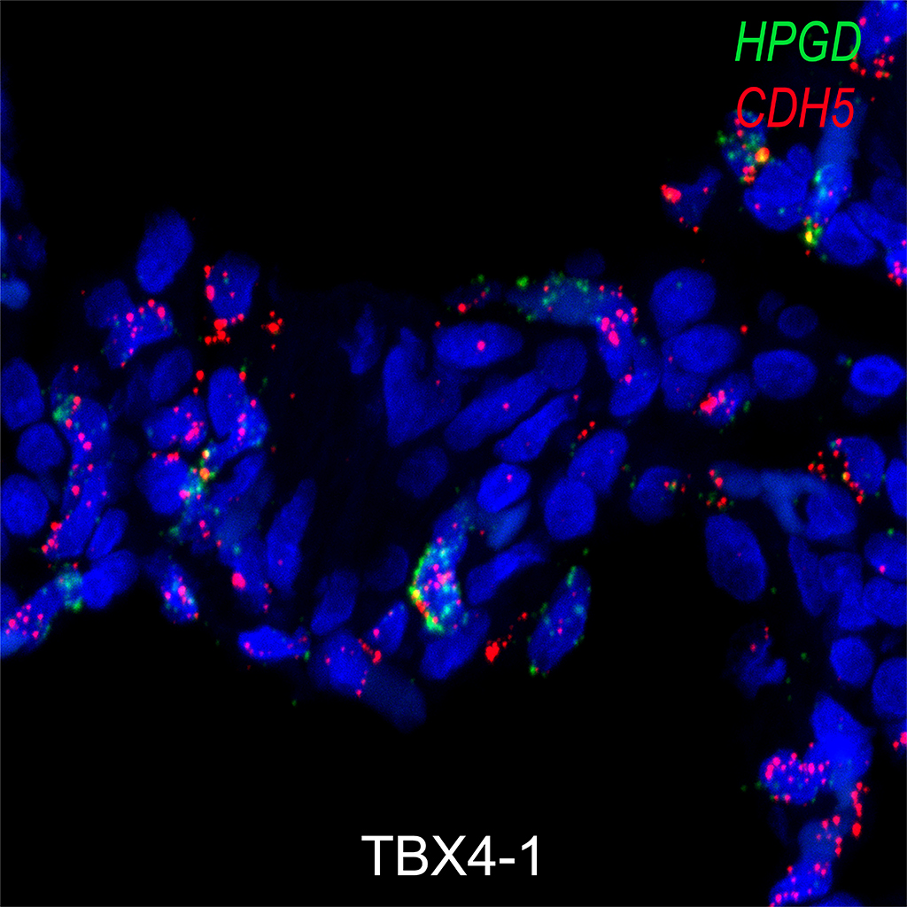
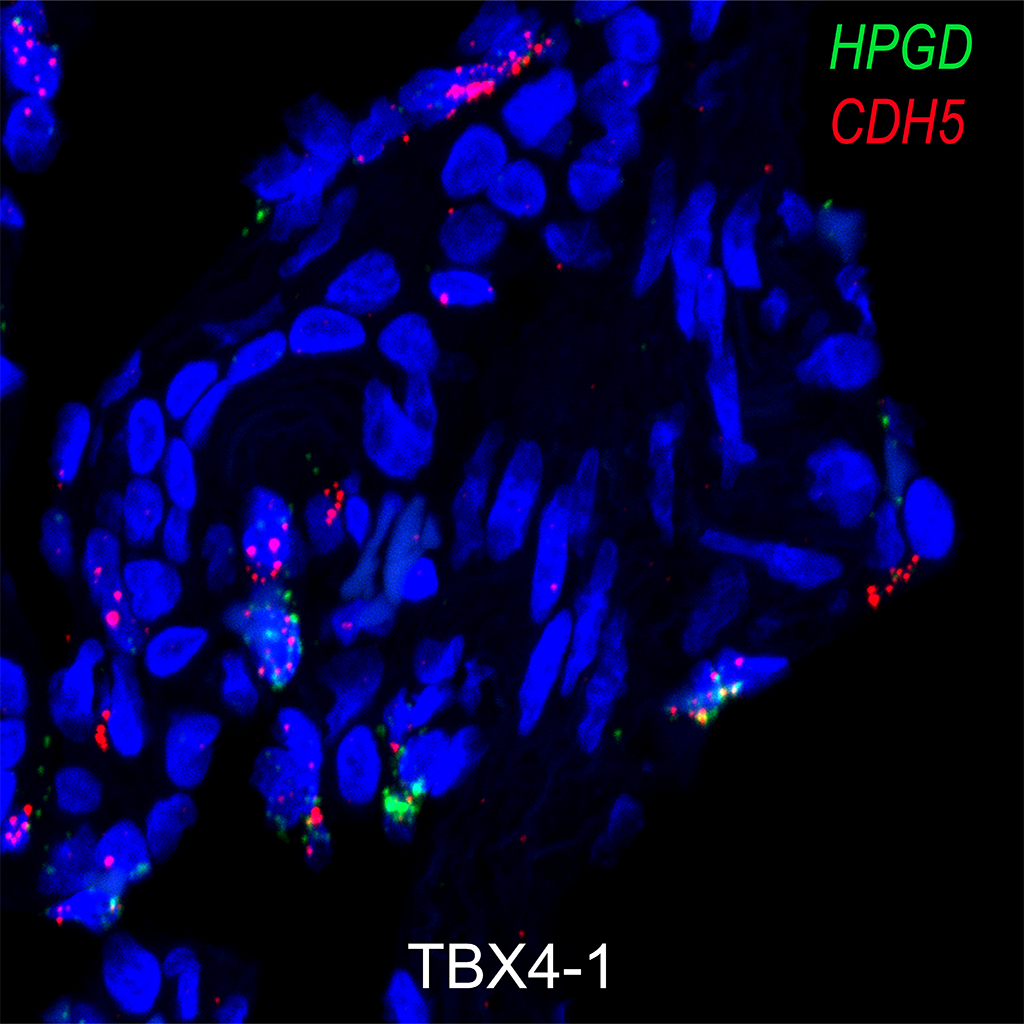
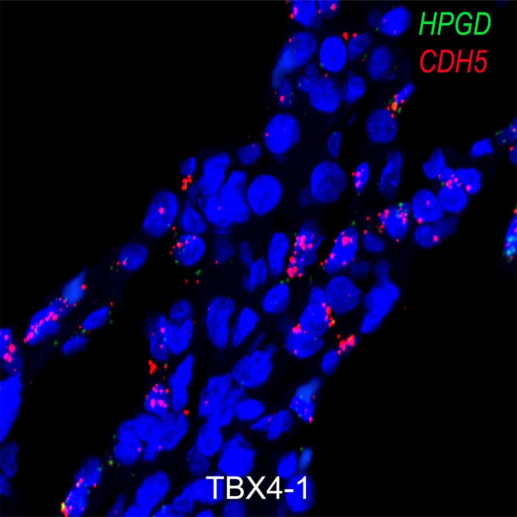
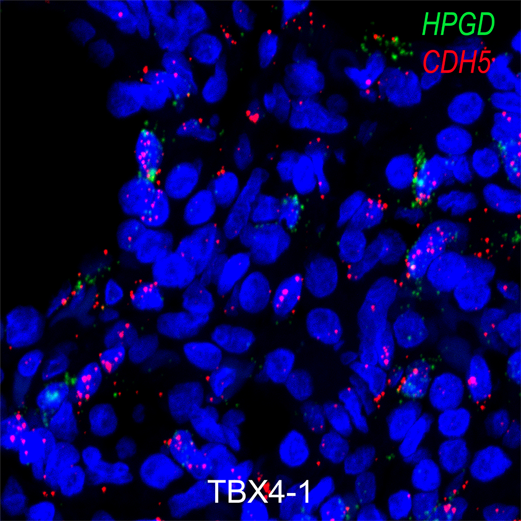
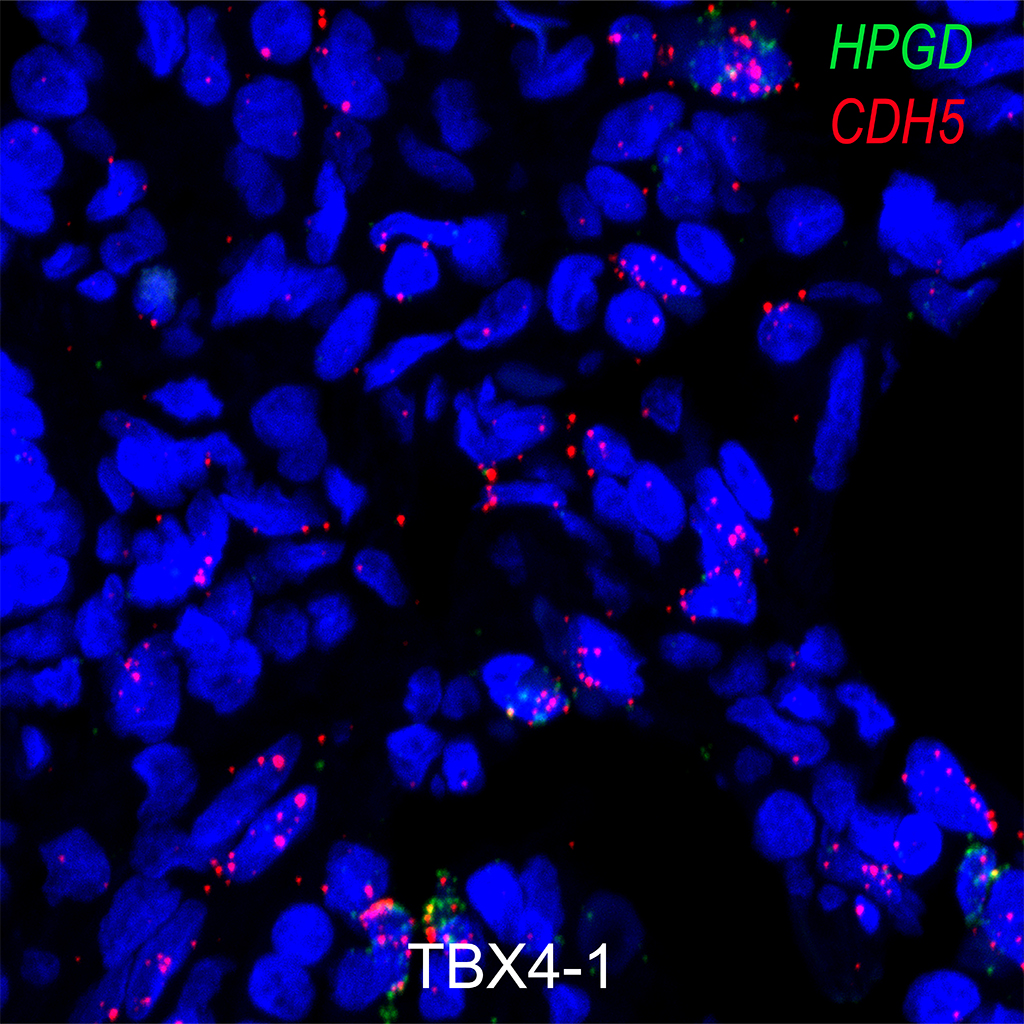
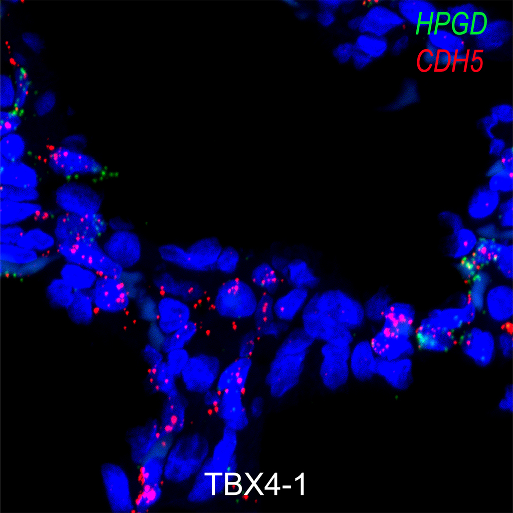
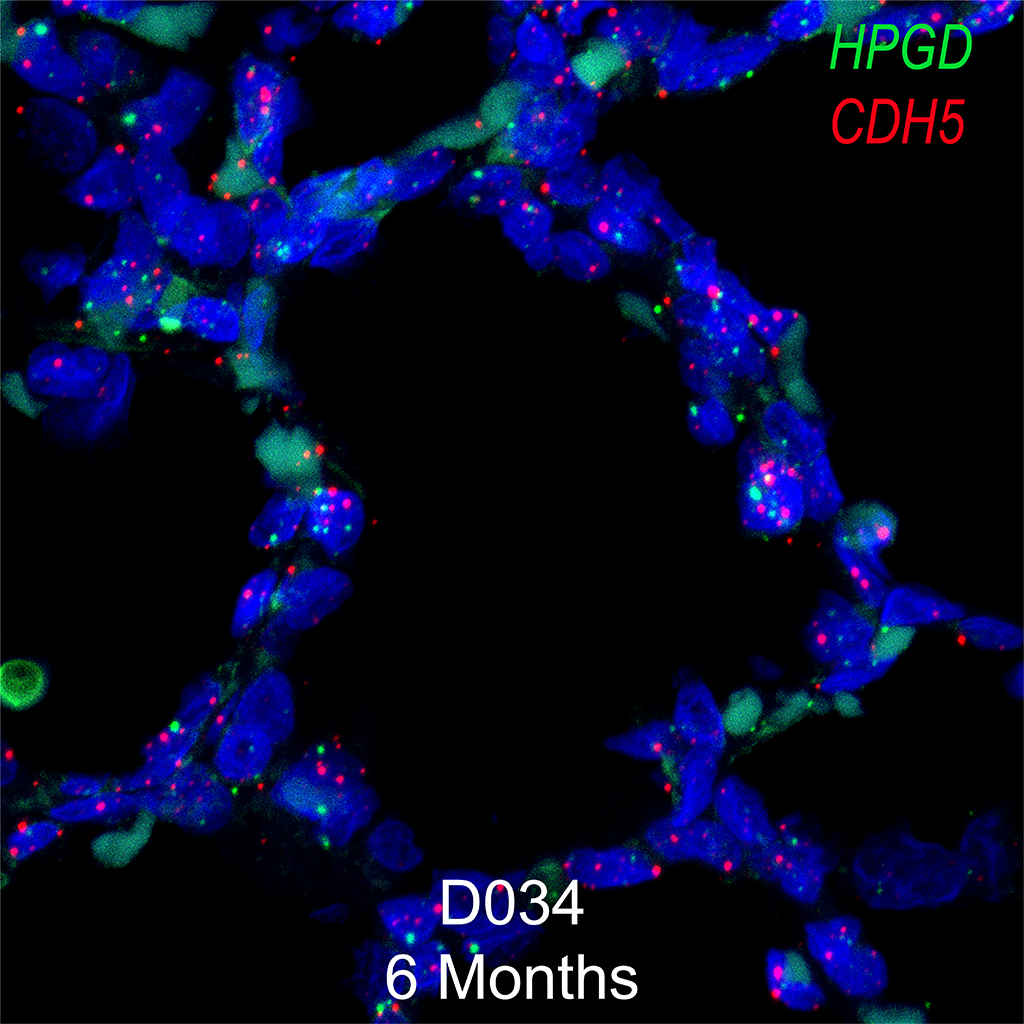
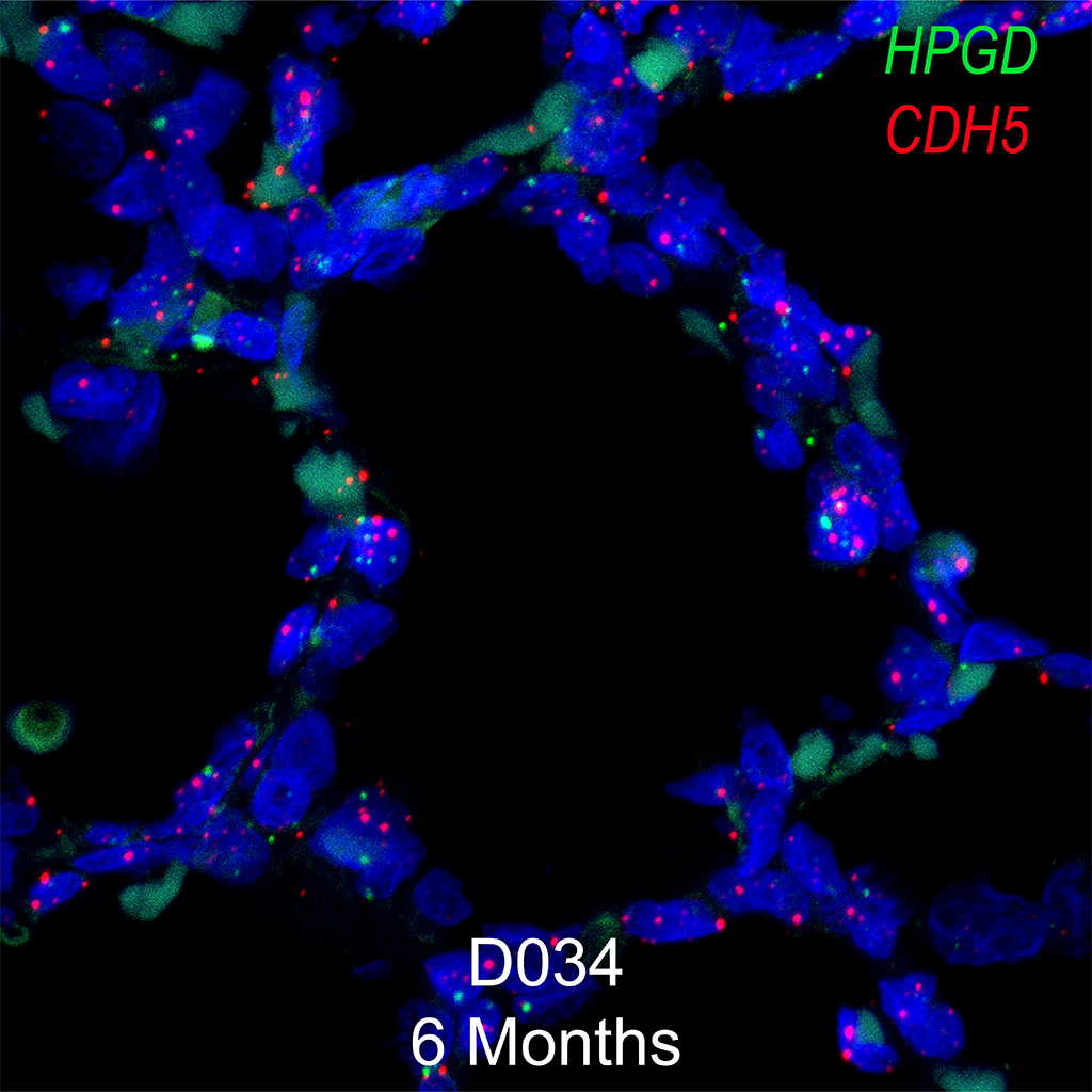
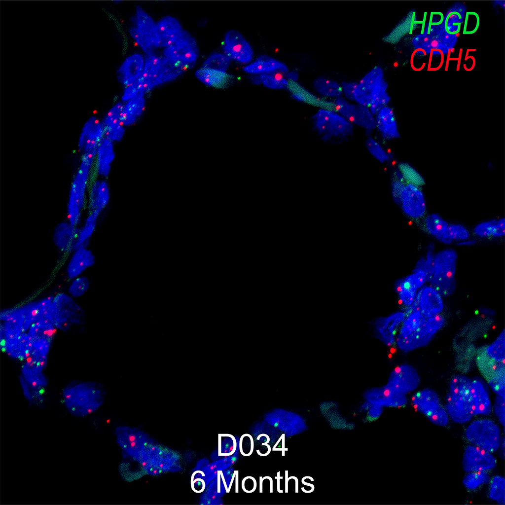
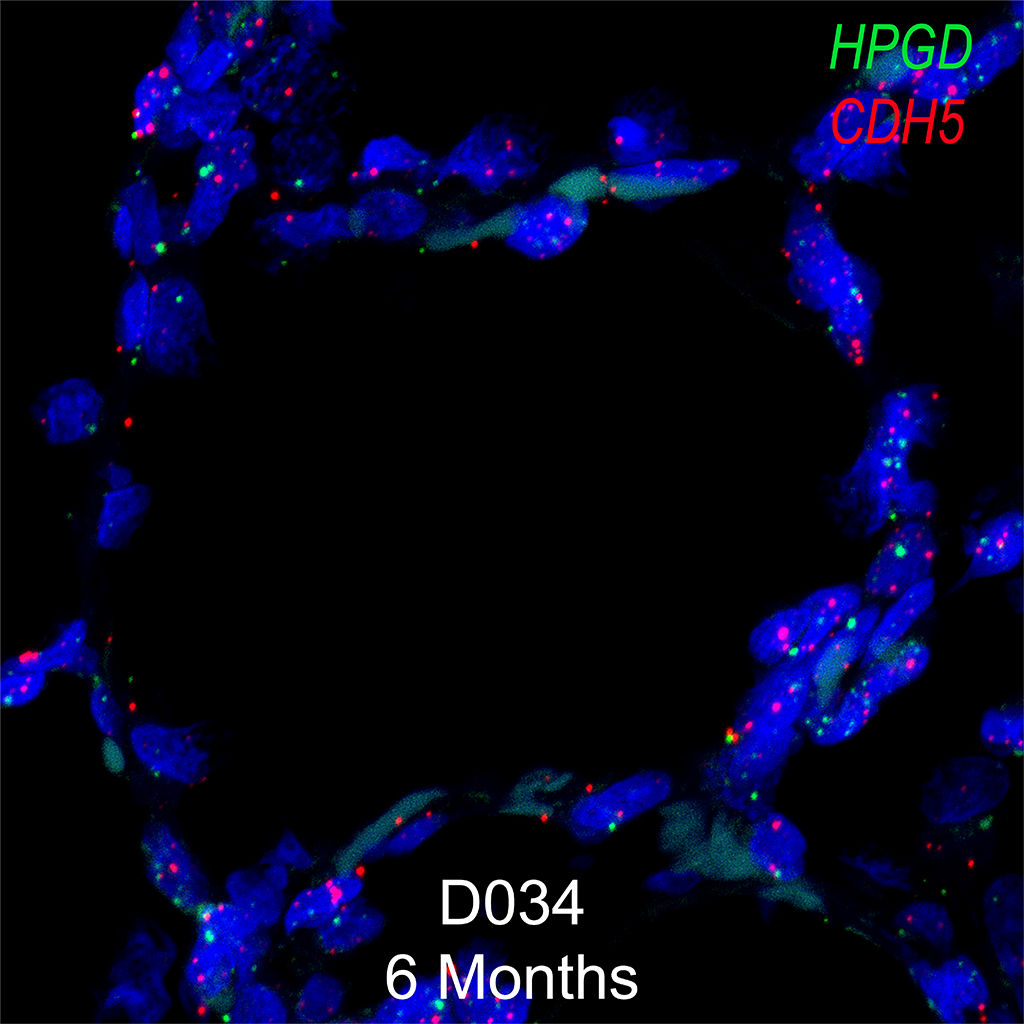
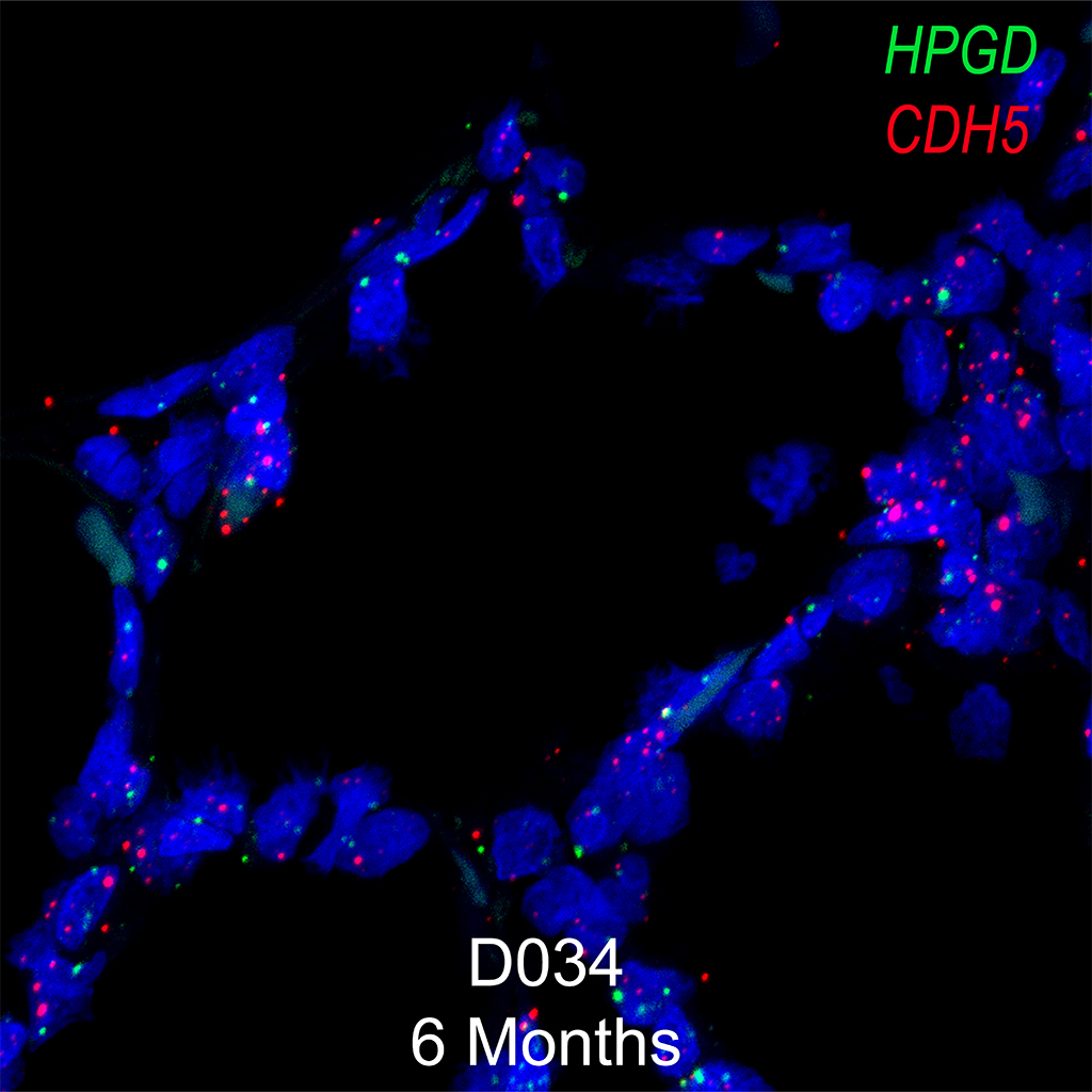
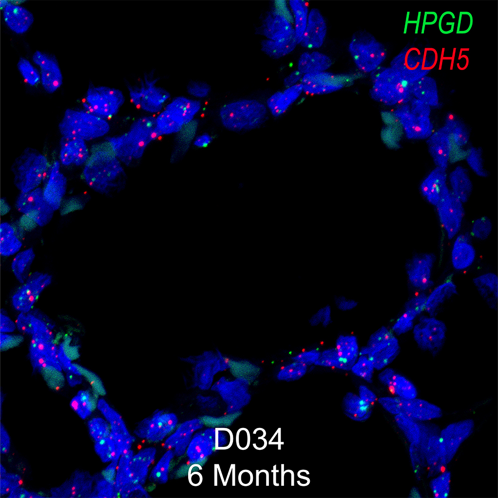
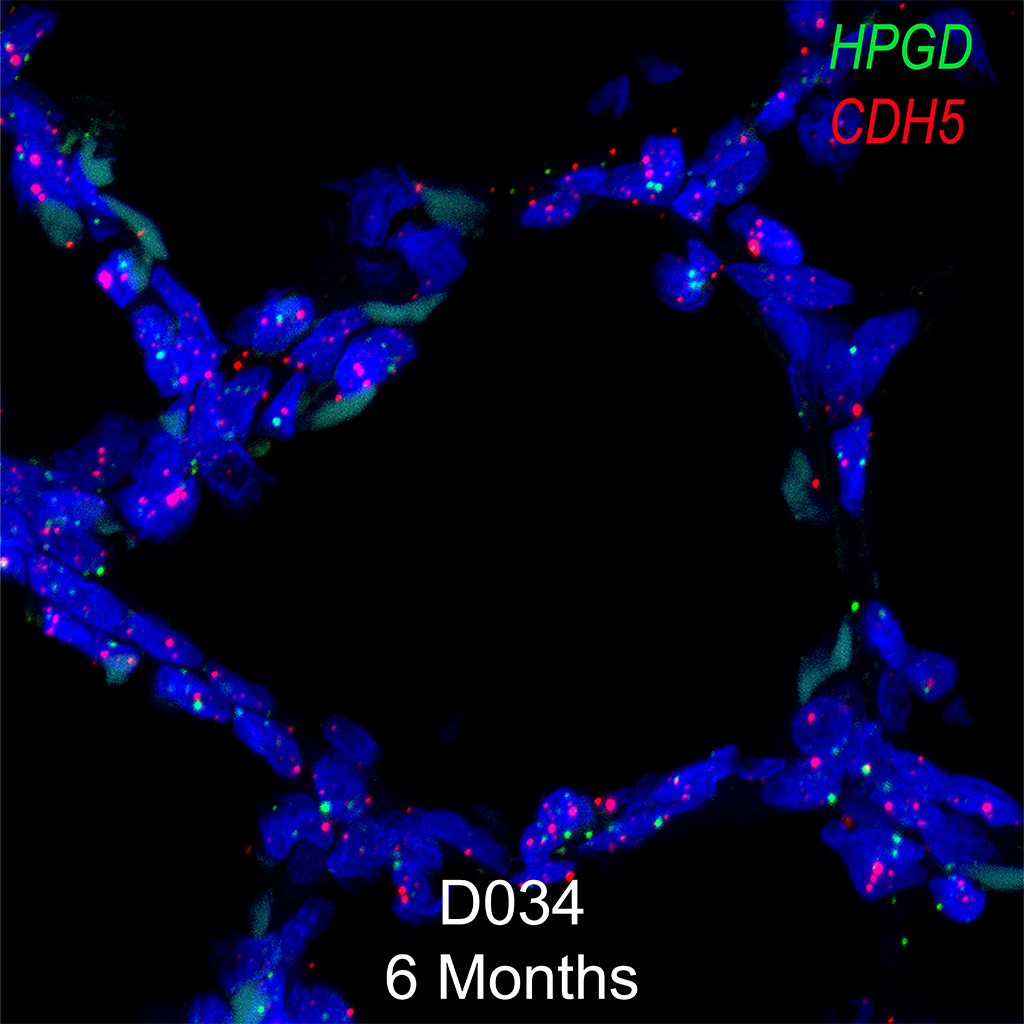
Immunofluorescence- NKX2.1, ABCA3, and ACTA2
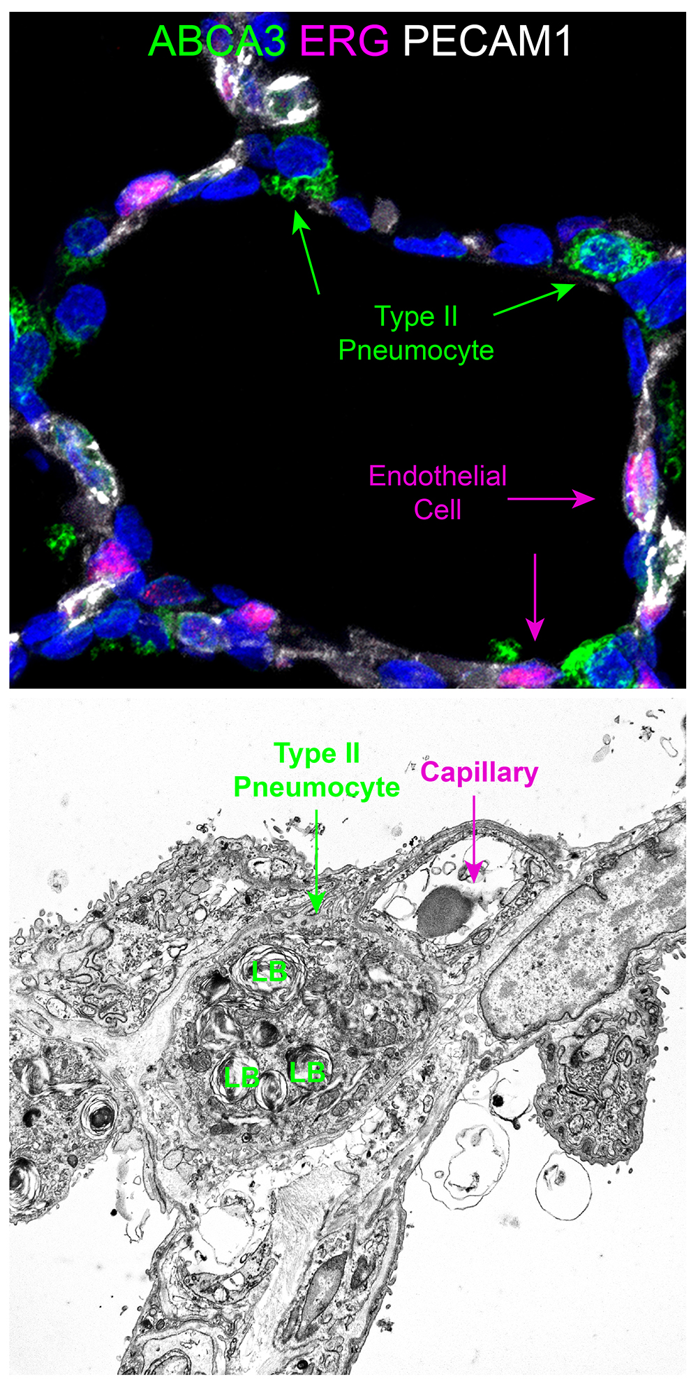
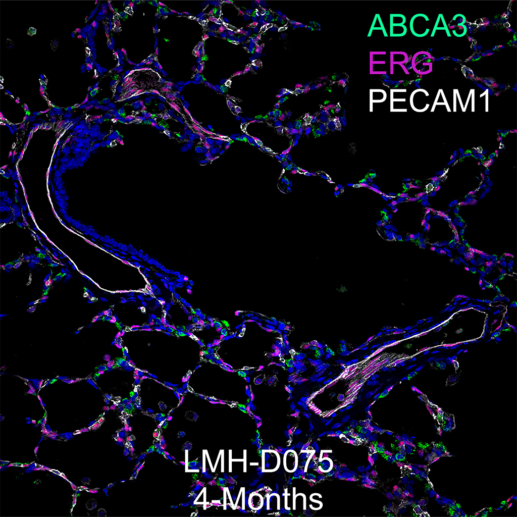
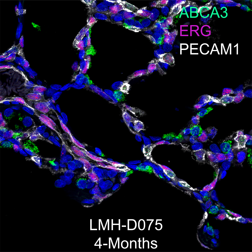
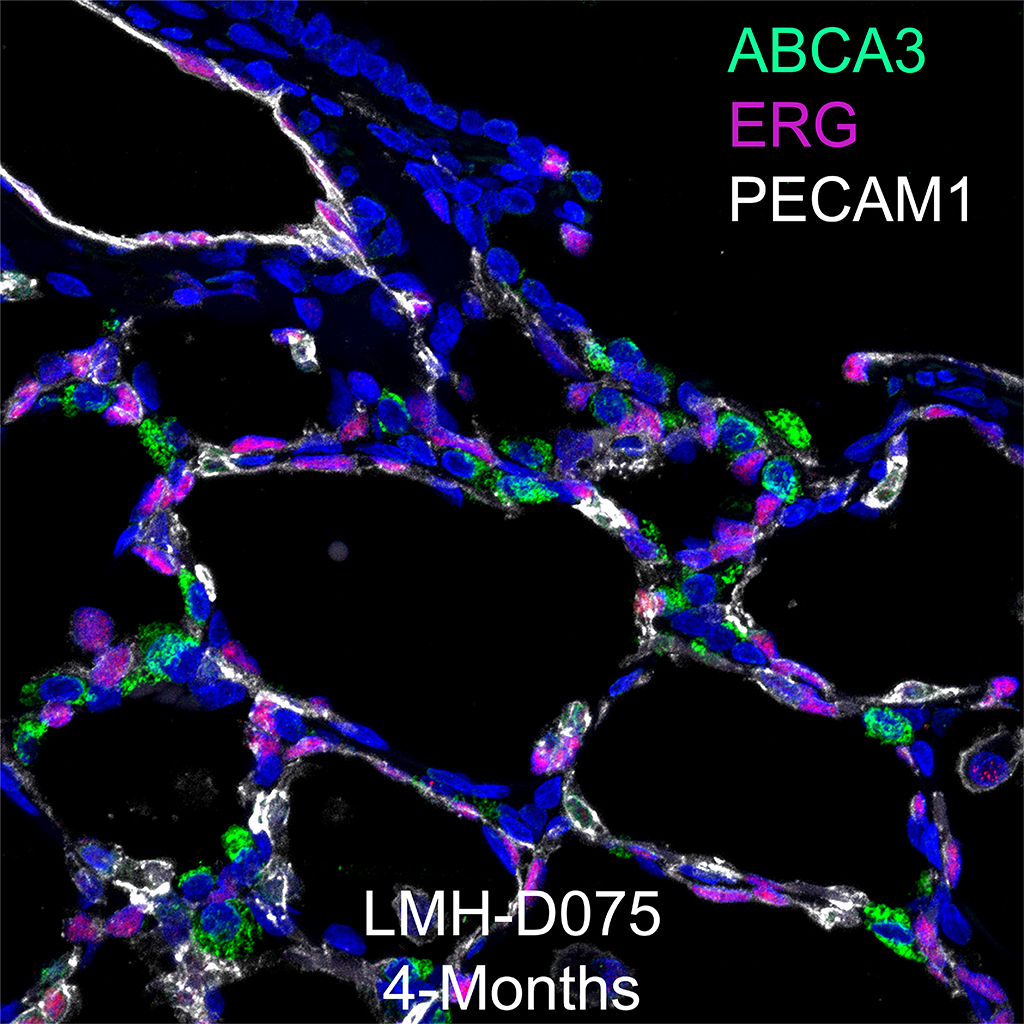
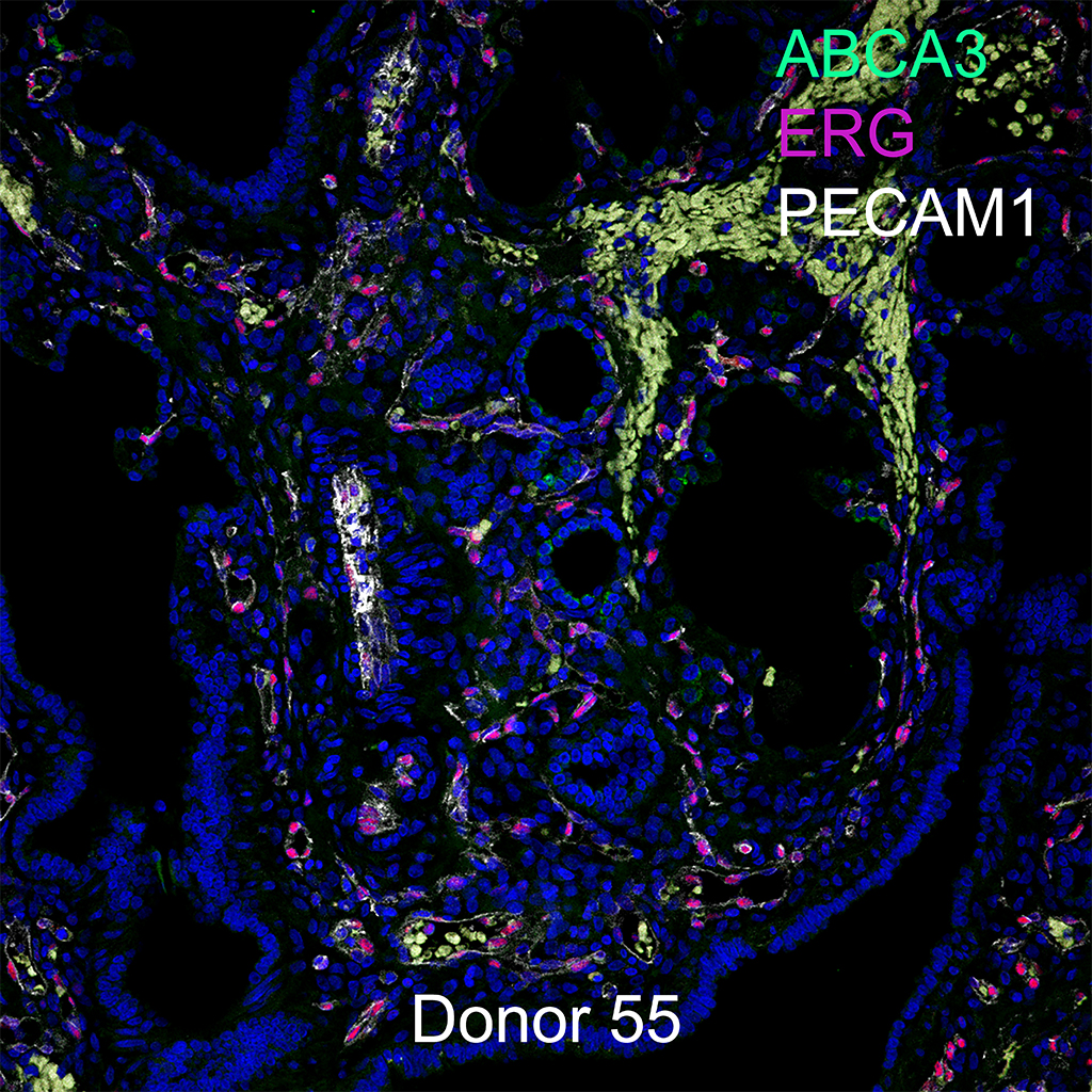
Purpose: Immunofluorescence on slides of 7µm frozen sections of C57BL6 PND01 lungs for NKX2.1, ABCA3, and ACTA2 with antigen retrieval.
Day 1
- For Frozen tissue, rinse 2X in PBS, then 5 min in 4% PFA/PBS, then rinse 1X in PBS.
- Briefly equilibrate slides in Antigen Retrieval Buffer.
- Antigen retrieval, pH 6.0 (times will vary according to microwave).
- 10 mM sodium citrate, pH 6.0, and heat in a microwave at 96oC. *We usually do a series of three runs (6-7 minutes each run) to equal the time/temp because of evaporation (refill coplin jars with dH2O).
- Microwave according to instructions on microwave.
- Cool on countertop, 15 min.
- Rinse with dH2O
- 1X PBS, 5 min.
- Block in 4% Goat serum/PBS-T, 2 hours at RT.
- For rabbit anti-NKX2.1 (RB TTF-1 1231, Lot# 0912A) dilute 1:1000, for guinea pig anti-ABCA3 (G985, BL 3/10/05, Seven Hills) dilute 1:100, for mouse anti-ACTA2 (α Smooth muscle actin, A5228, Sigma) dilute 1:2000 in blocking buffer. Spin down in µfuge for 10 minutes and incubate on tissue overnight @ 4oC.
Day 2
- Rinse slides in PBS-T 3X, 5 min.
- Apply secondary antibody, Goat Alexa Fluor 488 anti-rabbit IgG (A11034, Lot# 1298480, for anti- NKX2.1) at 1:200, Goat Alexa Fluor 555 anti-guinea pig IgG (A21435, Lot# 1571699, for anti-ABCA3) at 1:200, Donkey Alexa Fluor 647 anti-mouse IgG A31571, Lot# 1549801, anti-ACTA2) at 1:200, in blocking buffer. Spin down in µfuge for 10 min, apply to tissue and incubated at room temperature for 1 hour.
- Rinse in PBS-T 3X, 5 min.
- Dilute DAPI 1:2000 and apply to slides for 15 min.
- Wash in PBS-T 3X, 5 min.
- Rinse slides in 0.1M PB, 3X, 5 min.
- Add 1 drop of Prolong Gold anti-fade mounting medium (P36930).
- Coverslip with Gold Seal Coverslip (Cat# 3422 Electron Microscopy Sciences, 22 X 22 mm).
- Allow Prolong Gold to cure overnight at room temp in light sealed box.
- Store slides in light sealed box @ 4oC
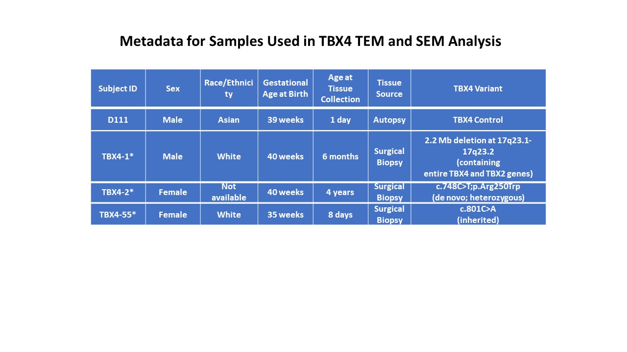
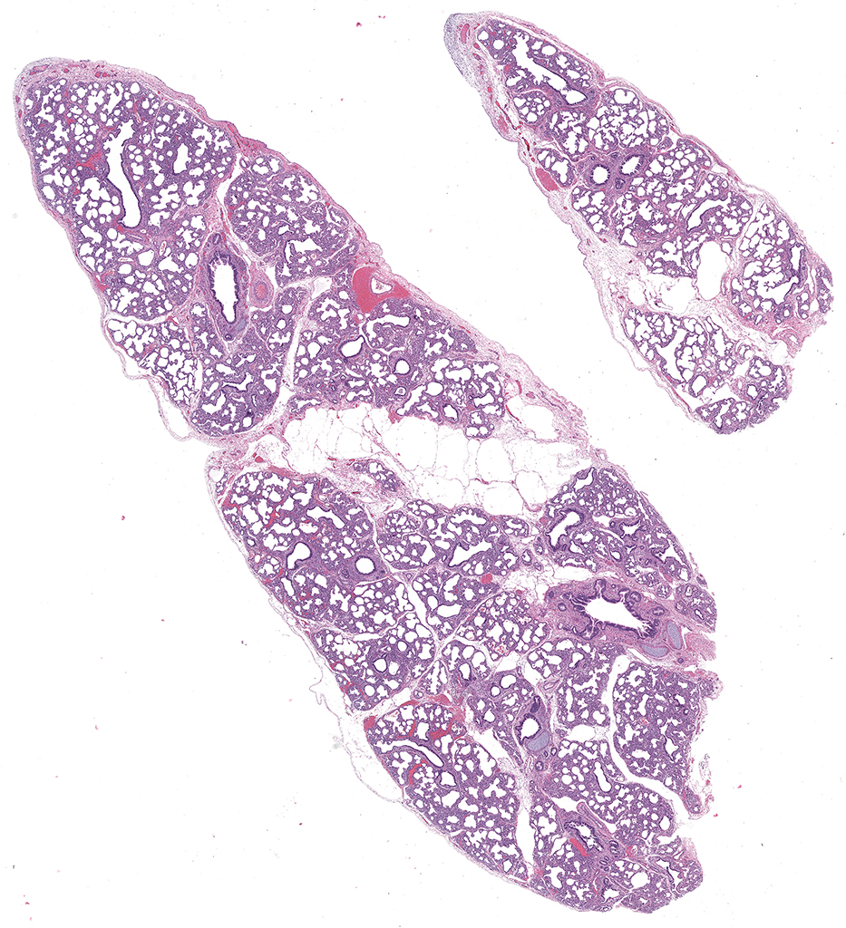
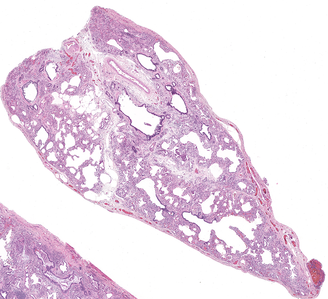
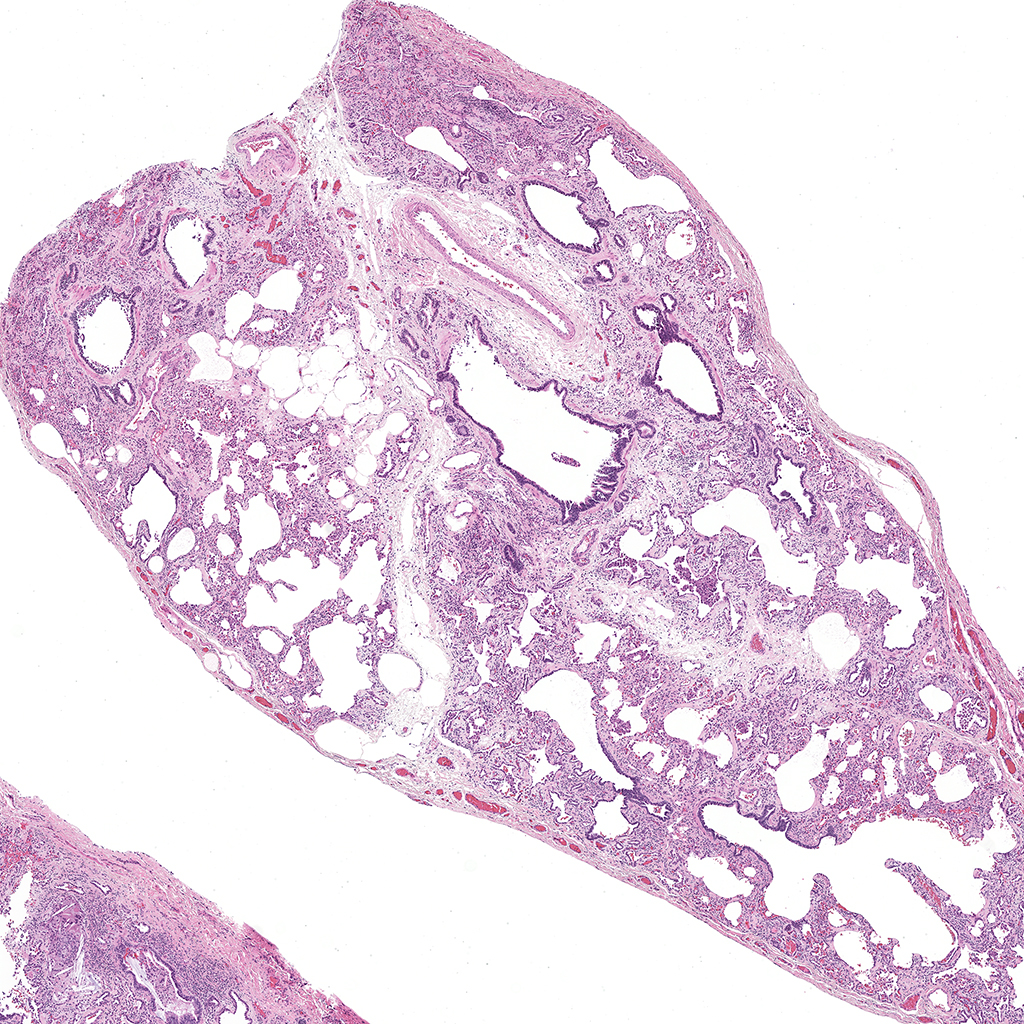
Tissue Used:
LMM.15.66A.6.50
PND01 C57BL6
Gender: Female
Weight= 1.14g
Appendix
Antigen Retrieval Solution
18ml Solution A- 0.1M Citric Acid (Sigma, C1909) pH 2.5
82ml Reagent B- 0.1M Sodium Citrate (Sigma, S4641 ) pH 8.2
1L dH2O
pH 6.0


