Gestational Day 150 Fetal Monkey SCGB1A1, NKX2.1, and TUBA4A Confocal Imaging
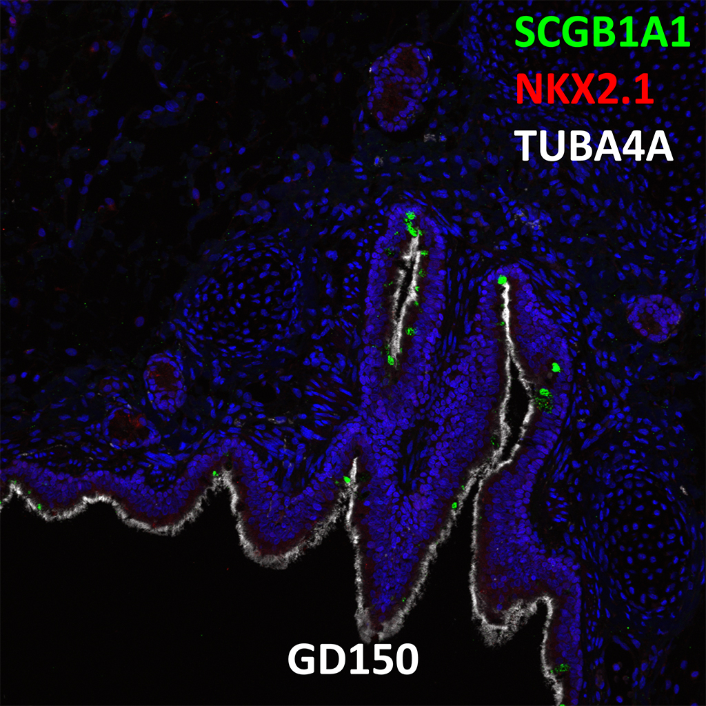
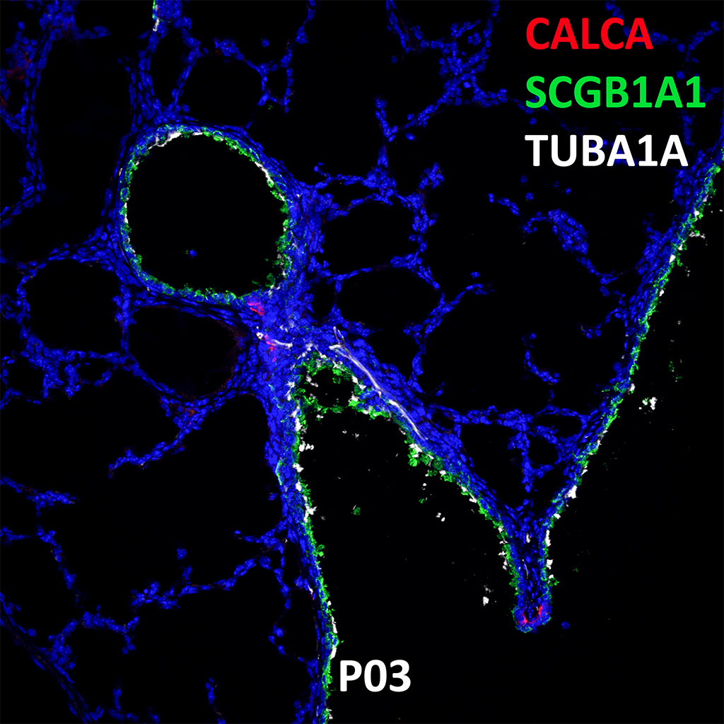
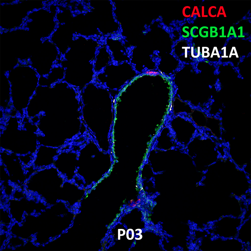
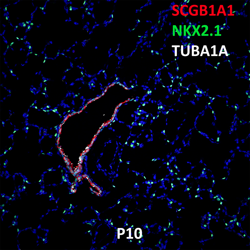
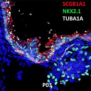
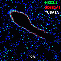
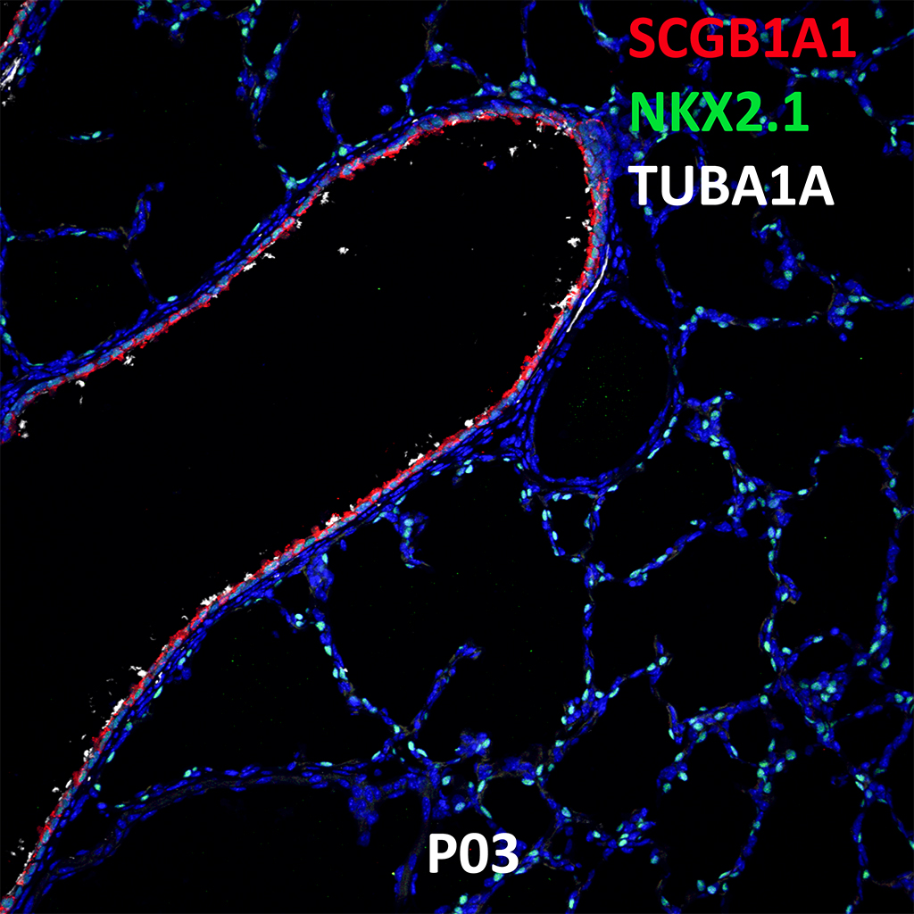
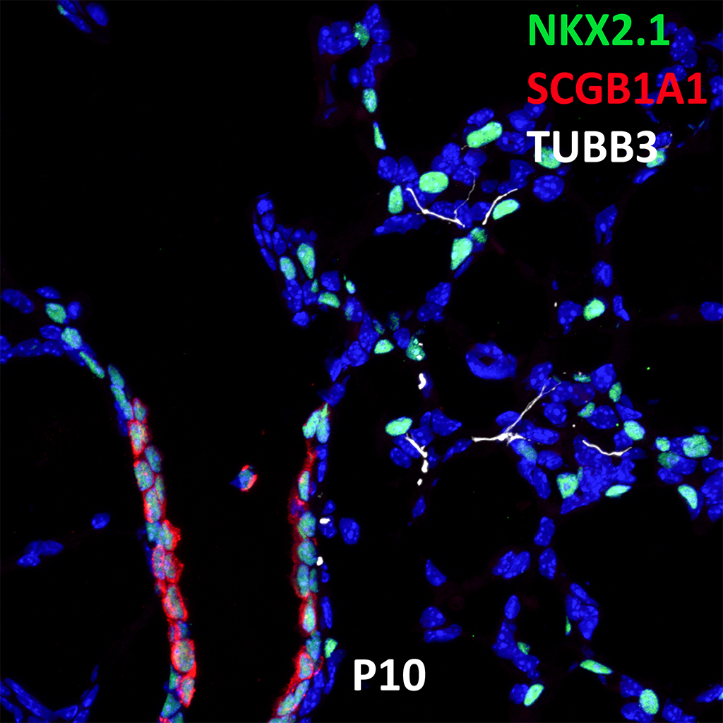
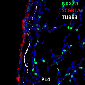
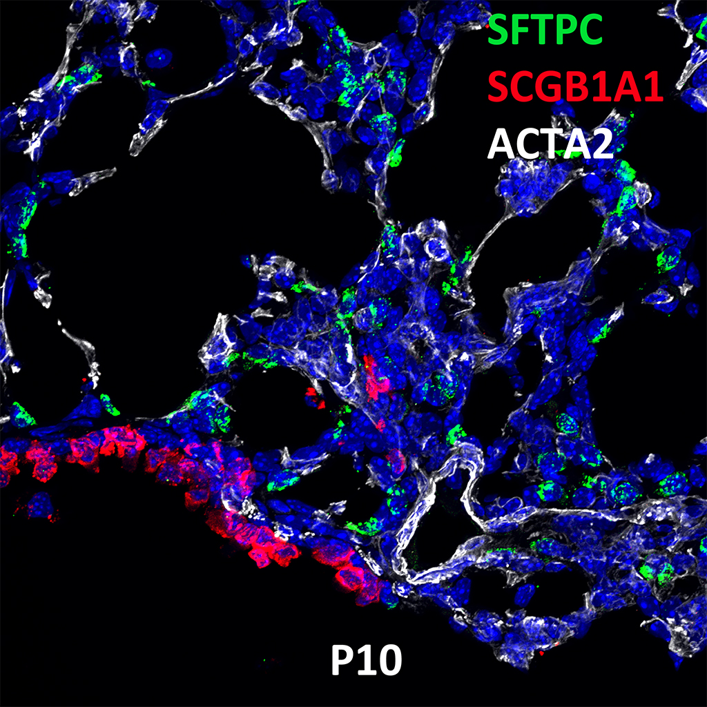
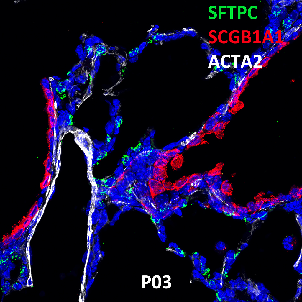
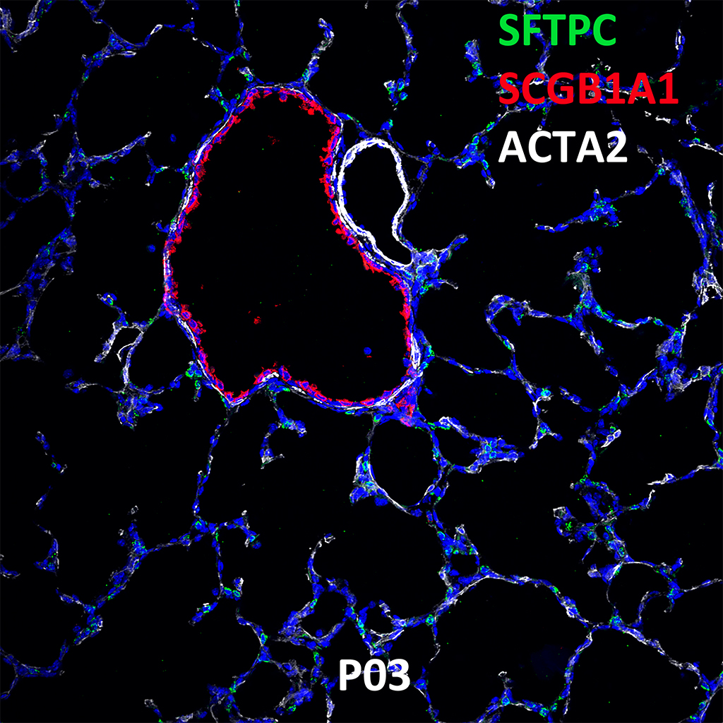
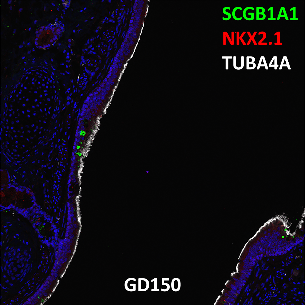
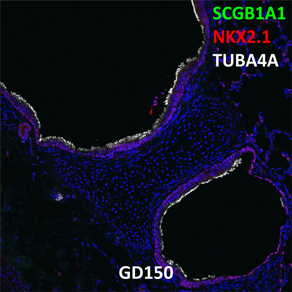
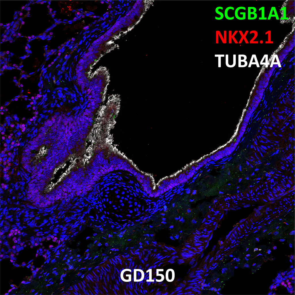
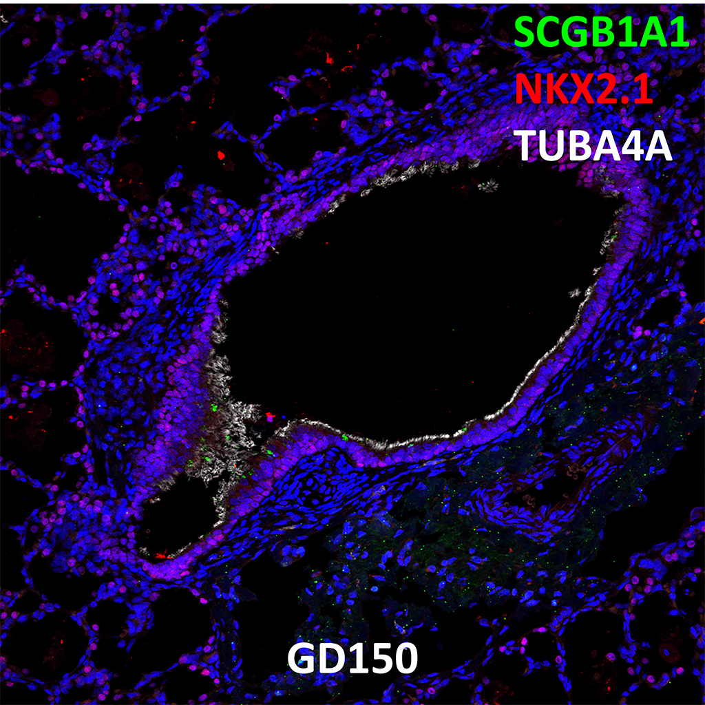
GD150 Fetal Monkey Lung Immunofluorescence and Confocal Imaging Showing Expression of SCGB1A1, NKX2.1, and TUBA4A
20X
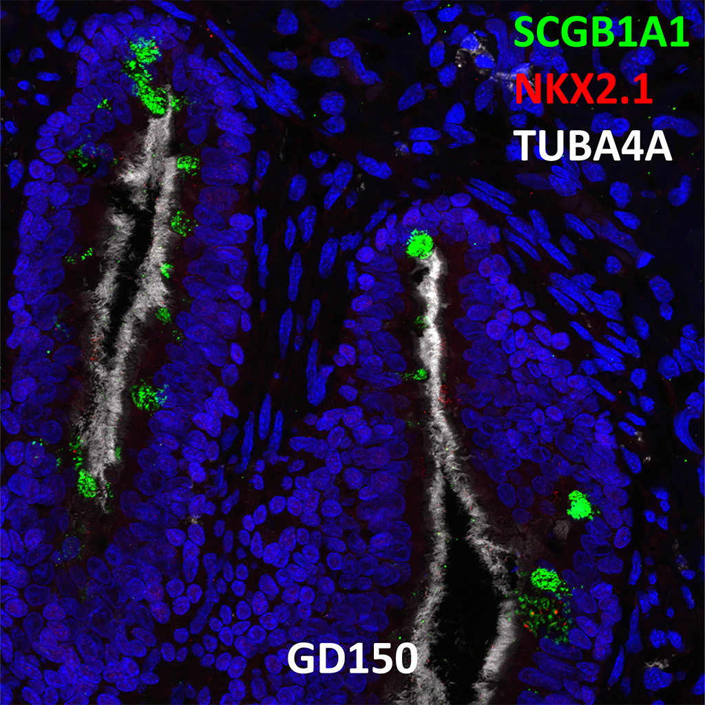
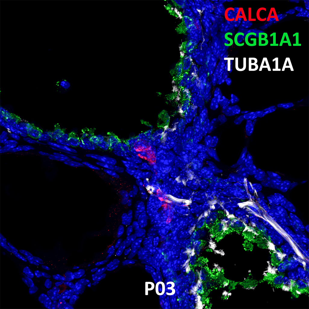
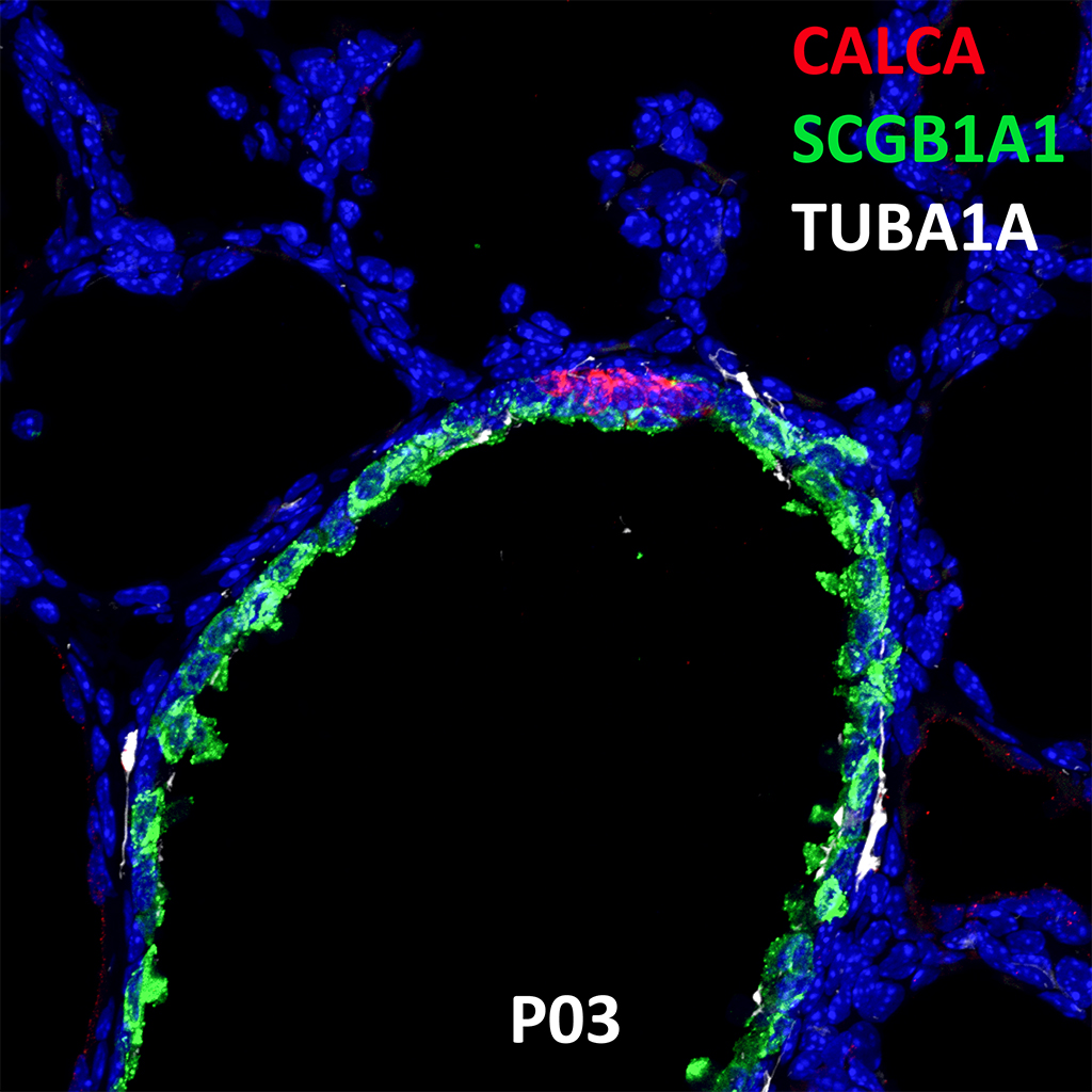
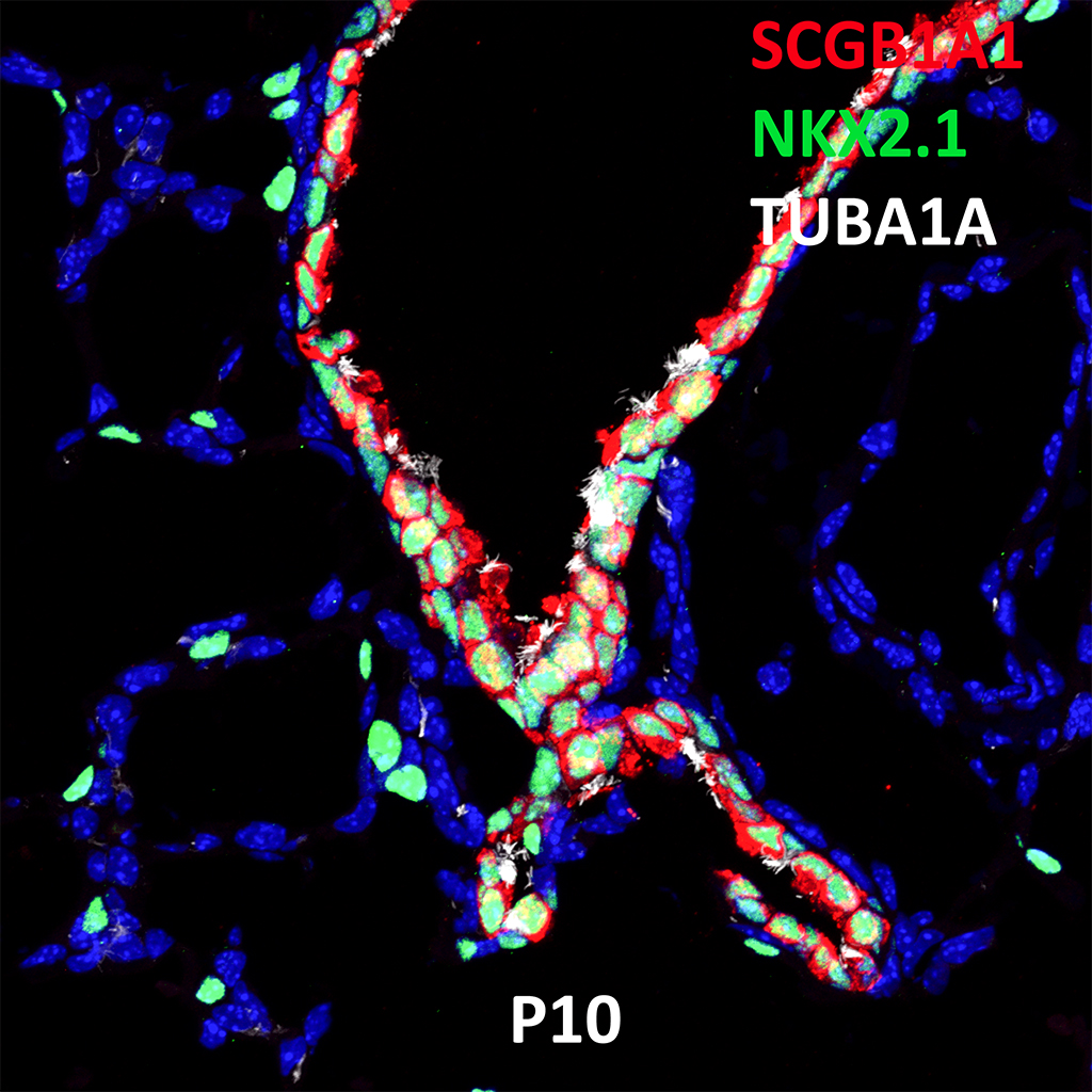
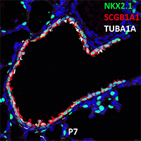
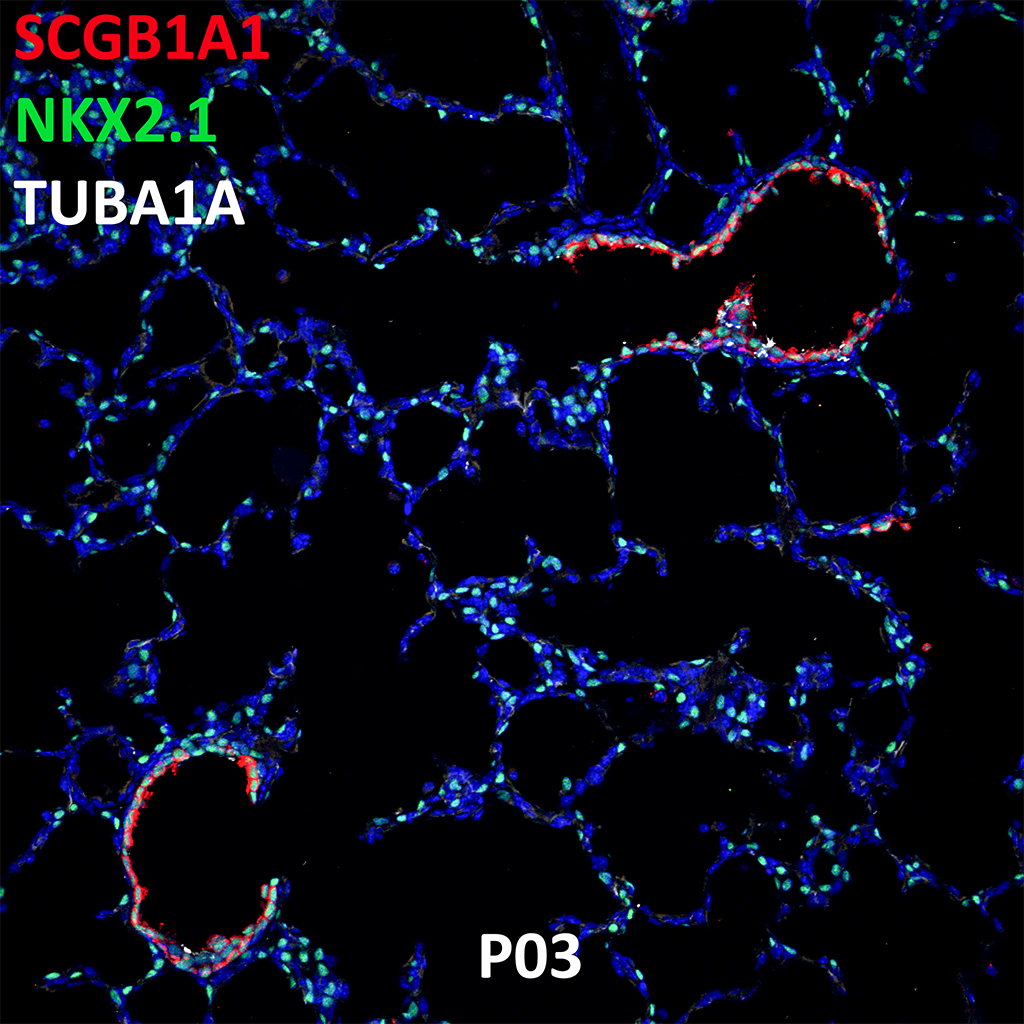
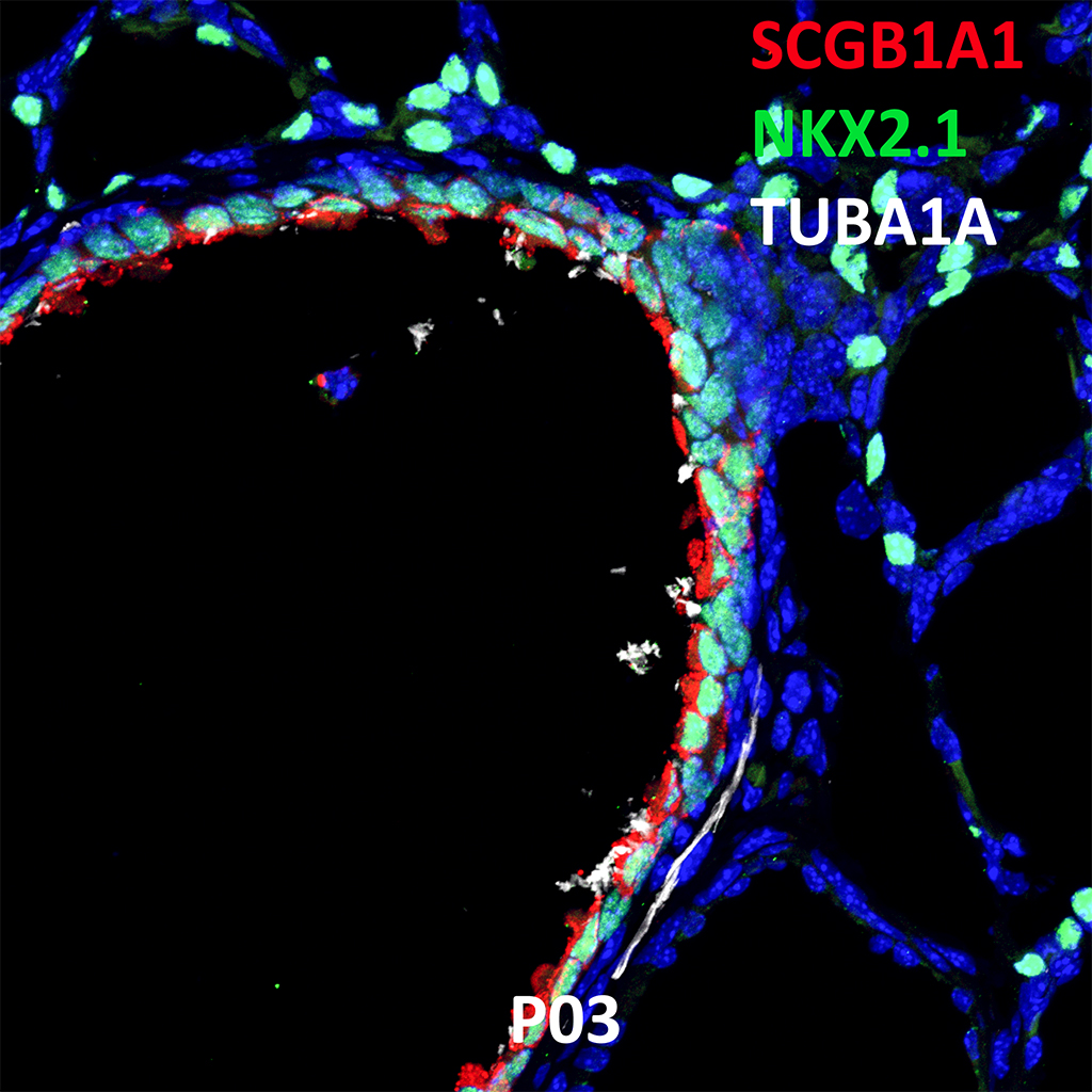
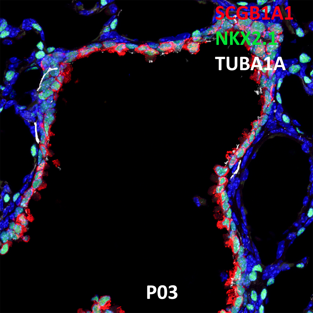
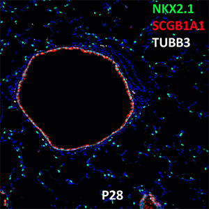
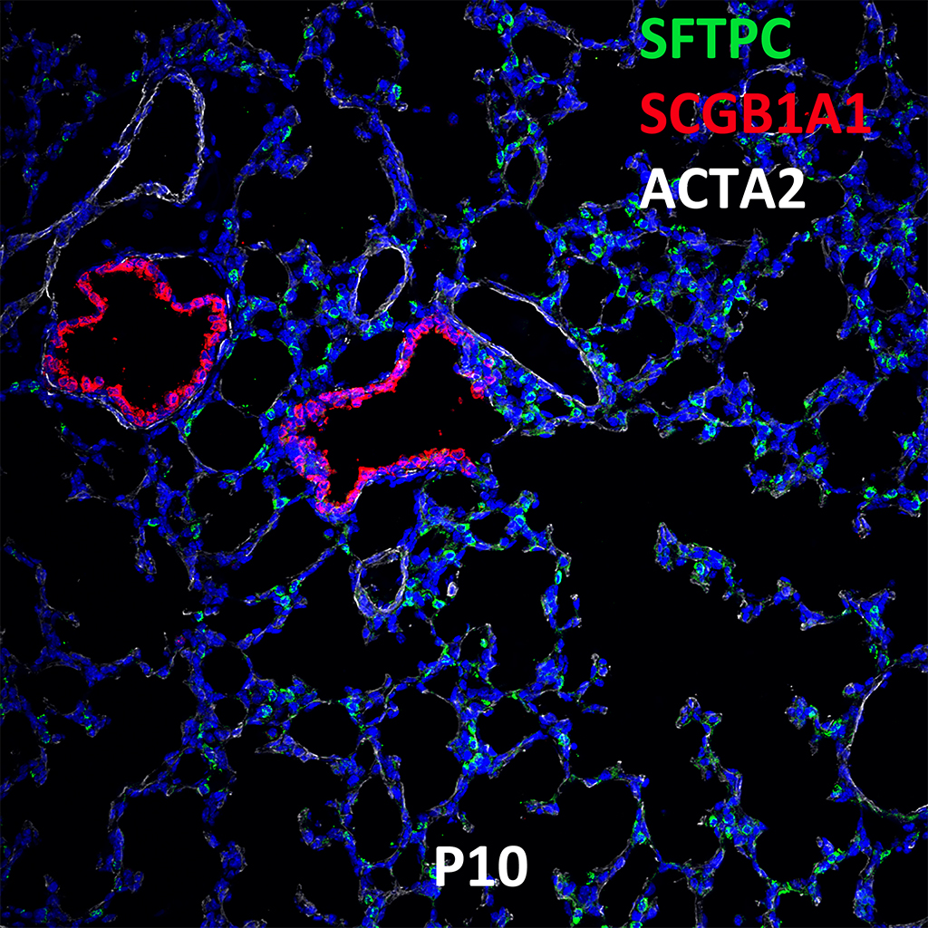
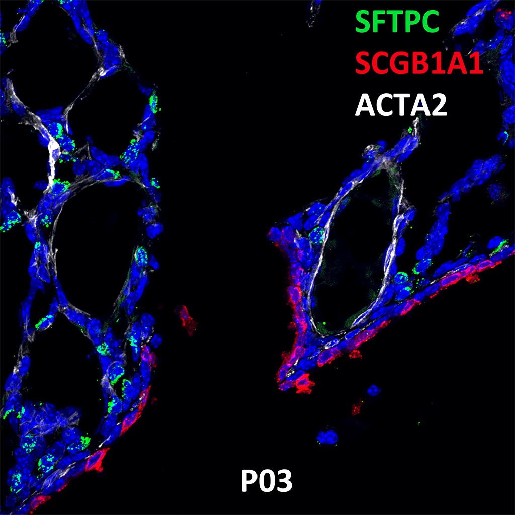
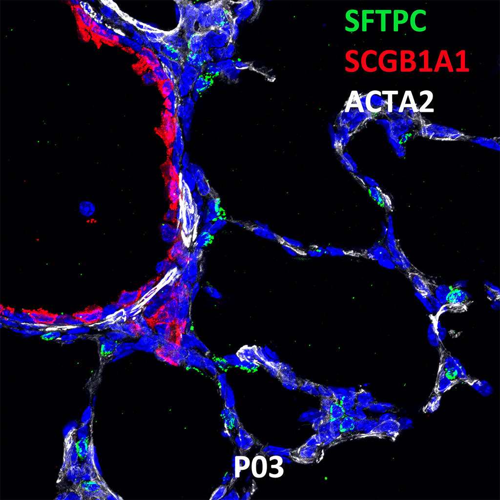
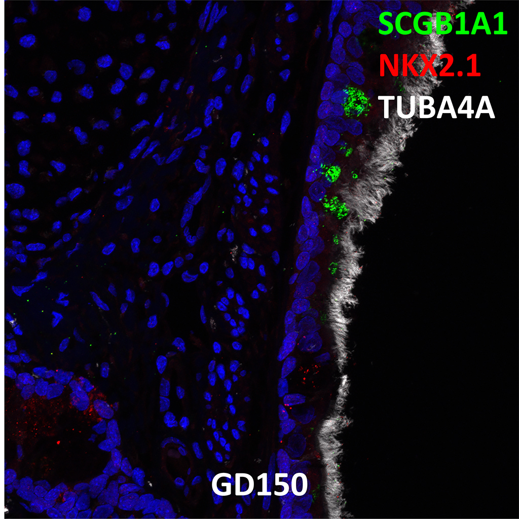
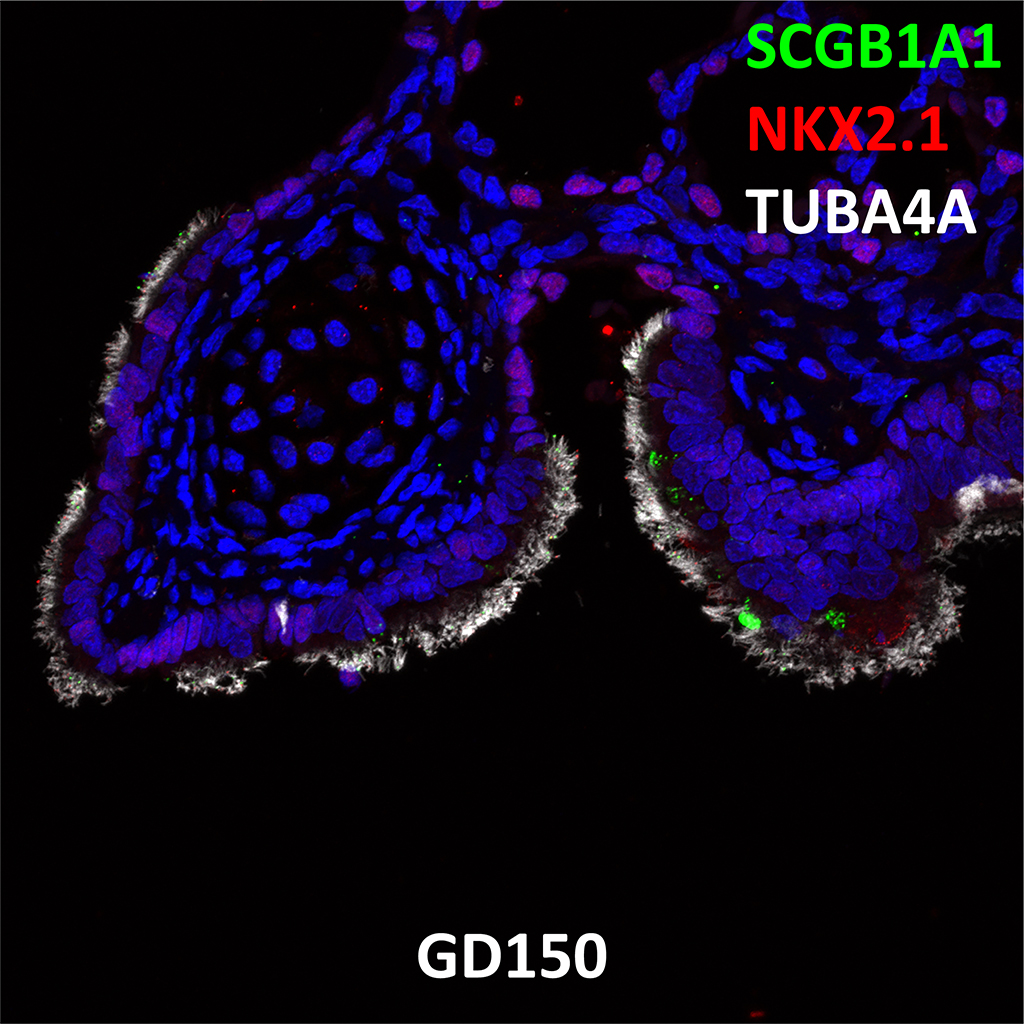
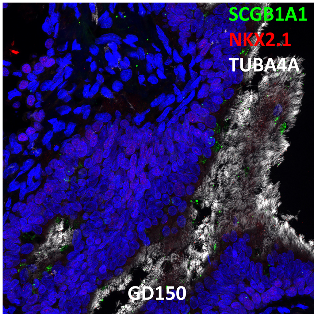
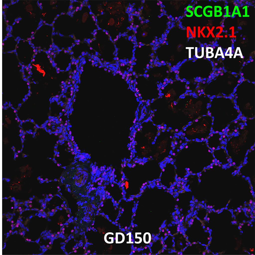
GD150 Fetal Monkey Lung Immunofluorescence and Confocal Imaging Showing Expression of SCGB1A1, NKX2.1, and TUBA4A
60X
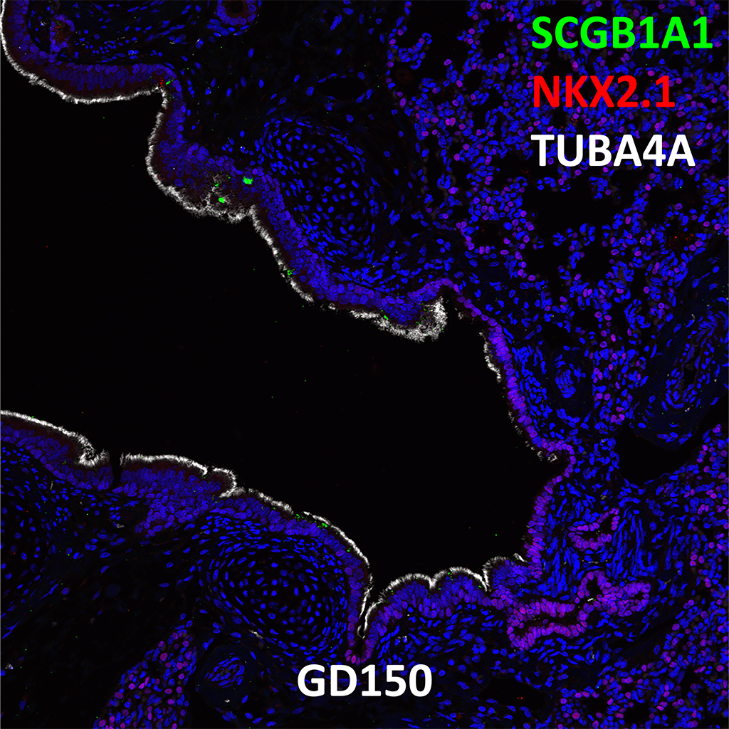
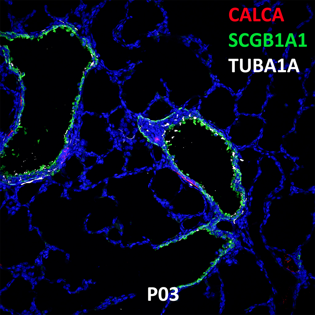
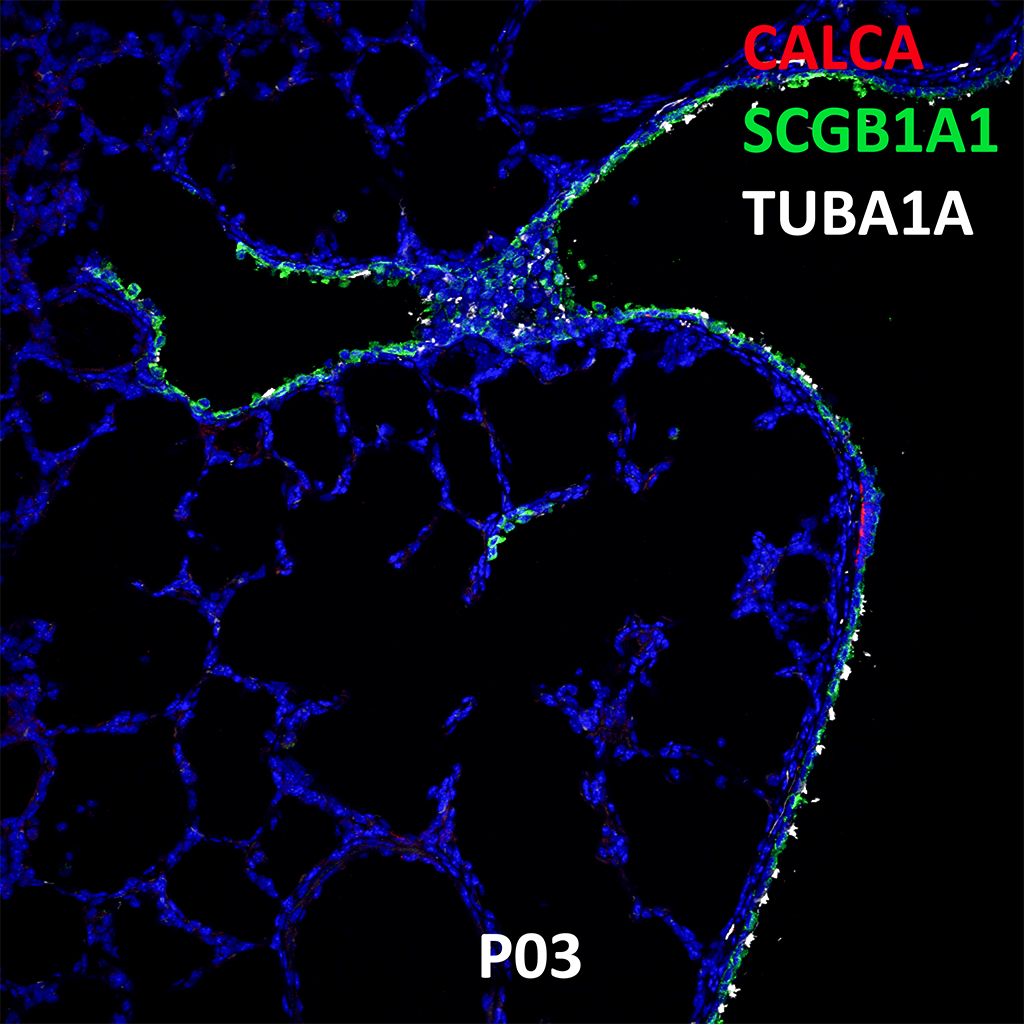
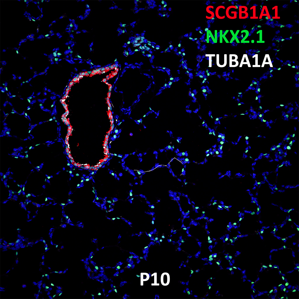
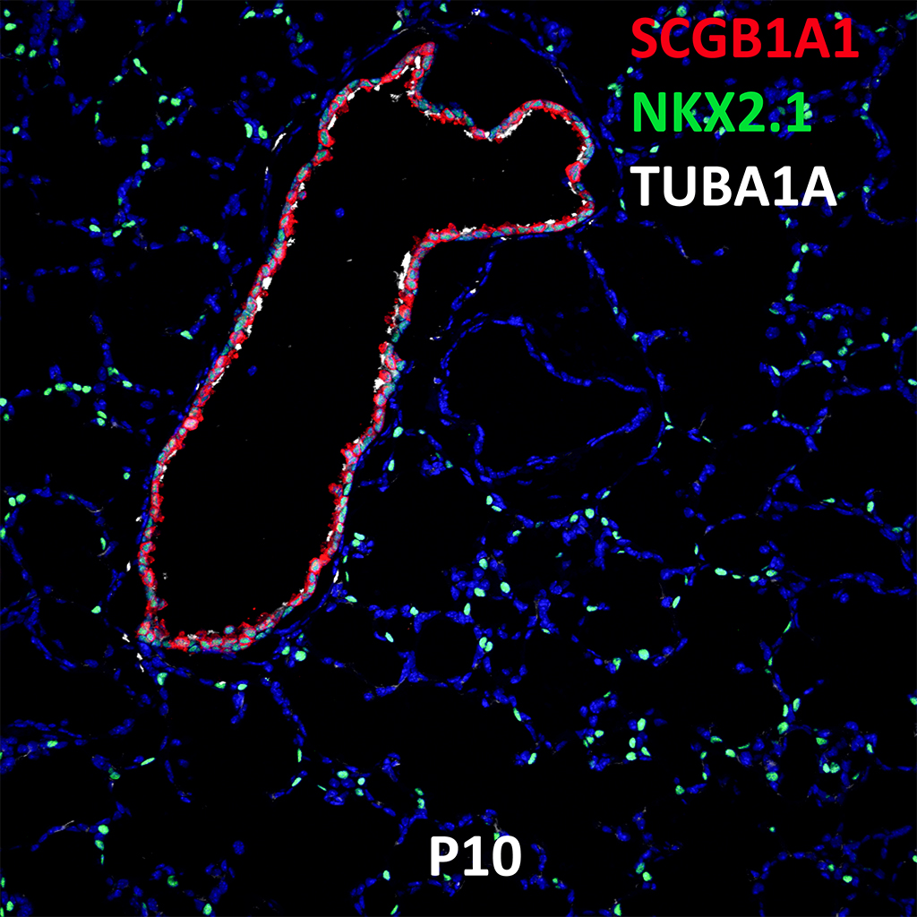
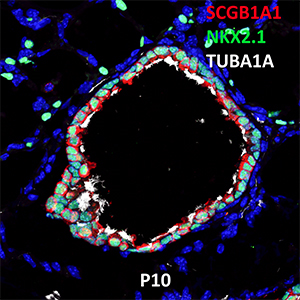
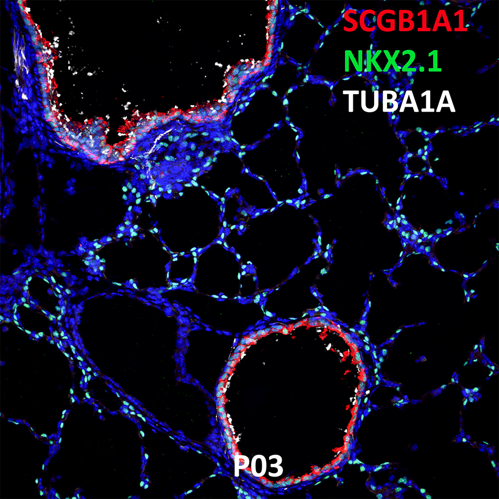
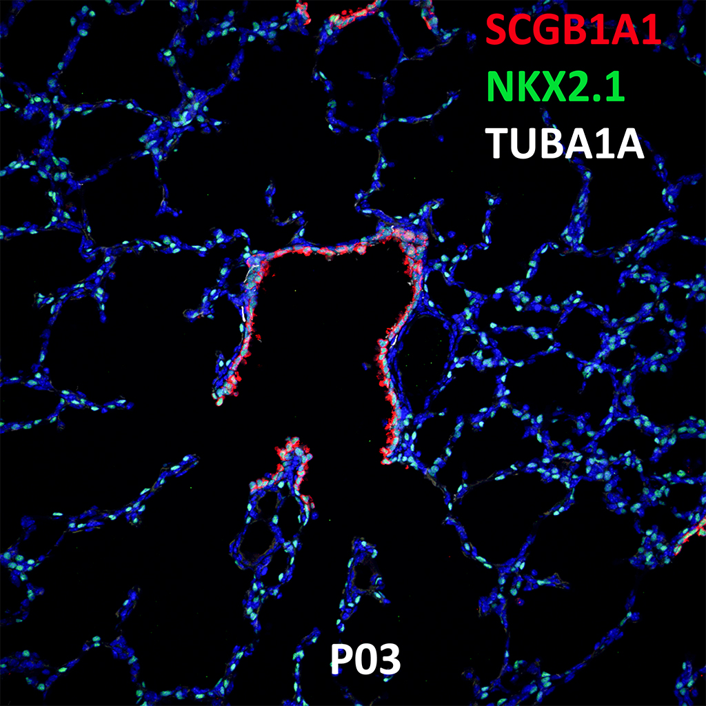
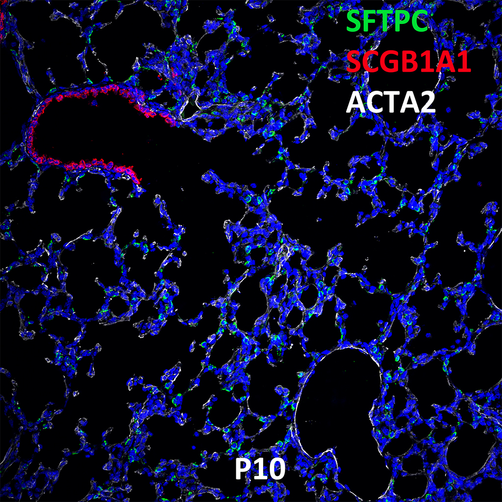
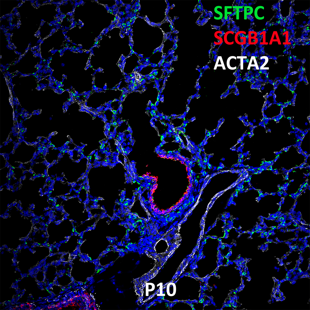
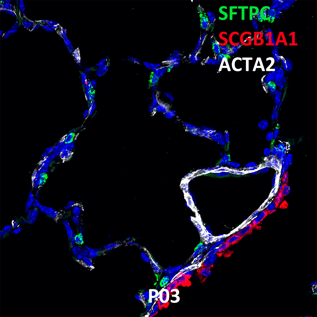
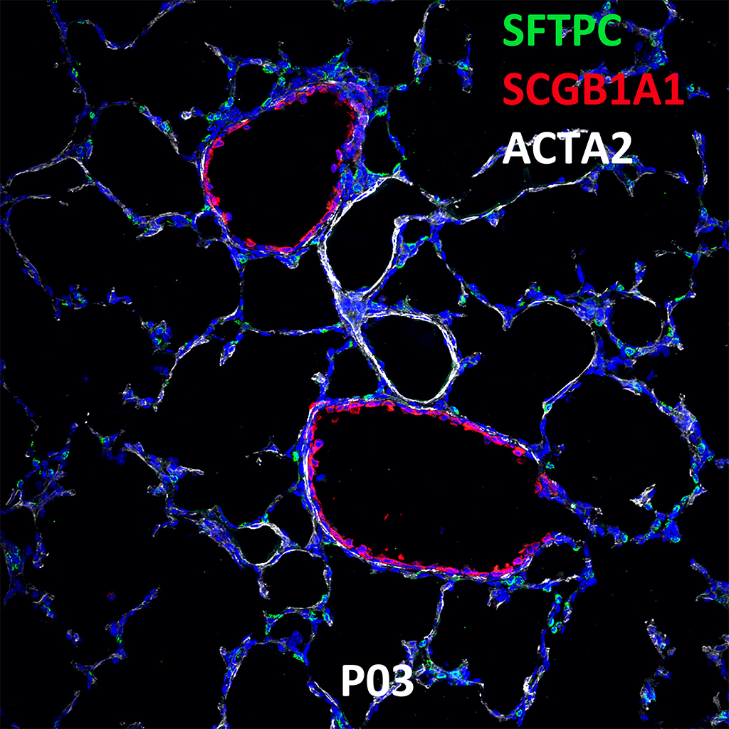
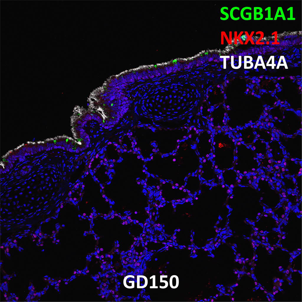
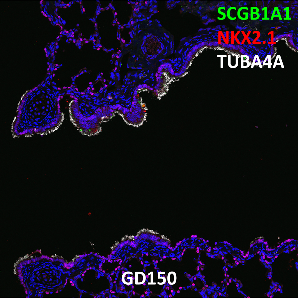
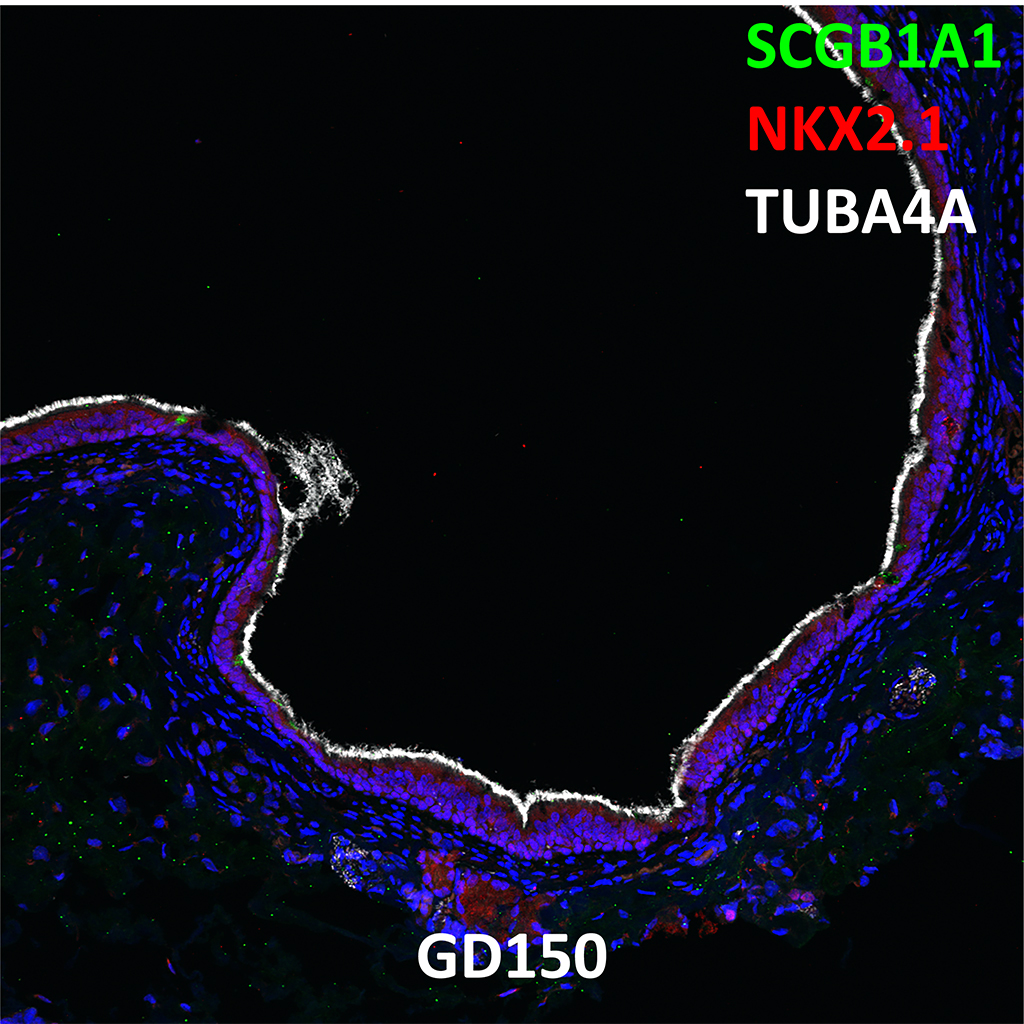
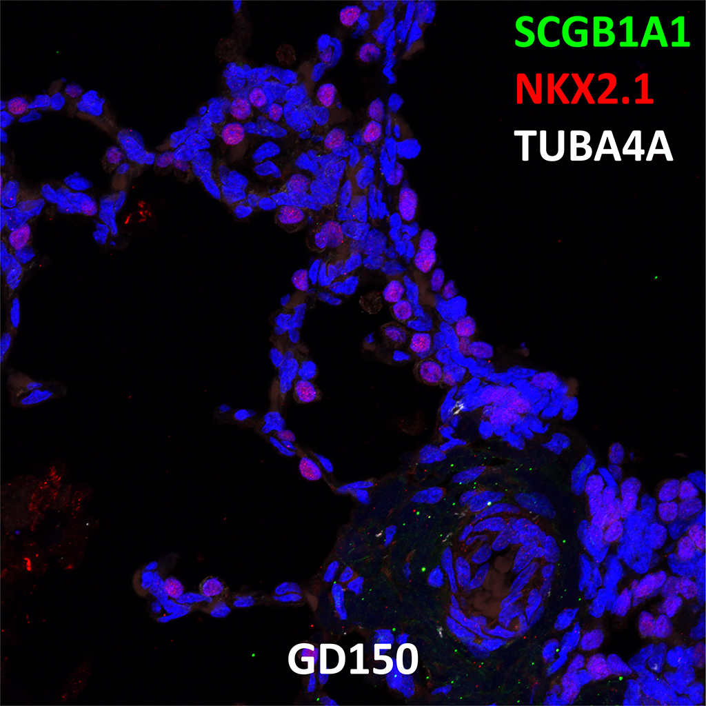
GD150 Fetal Monkey Lung Immunofluorescence and Confocal Imaging Showing Expression of SCGB1A1, NKX2.1, and TUBA4A
20X
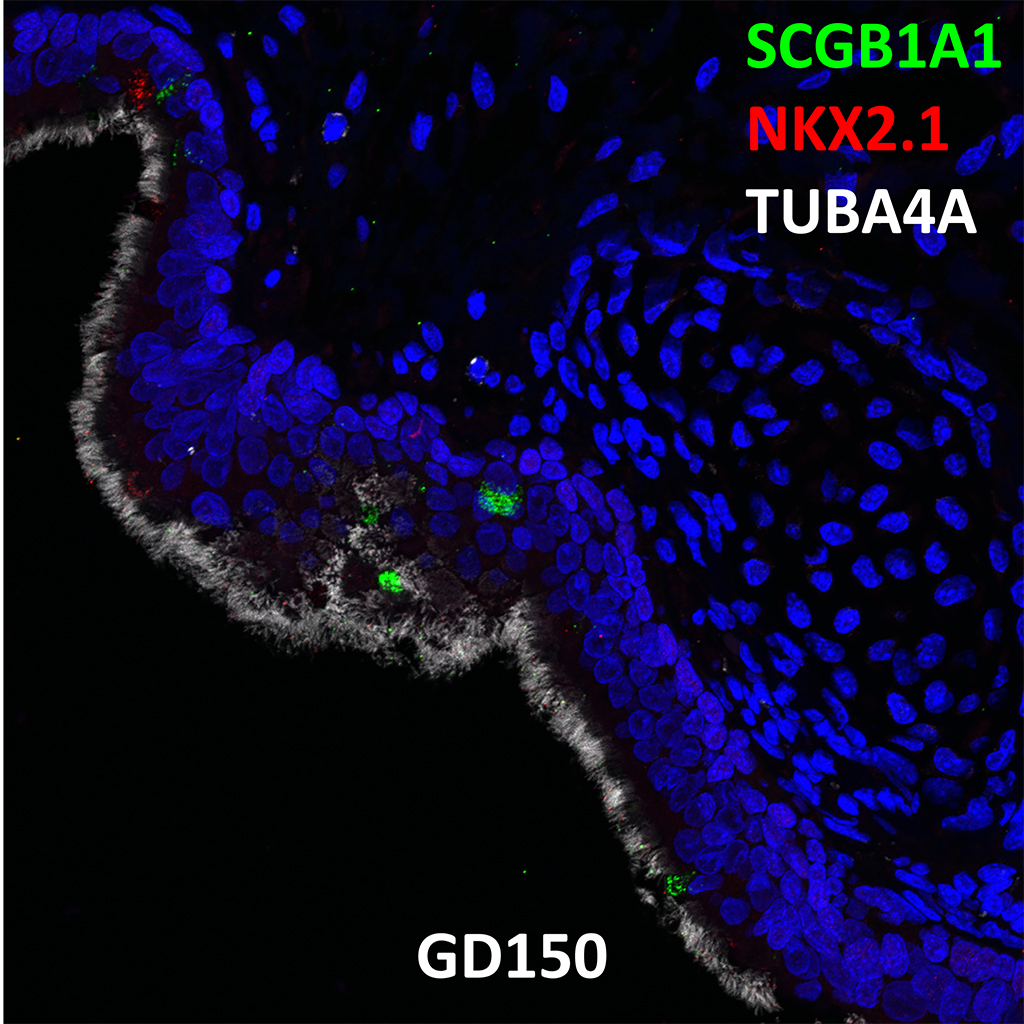
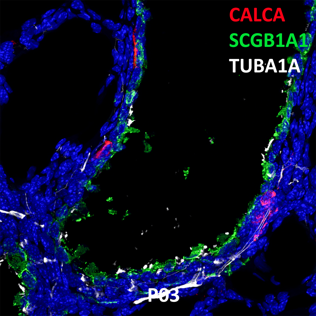
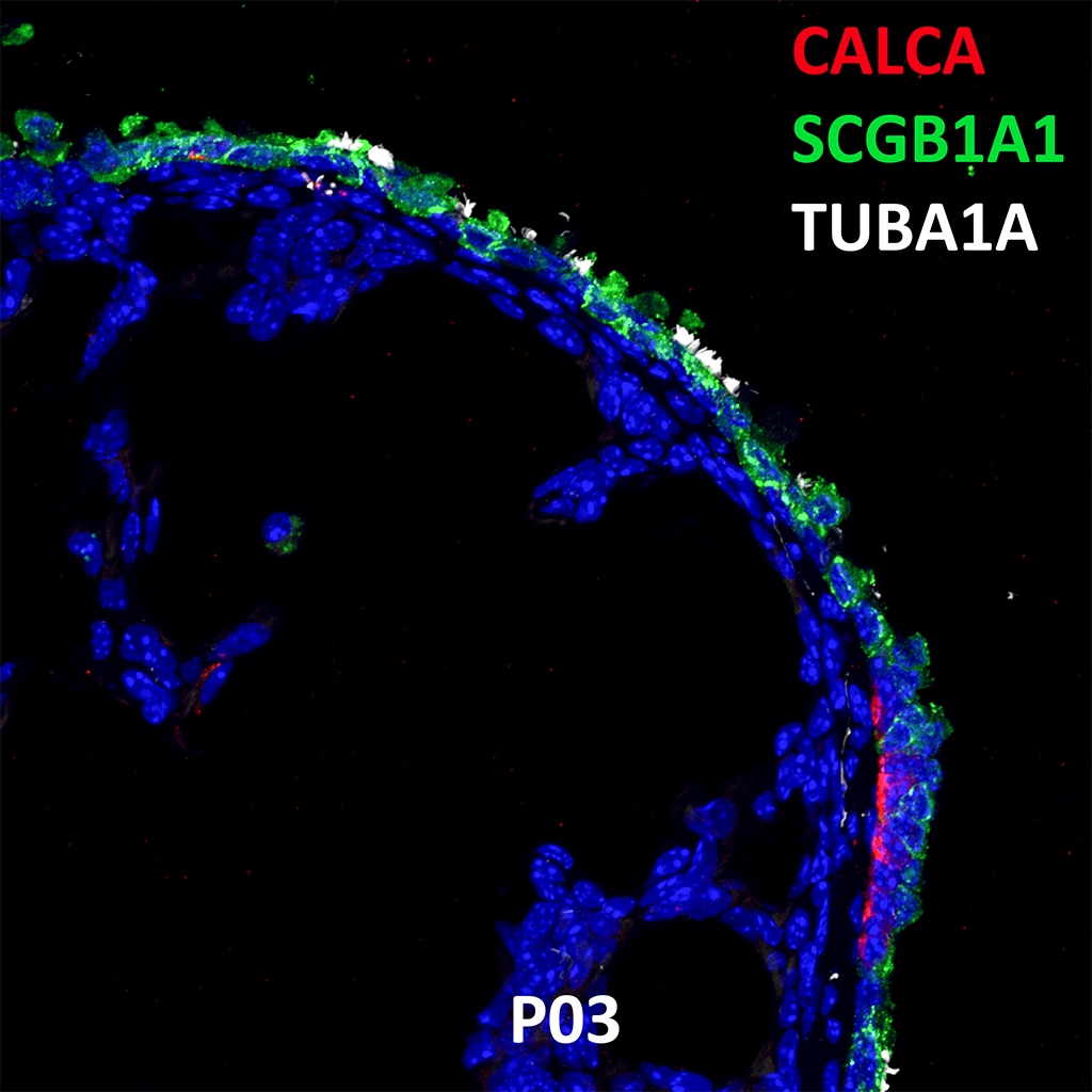
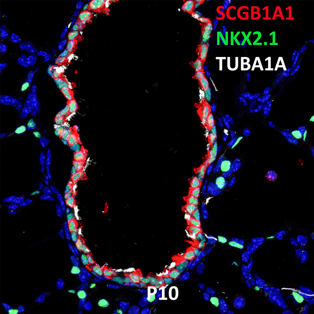
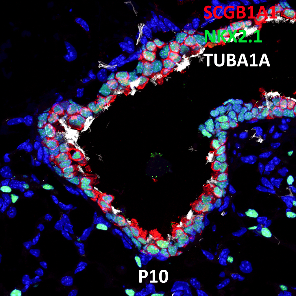
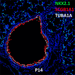
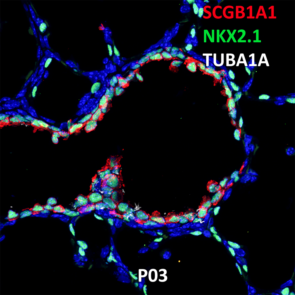
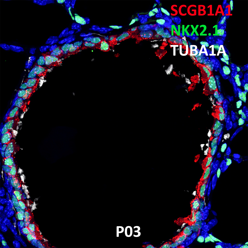
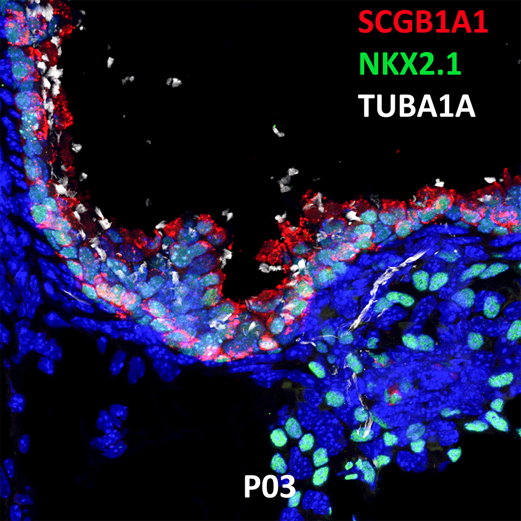
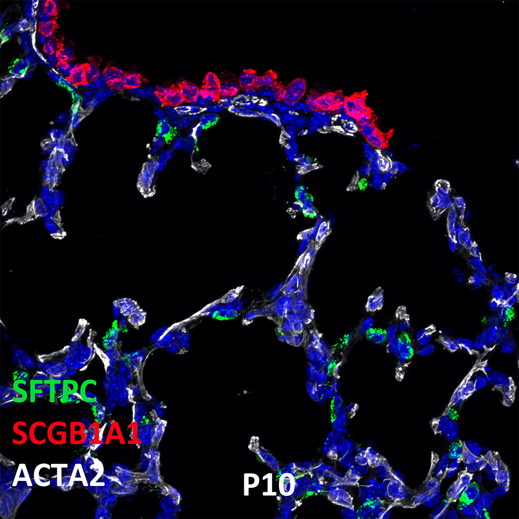
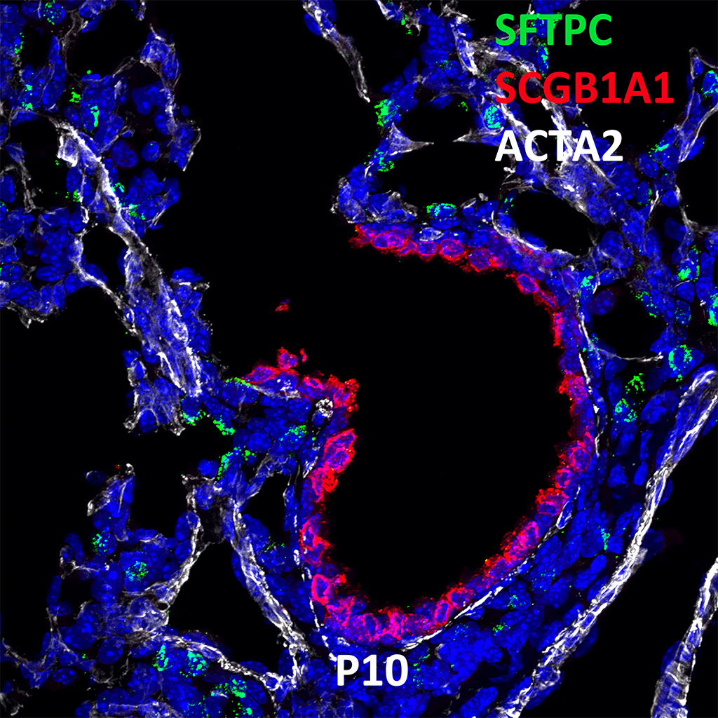
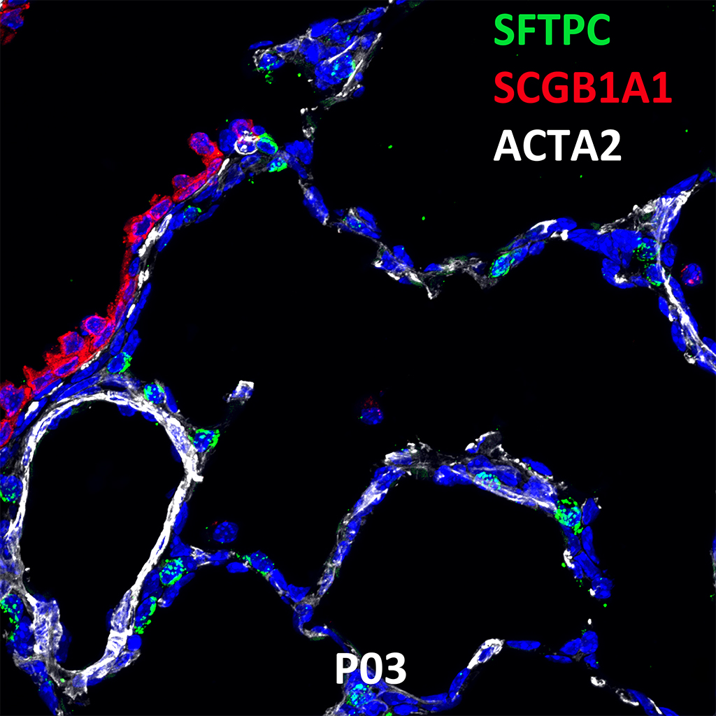
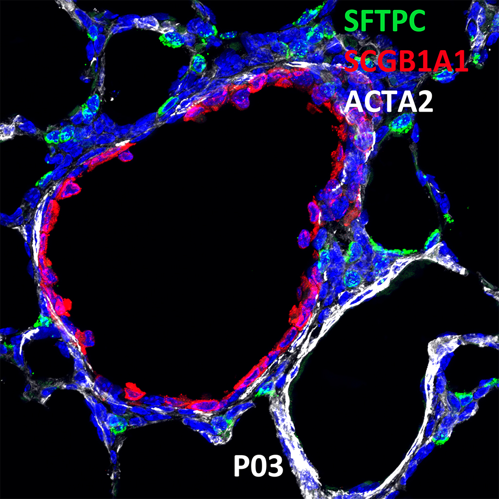
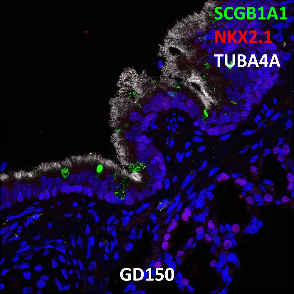
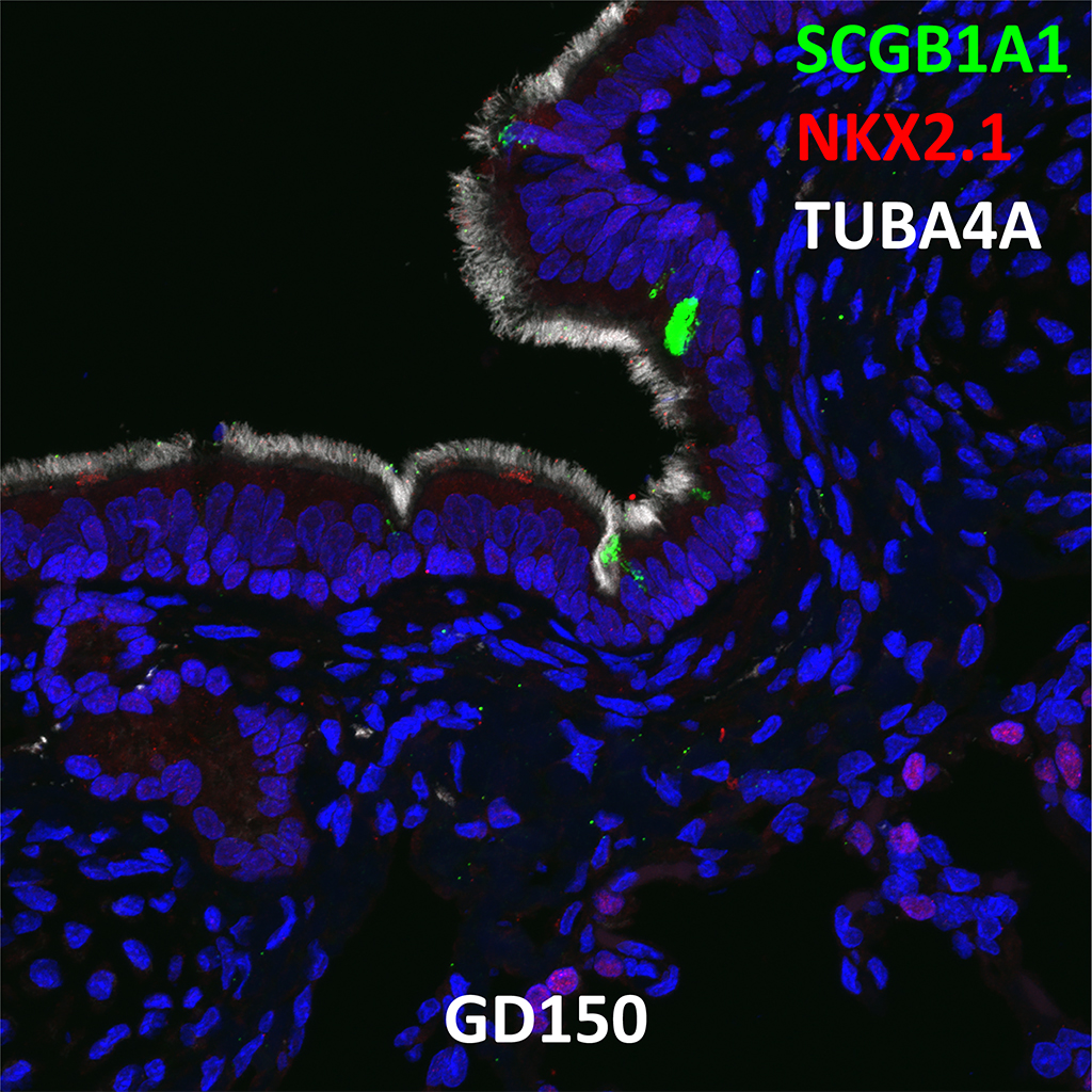
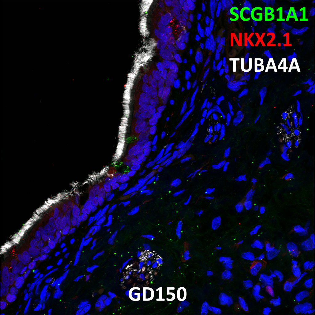
GD150 Fetal Monkey Lung Immunofluorescence and Confocal Imaging Showing Expression of SCGB1A1, NKX2.1, and TUBA4A
60X
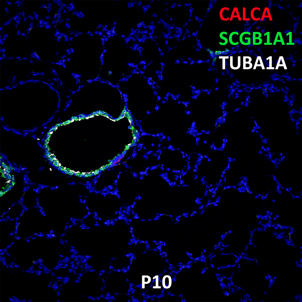
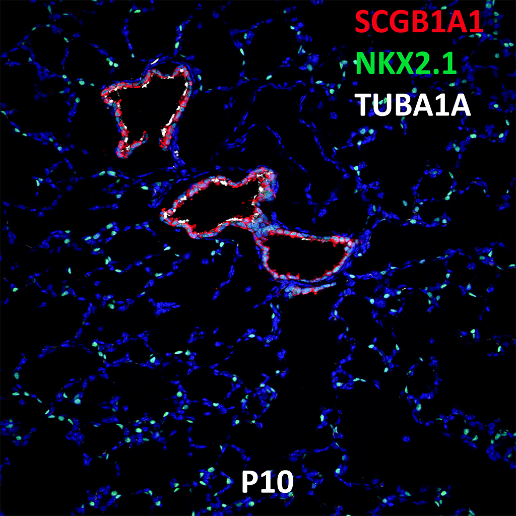
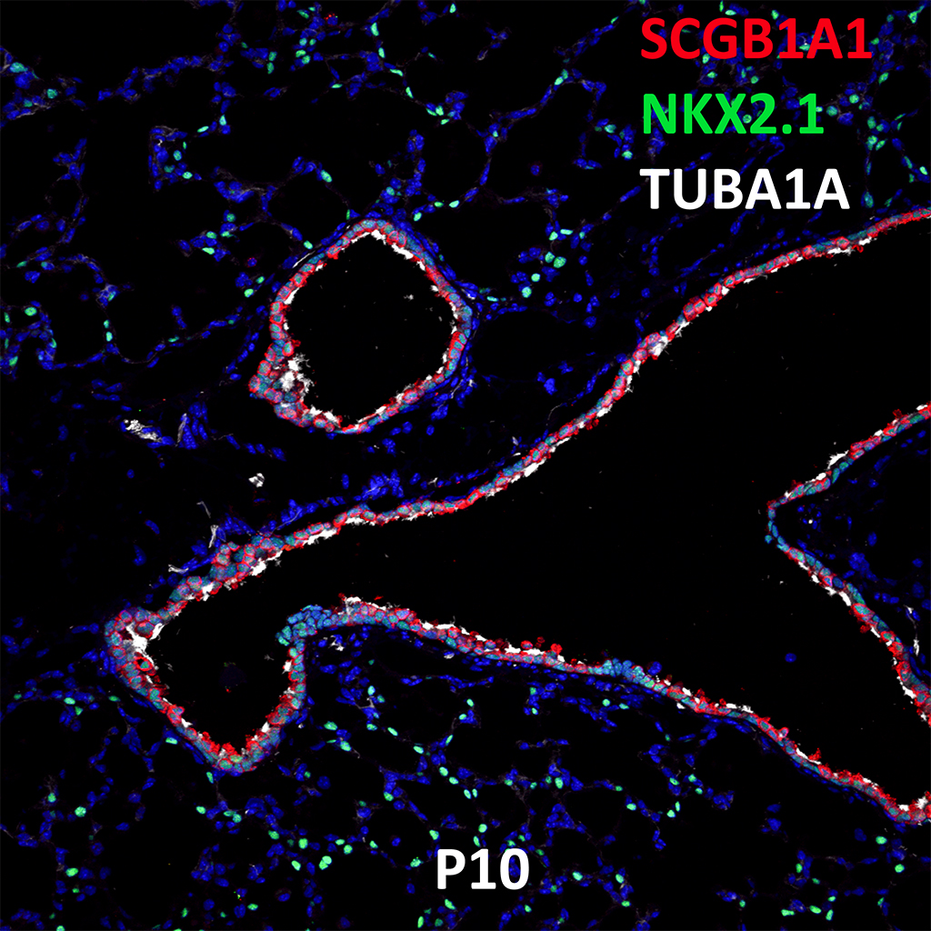
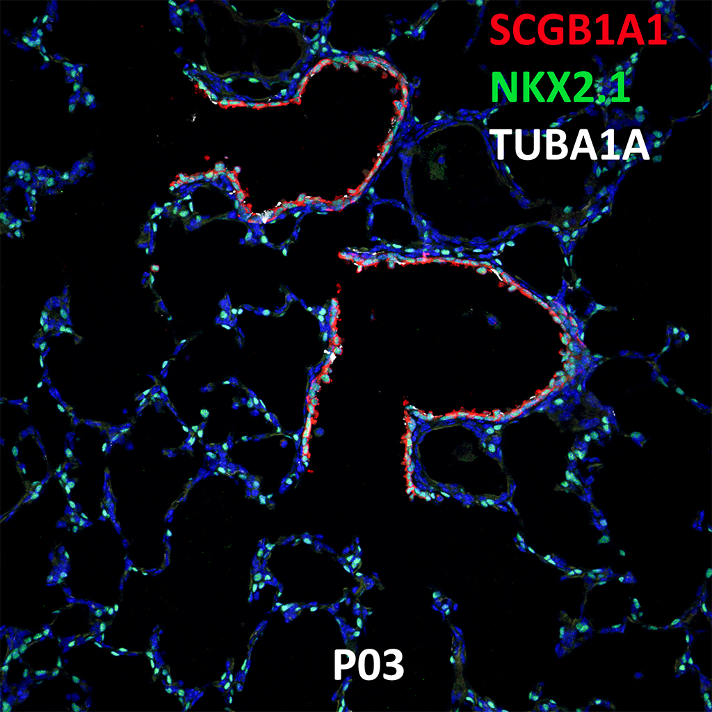
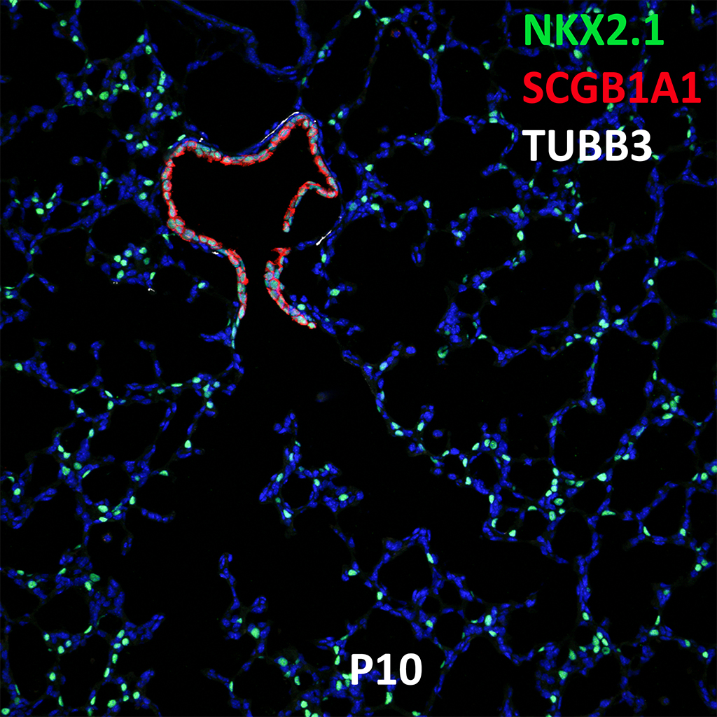
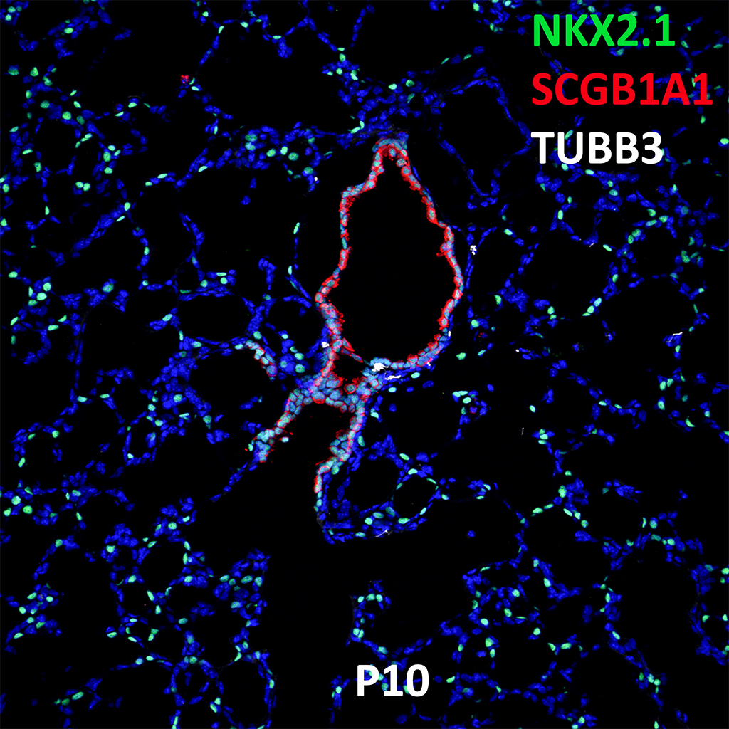
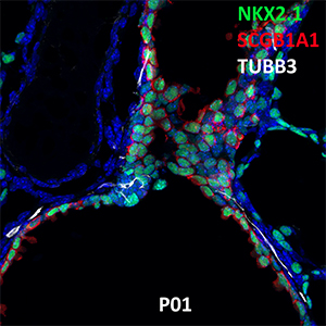
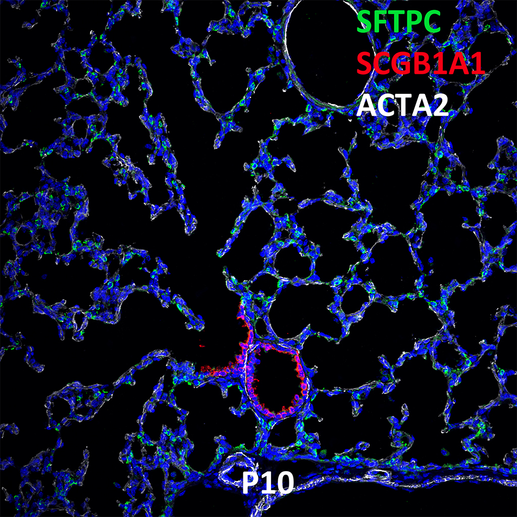
GD150 Fetal Monkey Lung Immunofluorescence and Confocal Imaging Showing Expression of SCGB1A1, NKX2.1, and TUBA4A
20X
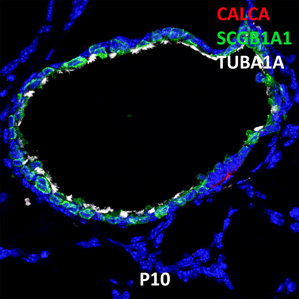
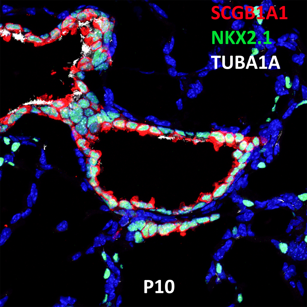
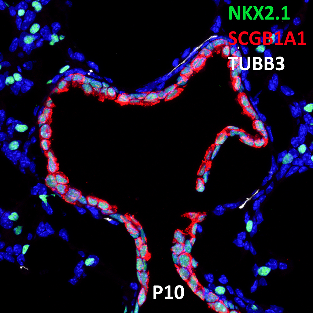
GD150 Fetal Monkey Lung Immunofluorescence and Confocal Imaging Showing Expression of SCGB1A1, NKX2.1, and TUBA4A
60X
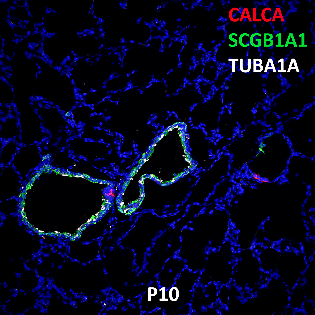
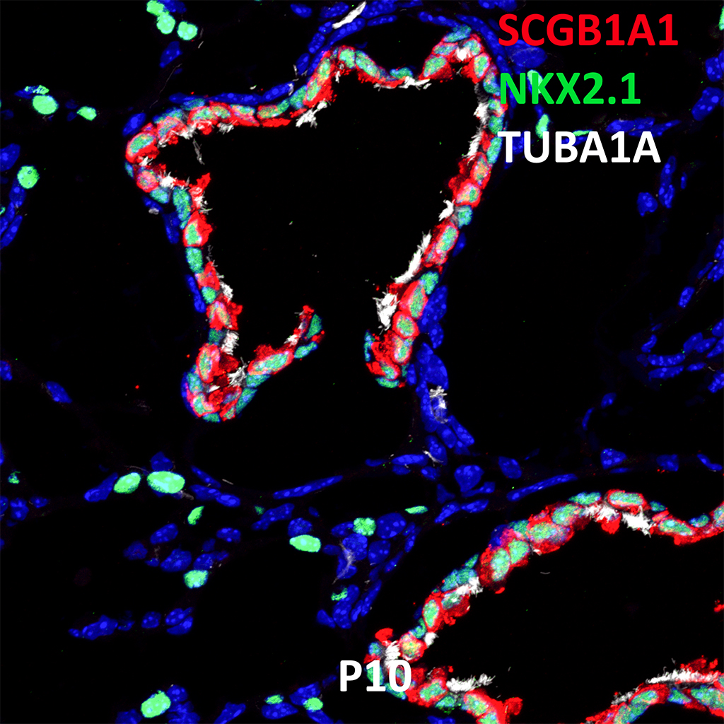
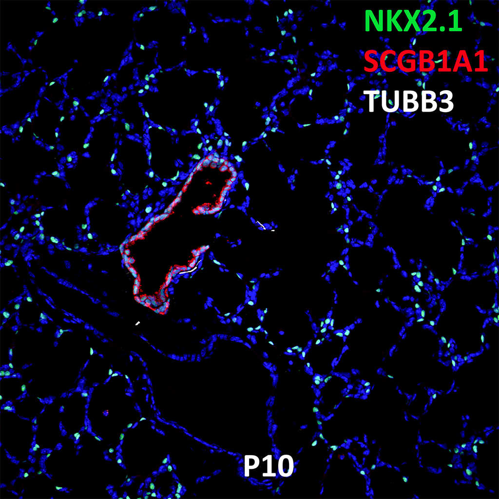
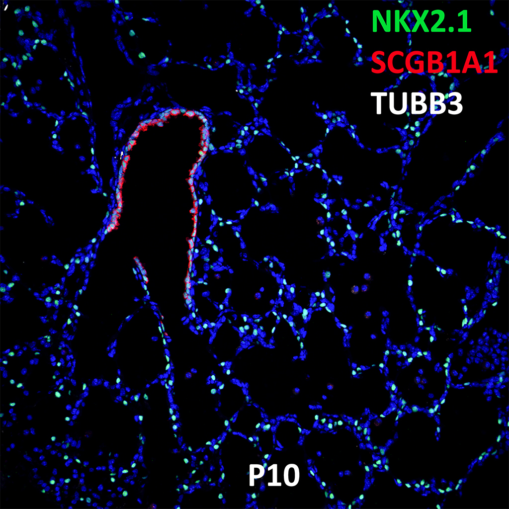
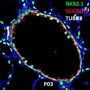
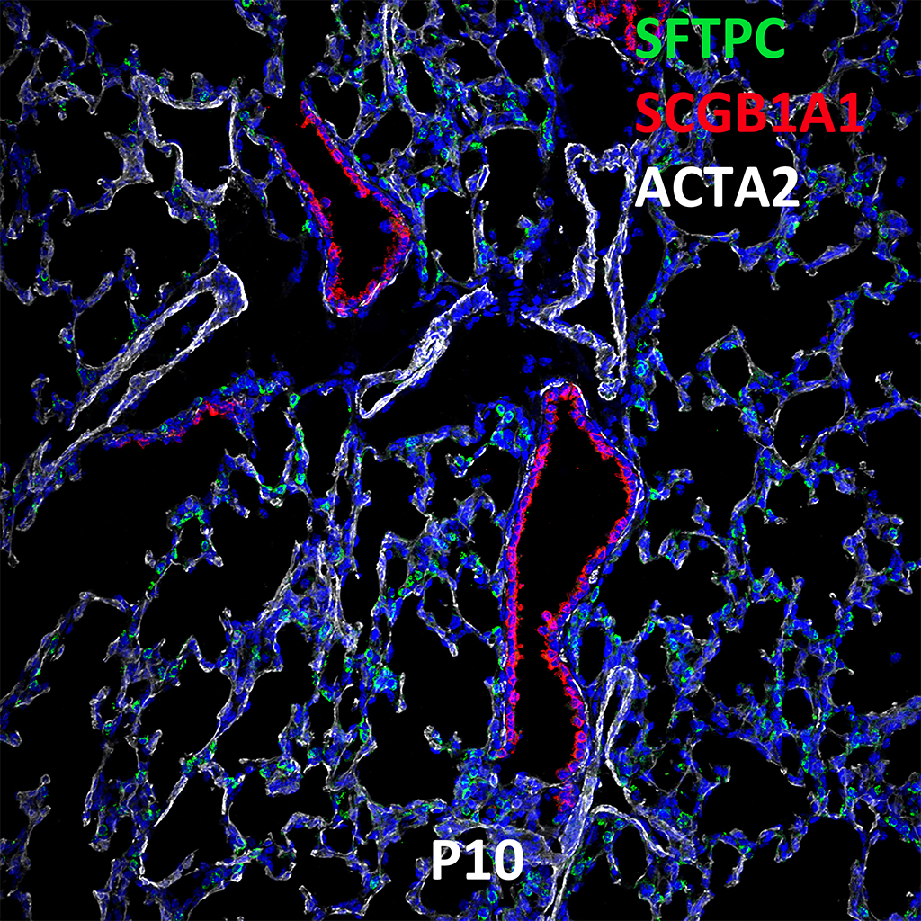
GD150 Fetal Monkey Lung Immunofluorescence and Confocal Imaging Showing Expression of SCGB1A1, NKX2.1, and TUBA4A
20X
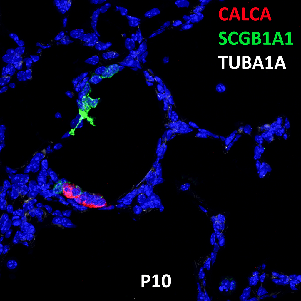
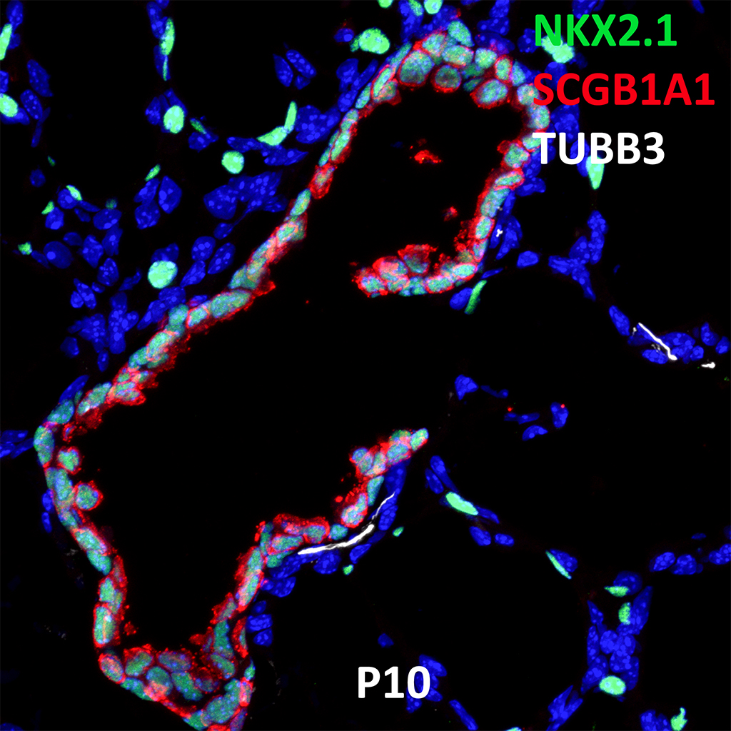
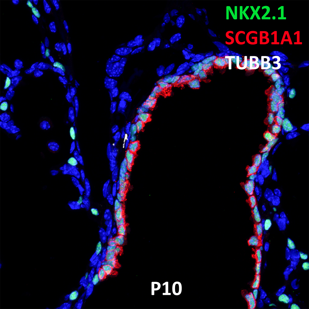
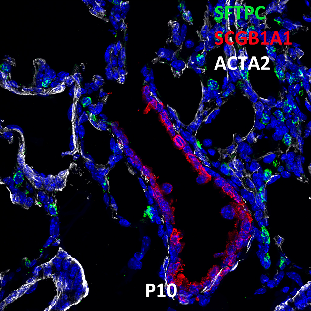
GD150 Fetal Monkey Lung Immunofluorescence and Confocal Imaging Showing Expression of SCGB1A1, NKX2.1, and TUBA4A
60X

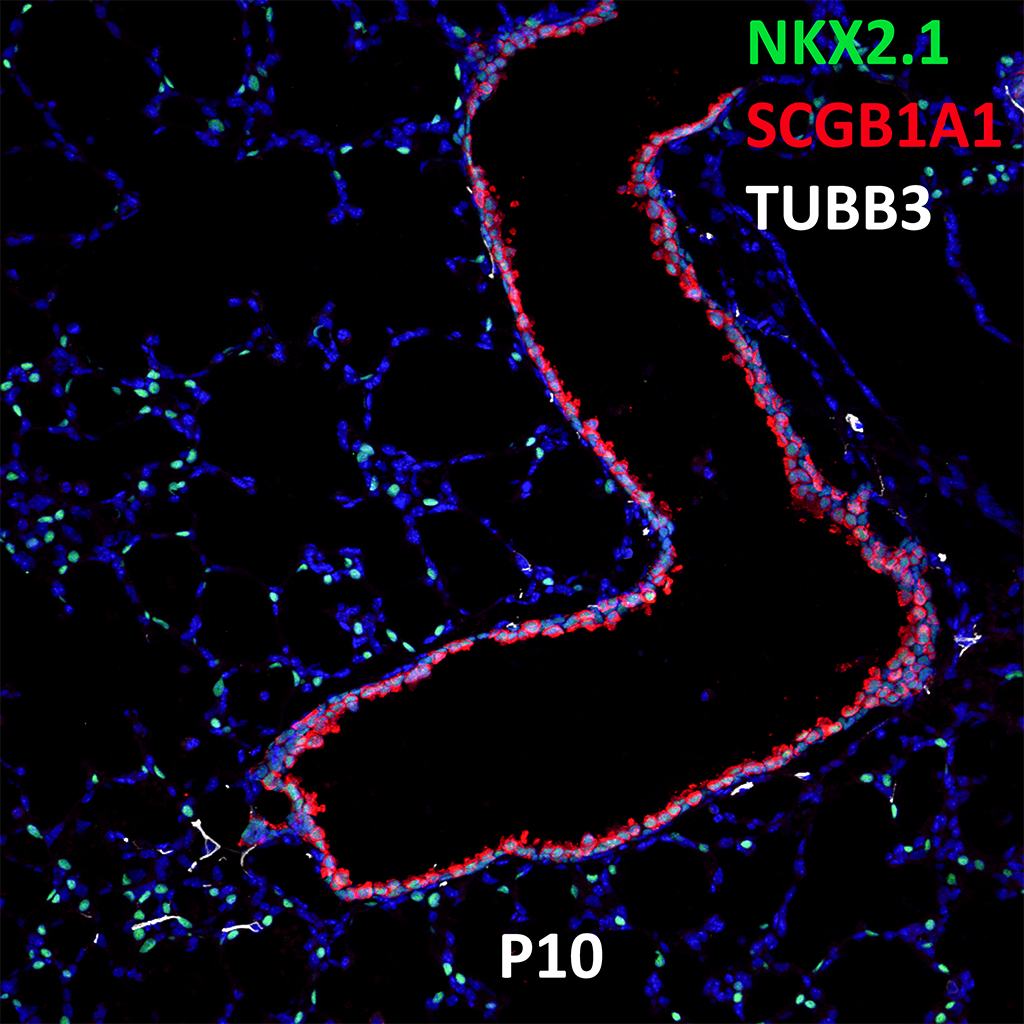
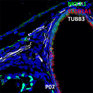
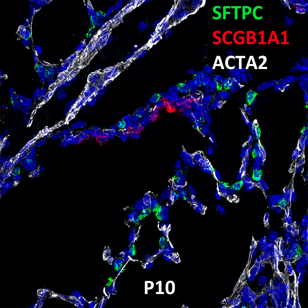
GD150 Fetal Monkey Lung Immunofluorescence and Confocal Imaging Showing Expression of SCGB1A1, NKX2.1, and TUBA4A
20X
GD150 Fetal Monkey Lung Immunofluorescence and Confocal Imaging Showing Expression of SCGB1A1, NKX2.1, and TUBA4A
60X
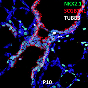
GD150 Fetal Monkey Lung Immunofluorescence and Confocal Imaging Showing Expression of SCGB1A1, NKX2.1, and TUBA4A
20X
GD150 Fetal Monkey Lung Immunofluorescence and Confocal Imaging Showing Expression of SCGB1A1, NKX2.1, and TUBA4A
60X
GD150 Fetal Monkey Lung Immunofluorescence and Confocal Imaging Showing Expression of SCGB1A1, NKX2.1, and TUBA4A
20X

GD150 Fetal Monkey Lung Immunofluorescence and Confocal Imaging Showing Expression of SCGB1A1, NKX2.1, and TUBA4A
60X
GD150 Fetal Monkey Lung Immunofluorescence and Confocal Imaging Showing Expression of SCGB1A1, NKX2.1, and TUBA4A
20X
GD150 Fetal Monkey Lung Immunofluorescence and Confocal Imaging Showing Expression of SCGB1A1, NKX2.1, and TUBA4A
60X
GD150 Fetal Monkey Lung Immunofluorescence and Confocal Imaging Showing Expression of SCGB1A1, NKX2.1, and TUBA4A
20X
GD150 Fetal Monkey Lung Immunofluorescence and Confocal Imaging Showing Expression of SCGB1A1, NKX2.1, and TUBA4A
60X
GD150 Fetal Monkey Lung Immunofluorescence and Confocal Imaging Showing Expression of SCGB1A1, NKX2.1, and TUBA4A
20X

GD150 Fetal Monkey Lung Immunofluorescence and Confocal Imaging Showing Expression of SCGB1A1, NKX2.1, and TUBA4A
60X
GD150 Fetal Monkey Lung Immunofluorescence and Confocal Imaging Showing Expression of SCGB1A1, NKX2.1, and TUBA4A
20X
GD150 Fetal Monkey Lung Immunofluorescence and Confocal Imaging Showing Expression of SCGB1A1, NKX2.1, and TUBA4A
60X
The requested content cannot be found